CELLS,TISSUES, ORGANS AND
SYSTEMS
CourseName: ANATOMY AND PHYSIOLOGY
Lecturer: Nr. DESHE HARRISON HANNANIAH
(RN,RNA,BNSC,BSC (HUMAN ANATOMY), MPH
(Epidemiology),PGDE in view)
Presented to; School of Paediatric Nursing set 2
Date: 23-01-2026
1
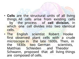
• Cells arethe structural units of all living
things. All cells arise from existing cells
by the process of cell division, in
which one cell divides into two identical
cells.
• The English scientist Robert Hooke
first observed plant cells with a crude
microscope in the late 1600s. Then, in
the 1830s two German scientists,
Matthias Schleiden and Theodor
Schwann, proposed that all living things
are composed of cells.
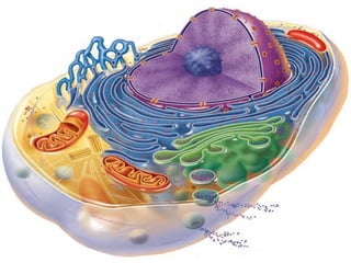
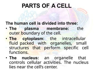
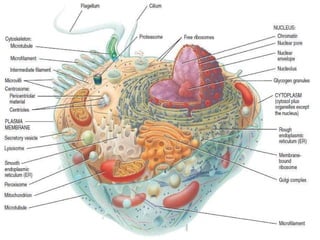
PARTS OF ACELL
The human cell is divided into three:
• The plasma membrane: the
outer boundary of the cell.
• The cytoplasm: the intracellular
fluid packed with organelles, small
structures that perform specific cell
functions.
• The nucleus: an organelle that
controls cellular activities. The nucleus
lies near the cell’s center.
8.
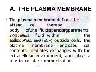
A. THE PLASMAMEMBRANE
• The plasma membrane defines the
extent cell
,
thereby
separating
intracellular fluid within
cells
of a
body of the fluid compartments
the
the
Extracellular fluid (ECF) outside cells. The
plasma membrane encloses cell
contents, mediates exchanges with the
extracellular environment, and plays a
role in cellular communication.
10.
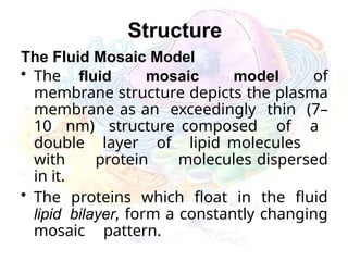
Structure
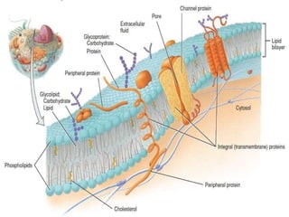
The Fluid MosaicModel
• The fluid mosaic model of
membrane structure depicts the plasma
membrane as an exceedingly thin (7–
10 nm) structure composed of a
double layer of lipid molecules
with protein molecules dispersed
in it.
• The proteins which float in the fluid
lipid bilayer, form a constantly changing
mosaic pattern.
11.
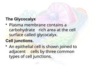
The Glycocalyx
• Plasmamembrane contains a
carbohydrate rich area at the cell
surface called glycocalyx.
Cell junctions.
• An epithelial cell is shown joined to
adjacent cells by three common
types of cell junctions.
12.
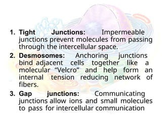
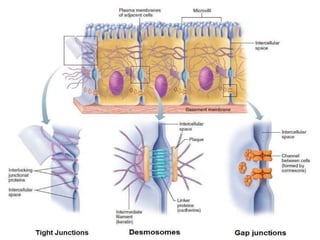
1. Tight Junctions:Impermeable
junctions prevent molecules from passing
through the intercellular space.
2. Desmosomes: Anchoring junctions
bind adjacent cells together like a
molecular “Velcro” and help form an
internal tension reducing network of
fibers.
3. Gap junctions: Communicating
junctions allow ions and small molecules
to pass for intercellular communication
14.
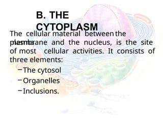
B. THE
CYTOPLASM
The cellularmaterial between the
plasma
membrane and the nucleus, is the site
of most cellular activities. It consists of
three elements:
–The cytosol
–Organelles
–Inclusions.
15.
1. The cytosol:it is the
viscous, semitransparent fluid in
which the other cytoplasmic elements
are suspended.
2. The organelles are the metabolic
machinery of the cell. Each type of
organelle carries out a specific
function for the cell.
3. The inclusions are chemical
substances that may or may not be
present, depending on cell type.
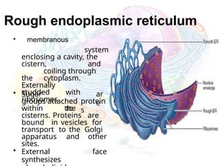
Rough endoplasmic reticulum
•membranous
system
enclosing a cavity, the
cistern, and
coiling through
the cytoplasm.
Externally
studded with
ribosomes.
ar
e
• Sugar
groups attached
to
protein
s
within the
cisterns. Proteins are
bound in vesicles for
transport to the Golgi
apparatus and other
sites.
• External face
synthesizes
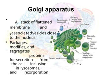
Golgi apparatus
•
A stackof flattened
membrane and
sassociatedvesicles close
to the nucleus.
• Packages,
modifies, and
segregates
proteins
for secretion from
the cell, inclusion
in lysosomes,
and incorporation
21.
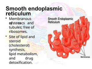
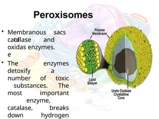
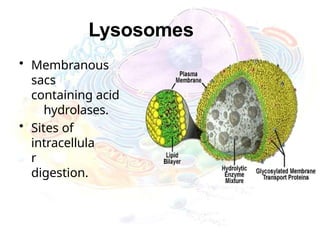
Peroxisomes
• Membranous sacs
of
catalaseand
oxidas enzymes.
e
• The enzymes
detoxify a
number of toxic
substances. The
most important
enzyme,
catalase, breaks
down hydrogen
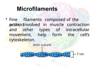
Microfilaments
• Fine filamentscomposed of the
protein
actin. Involved in muscle contraction
and other types of intracellular
movement, help form the cell’s
cytoskeleton.
25.
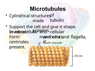
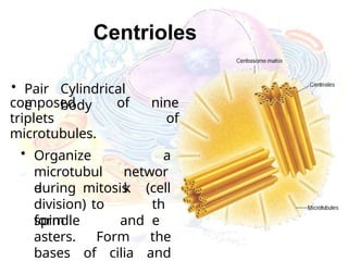
Centrioles
• Pair
e
Cylindrical
body
composed ofnine
triplets of
microtubules.
• Organize
microtubul
e
a
networ
k
during mitosis
division) to
form
(cell
th
e
spindle and
asters. Form the
bases of cilia and
26.
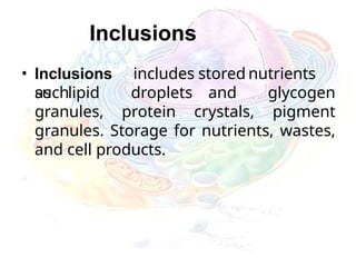
Inclusions
• Inclusions includesstored nutrients
such
as lipid droplets and glycogen
granules, protein crystals, pigment
granules. Storage for nutrients, wastes,
and cell products.
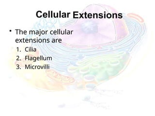
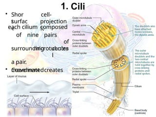
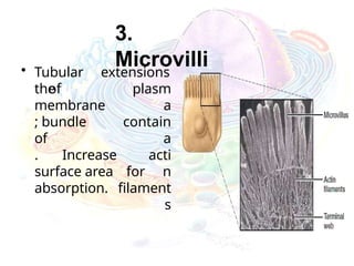
1. Cili
• Shor
t
cell-
surfac
e
projection
s;
eachcilium composed
of nine pairs
of
microtubules
centra
l
surrounding
a pair.
• Coordinated
movement creates
a
unidirection
al that
curren
t
propel
s
substances across
cell surfaces.
29.
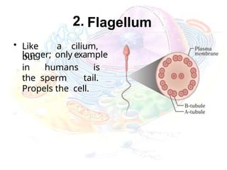
2. Flagellum
• Likea cilium,
but
longer; only example
in humans is
the sperm tail.
Propels the cell.
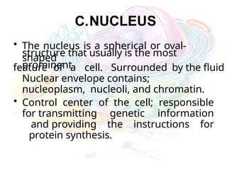
C.NUCLEUS
• The nucleusis a spherical or oval-
shaped
structure that usually is the most
prominent
feature of a cell. Surrounded by the fluid
Nuclear envelope contains;
nucleoplasm, nucleoli, and chromatin.
• Control center of the cell; responsible
for transmitting genetic information
and providing the instructions for
protein synthesis.
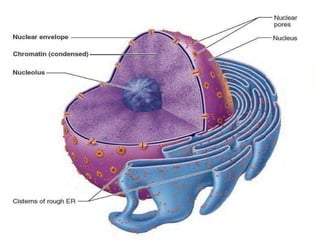
Nuclear Envelope
• Thenuclear envelope is a
double-
layered membrane perforated
with pores, which control the flow of
material going in and out of the
nucleus.
• The outer layer is connected to
the endoplasmic reticulum,
communicating with the cytoplasm
of the cell. The exchange of the
large molecules (protein and RNA)
between the nucleus and
cytoplasm happens here.
35.
Nucleoplasm
• A jelly-like(made mostly of water)
matrix within the nucleus
• All the other materials “float” inside
• Helps the nucleus keep its shape
• Serves as the medium for the
transportation of important
molecules within the nucleus.
36.
Chromatin
• Chromatin appearsas a fine,
unevenly
stained network, but special
techniques reveal it as a system of
bumpy threads weaving through the
nucleoplasm.
– Chromatin is composed of approximately
– 30% DNA, our genetic material
– 60% globular histone proteins
which package and regulate the DNA
– 10% RNA chains, newly formed or forming
37.
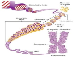
Chromosomes
• Chromosomes containDNA in a condensed
form attached to a histone protein.
• Chromatin is comprisedof DNA. There are
two types based on function.
38.
Functions
• The nucleusis often compared to
the “command center,” as it
controls all functions of the cell.
• It is important in regulating the
actions of the cells.
• It plays an important part in
creating the cell’s proteins.
• It is involved in important
processes dealing with DNA
and other genetic molecules.
39.
DNA
• DNA ordeoxyribonucleic acid,
contains the information needed for
the creation of proteins (which
include enzymes and hormones)
and is stored in the nucleus, as
already said, in the form of chromatin
or chromosomes.
• The nucleus is the site of DNA
duplication, which is needed for
cell division (mitosis) and organism
reproduction and growth.
41.
RNA
• RNA aremade from the DNA template:
1. Messenger RNA (mRNA) directs
the synthesis of a protein.
2. Ribosomal RNA (rRNA) joins with
ribosomal proteins to make ribosomes.
3. Transfer RNA (tRNA) binds to an amino
acid and holds it in place on a ribosome
until it is incorporated into a protein
during translation
42.
Proteins and CellRegulation
• The nucleus oversees cells’ functions
and regulatory mechanisms for keeping
the cell healthy and alive.
• The nucleus controls growth of the
cell through the synthesis of structural
proteins, energy and nutrient
metabolism.
• The nucleus regulates the secretion
of ribosomes, which are made in the
nucleolus and are the sites of gene
transcription.
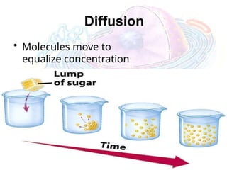
Passive Transport
• Noenergy
required
• Move due to gradient
– differences in concentration,
pressure, charge
• Move to equalize gradient
– High moves toward low
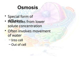
Osmosis
• Special formof
diffusion
• Fluid flows from lower
solute concentration
• Often involves movement
of water
– Into cell
– Out of cell
48.
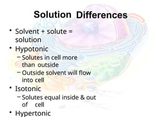
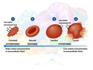
Solution Differences
• Solvent+ solute =
solution
• Hypotonic
– Solutes in cell more
than outside
– Outside solvent will flow
into cell
• Isotonic
– Solutes equal inside & out
of cell
• Hypertonic
50.
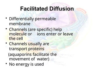
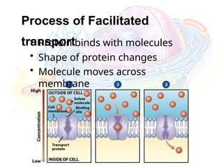
Facilitated Diffusion
• Differentiallypermeable
membrane
• Channels (are specific) help
molecule or ions enter or leave
the cell
• Channels usually are
transport proteins
(aquaporins facilitate the
movement of water)
• No energy is used
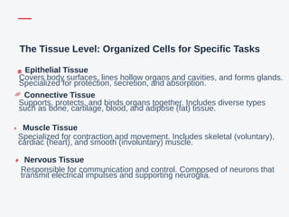
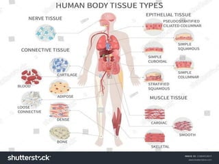
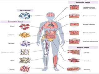
The Tissue Level:Organized Cells for Specific Tasks
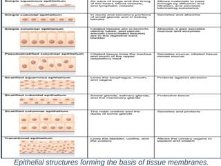
Epithelial Tissue
Covers body surfaces, lines hollow organs and cavities, and forms glands.
Specialized for protection, secretion, and absorption.
Connective Tissue
Supports, protects, and binds organs together. Includes diverse types
such as bone, cartilage, blood, and adipose (fat) tissue.
Muscle Tissue
Specialized for contraction and movement. Includes skeletal (voluntary),
cardiac (heart), and smooth (involuntary) muscle.
Nervous Tissue
Responsible for communication and control. Composed of neurons that
transmit electrical impulses and supporting neuroglia.
59.
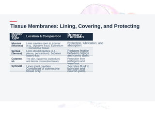
Tissue Membranes: Lining,Covering, and Protecting
Membr
ane
Type
Location & Composition Primary
Function
Mucous
(Mucosa)
Lines cavities open to exterior
(e.g., digestive tract). Epithelium
+ Connective tissue.
Protection, lubrication, and
absorption.
Serous
(Serosa)
Lines closed cavities (e.g.,
pleura, pericardium). Secretes
watery fluid.
Reduces friction
between organs
and cavity walls.
Cutaneo
us
The skin. Epidermis (epithelium)
and dermis (connective tissue).
Protection from
pathogens and
water loss.
Synovial Lines joint cavities.
Composed of connective
tissue only.
Secretes fluid to
lubricate and
nourish joints.
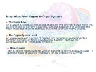
Integration: From Organsto Organ Systems
The Organ Level
An organ is a structure composed of at least two different tissue types that
work together to perform a specific, complex function. For example, the
heart integrates muscle, nervous, epithelial, and connective tissues.
The Organ System Level
An organ system is a group of organs that cooperate to accomplish a
common purpose or major physiological function, such as the
Cardiovascular or Respiratory systems.
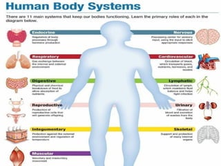
Homeostasis
The 11 major organ systems work in concert to maintain homeostasis—a
dynamic state of balance necessary for the organism's survival.
66.
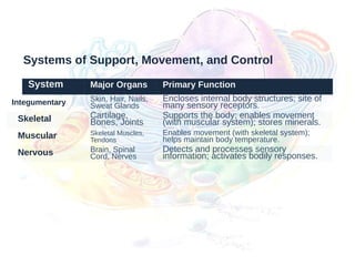
Systems of Support,Movement, and Control
System Major Organs Primary Function
Integumentary Skin, Hair, Nails,
Sweat Glands
Encloses internal body structures; site of
many sensory receptors.
Skeletal Cartilage,
Bones, Joints
Supports the body; enables movement
(with muscular system); stores minerals.
Muscular Skeletal Muscles,
Tendons
Enables movement (with skeletal system);
helps maintain body temperature.
Nervous Brain, Spinal
Cord, Nerves
Detects and processes sensory
information; activates bodily responses.
68.
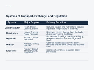
Systems of Transport,Exchange, and Regulation
System Major Organs Primary Function
Cardiovascular Heart, Blood
Vessels, Blood
Delivers oxygen and nutrients to tissues;
equalizes temperature in the body.
Respiratory Lungs, Trachea,
Nasal Passage
Removes carbon dioxide from the body;
delivers oxygen to the blood.
Digestive Stomach, Liver,
Intestines
Processes food for use by the body;
removes wastes from undigested
food.
Urinary Kidneys, Urinary
Bladder
Controls water balance in the body;
removes wastes from blood and excretes
them.
Endocrine
Pituitary Gland,
Thyroid Gland,
Pancreas
Secretes hormones; regulates bodily
processes.
69.
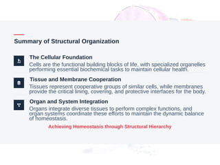
Summary of StructuralOrganization
The Cellular Foundation
Cells are the functional building blocks of life, with specialized organelles
performing essential biochemical tasks to maintain cellular health.
Tissue and Membrane Cooperation
Tissues represent cooperative groups of similar cells, while membranes
provide the critical lining, covering, and protective interfaces for the body.
Organ and System Integration
Organs integrate diverse tissues to perform complex functions, and
organ systems coordinate these efforts to maintain the dynamic balance
of homeostasis.
Achieving Homeostasis through Structural Hierarchy
70.
Sources for ThisPresentation
This presentation is based on the comprehensive academic text provided by OpenStax, ensuring all
anatomical and physiological information is accurate and peer-reviewed.
[1] Betts, J. G., Young, K. A., Wise, J. A., Johnson, E., Poe, B., Kruse, D. H., Korol, O., Johnson, J. E., Womble, M., & DeSaix, P. (2022).
Anatomy and Physiology 2e. OpenStax.
https://openstax.org/books/anatomy-and-physiology-2e/pages/1-introduction


![Sources for This Presentation
This presentation is based on the comprehensive academic text provided by OpenStax, ensuring all
anatomical and physiological information is accurate and peer-reviewed.
[1] Betts, J. G., Young, K. A., Wise, J. A., Johnson, E., Poe, B., Kruse, D. H., Korol, O., Johnson, J. E., Womble, M., & DeSaix, P. (2022).
Anatomy and Physiology 2e. OpenStax.
https://openstax.org/books/anatomy-and-physiology-2e/pages/1-introduction](https://image.slidesharecdn.com/celltissueorgansystemanatomy-260127095155-93f8d44f/85/CELL-TISSUE-ORGAN-SYSTEMANATOMY-power-point-PPT-70-320.jpg)


![CASE_PRESENTATION_ON_subdural_hematoma(SDH)[1 FINAL PPT]-1.pptx](https://cdn.slidesharecdn.com/ss_thumbnails/casepresentationonsubduralhematomasdh1finalppt-1-260129172522-d405d375-thumbnail.jpg?width=640&height=640&fit=bounds)


