At the endof this lesson
Define cell theory and a cell.
Give two types of cells
Give the major differences between a typical plant cell and a
typical animal cell
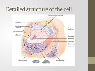
Give the detailed structure of the cell and its functions.
mention the organization of cells
3.
Definition of celltheory
Cell theory is one of the important
concepts in biology which states that the
basic unit of structure and function in
living organism is the cell.
The theory was proposed by Schleiden
(a botanist) and Schwann (a zoologist) in
1838 and 1839 respectively.
4.
Definition of thecell
It is the basic unit of life.
Or it is the basic unit of the living
organism.
The cells are very small and they are
observed using the microscope which
can be light microscope and electron
microscope
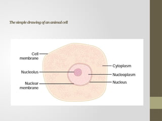
The cell
There twotypes of cells prokaryotic and eukaryotic cells.
Has two major parts; the nucleus and the cytoplasm.
The nucleus is separated from the cytoplasm by a nuclear
membrane, and the cytoplasm is separated from the surrounding
fluids by a cell membrane (plasma membrane)
Plant cells have cell walls on the outside.
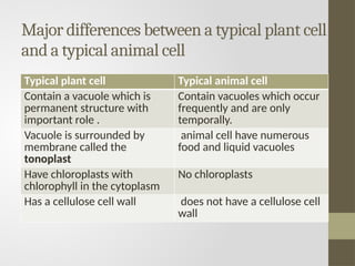
Major differences betweena typical plant cell
and a typical animal cell
Typical plant cell Typical animal cell
Contain a vacuole which is
permanent structure with
important role .
Contain vacuoles which occur
frequently and are only
temporally.
Vacuole is surrounded by
membrane called the
tonoplast
animal cell have numerous
food and liquid vacuoles
Have chloroplasts with
chlorophyll in the cytoplasm
No chloroplasts
Has a cellulose cell wall does not have a cellulose cell
wall
9.
Cell wall, membranesystems,
organelles
Cell wall
it is found in plants, bacteria and fungi
It surrounds the cell and consists of
cellulose as well as chitin.
It surrounds the plasma membrane and
prevents the osmotic bursting of the cell
In bacteria, the cell wall is composed of
peptidoglycan
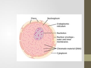
nucleus
The nucleus consistof the nucleoplasm bound by two
membranes known as the nuclear envelope, which has
perforations called nuclear pores.
Functions of the nucleus are to
1. Control all cell activity
2. Produce RNA
3. Produce ribosomes
4. Contain DNA that is essential for inheritance
5. Undergo nuclear division so that cell replication can occur.
13.
Cell surface membrane
it is a partially permeable barrier controlling exchange between the
cell and its environment.
An animal cell is surrounded by the cell surface membrane.
cytoplasm
Cytoplasm is the living material between
the nucleus and the cell surface membrane.
Inside cell surface membrane is a jelly like liquid called cytoplasm. It
contains the nucleus, the two together are called protoplasm. The
cytoplasm contains the structures called organelles
14.
Cytoplasm continuation
The cytoplasmis filled with both tiny and large
dispersed particles.
The clear fluid portion of the cytoplasm in which
particles are dispersed is called cytosol; this contains
mainly dissolved proteins, electrolytes and glucose.
The cytoplasm contains neutral fat globules,
glycogen granules, ribosomes, secretory vesicles and
five especially important organelles: the
endoplasmic reticulum, the Golgi apparatus,
mitochondria, Lysososmes and peroxisomes.
An organelle isa distinct part of the cell which
has a particular structure and function.
the only organelle found in animal cells absent
from plant cells is the centriole
These organelles are briefly explained as follows:
Ribosomes
These are very small organelles consisting of a
large and a small subunit.
The main function of ribosomes is to assist in
protein synthesis by forming polysomes along
mRNA.
17.
There twotypes of the ribosomes that
differ in size and mass; the 80S that are found
in eukaryotic cells and 70S that is found in
prokaryotic cells. Each ribosome is composed
of two units, one larger than the other, which
contain proteins and ribosomal RNA.
18.
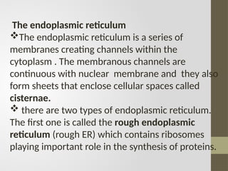
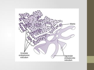
The endoplasmic reticulum
Theendoplasmic reticulum is a series of
membranes creating channels within the
cytoplasm . The membranous channels are
continuous with nuclear membrane and they also
form sheets that enclose cellular spaces called
cisternae.
there are two types of endoplasmic reticulum.
The first one is called the rough endoplasmic
reticulum (rough ER) which contains ribosomes
playing important role in the synthesis of proteins.
20.
Rough endoplasmic reticulumisolates and
transports once they are made. Some proteins
such a digestive enzymes and hormones are not
used inside the cell that makes, so they have to
be secreted that is moved out of the cell without
interfering with cell activities.
Rough ER has a large surface area for the
synthesis of all these proteins, and it stores them
both within the cell and from the inside to the
outside
21.
The second typeof endoplasmic reticulum
is the smooth endoplasmic reticulum (SER)
which has no ribosomes. Smooth ER is
abundant in cells that secrete steroid or lipid
substances such as those found in the
sebaceous glands of the skin.
Another example there a lot of SERs in the
testes which make the steroid hormone
testerone and in the liver which metabolizes
cholesterol among other lipids.
22.
The general functionsof endoplasmic
reticulum are:
a) To provide large surface area for biological
or chemical reactions.
b) To manufacture proteins especially enzymes.
c) To provide channels for the transport and
exchange of materials such as proteins
throughout the cell.
d) To synthesize lipids and steroids.
e) To form a structural skeleton for maintaining
cellular shape.
23.
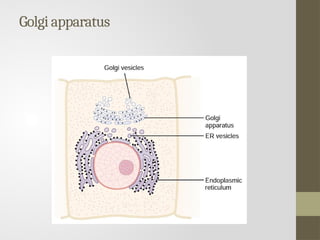
Golgi apparatus
theGolgi apparatus is composed of series of
membranes that enclose flattened fluid spaces
called cisternae.
The function of the Golgi apparatus are to:
a) Manufacture glycoproteins such as mucus,
which are required in secretions.
b) Secrete carbohydrates, such as for the
synthesis of new cells.
c) Transport, modify and store materials such as
lipids.
24.
d) Formation ofLysososmes
e) The Golgi body also seems to be
involved in producing materials for plant
cell walls and cuticles.
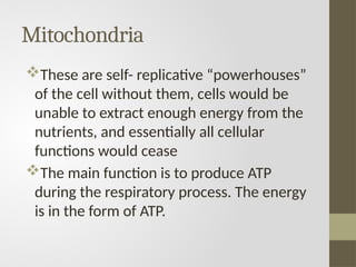
Mitochondria
These are self-replicative “powerhouses”
of the cell without them, cells would be
unable to extract enough energy from the
nutrients, and essentially all cellular
functions would cease
The main function is to produce ATP
during the respiratory process. The energy
is in the form of ATP.
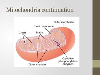
The mitochondrion issurrounded by an
envelope of two membranes, the inner being
folded to form cristae. it also contains a
matrix with few ribosomes, a circular DNA
molecule and phosphate granules.
In aerobic respiration, cristae are the sites
for oxidative phosphorylation and election
transport chain and matrix is the site of kreb
cycle enzymes.
29.
The cells thatrequire very little energy such as
fat storage cells have very little energy. Also any
cell with an energy demanding function like
muscle cells will contain large numbers of
mitochondria.
Mitochondria are also surrounded by an outer
and inner membrane.
They also contain their own genetic material so
that when cells divides the mitochondria
replicates themselves under the control of the
nucleus
30.
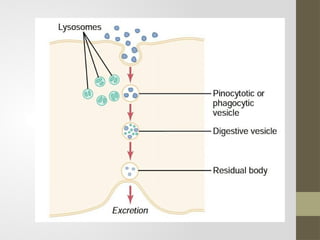
Lysosomes
Lysososmes are smalland spherical structures
containing digestive enzymes. They appear as
spherical bodies in the cytoplasm of most cells and
they contain a powerful mix digestive enzymes.
It is bounded by the single membrane
There concerned with breakdown of structures or
molecules. For example, to get rid of old organelles,
digest bacteria taken in by phagocytosis.
31.
Lysosome continuation
• Alysosome may fuse with the outer membrane to
release its enzymes outside the cell as
extracellular enzymes, for example to destroy the
bacteria.
• Lysosomes can also self destruct, if an entire cell is
damaged or wearing out, its lysosome may
rupture releasing their enzymes to destroy the
entire contents of the cell. This is called apoptosis.
The unprogrammed cell due to cell injury is called
Necrosis
32.
vacuole
The vacuole isa single membrane bound
sac that is filled with cell sap which contains
substances such as water, sugars, mineral
salts, oxygen, carbon dioxide, amino acids
and waste products.
In animals there just numerous food
vacuoles and in plants the vacuole is usually
large, central and its membrane is known as
the tonoplast.
33.
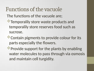
Functions of thevacuole
The functions of the vacuole are;
Temporalily store waste products and
temporalily store reserves food such as
sucrose.
Contain pigments to provide colour for its
parts especially the flowers.
Provide support for the plants by enabling
water molecules to pass through via osmosis
and maintain cell turgidity.
34.
cytoskeleton
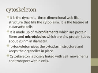
It is thedynamic, three dimensional web like
structure that fills the cytoplasm. It is the feature of
eukaryotic cells.
It is made up of microfilaments which are protein
fibres and microtubules which are tiny protein tubes
about 20 nm in diameter.
cytoskeleton gives the cytoplasm structure and
keeps the organelles in place.
Cytoskeleton is closely linked with cell movements
and transport within cells.
35.
Microvilli
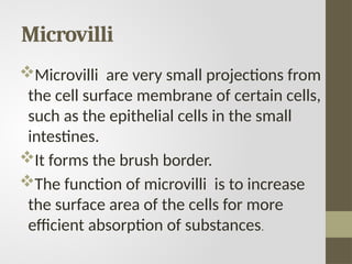
Microvilli are verysmall projections from
the cell surface membrane of certain cells,
such as the epithelial cells in the small
intestines.
It forms the brush border.
The function of microvilli is to increase
the surface area of the cells for more
efficient absorption of substances.
36.
chloroplast
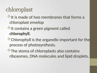
It is madeof two membranes that forms a
chloroplast envelop
It contains a green pigment called
chlorophyll.
Chlorophyll is the organelle important for the
process of photosynthesis.
The stoma of chloroplasts also contains
ribosomes, DNA molecules and lipid droplets.
37.
Cilia and flagella
Thefunction of the cilia and the flagella is
primarily for moving the organism or moving
substances along the lining of cellular layers
such as the respiratory.
Another cell which has the flagella is the
spermatozoon.
38.
centrioles
Centrioles are apair of cylindrical
structures that are found in the
centrosome which is just outside the
nucleus.
The centrioles are involved in cell division
When a cell divides the centrioles pulls
apart to produce a spindle of microtubules
which are involved in the movement of the
chromosomes.
39.
The organization ofcells
Tissues are groups of similar cells that all
develop from the same kind of cell.
Examples of tissues are; epithelial tissue,
connective tissue, muscle tissue and
nervous tissue.
Organs is made up of a group of tissues
that are grouped in to a structure so that
they can work together. The example of
organs are brain, lungs, and liver.
40.
System is wherea number of organs
work together to carry out large scale
functions. For example the digestive
system includes the organs of the
stomach, pancreas, small and large
intestines.
Organism is made by the group of
system. The example of the organism is
the human being.
41.
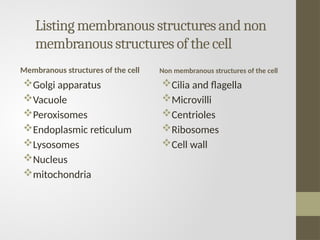
Listing membranous structuresand non
membranous structures of the cell
Membranous structures of the cell
Golgi apparatus
Vacuole
Peroxisomes
Endoplasmic reticulum
Lysosomes
Nucleus
mitochondria
Non membranous structures of the cell
Cilia and flagella
Microvilli
Centrioles
Ribosomes
Cell wall
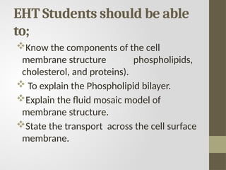
EHT Students shouldbe able
to;
Know the components of the cell
membrane structure phospholipids,
cholesterol, and proteins).
To explain the Phospholipid bilayer.
Explain the fluid mosaic model of
membrane structure.
State the transport across the cell surface
membrane.
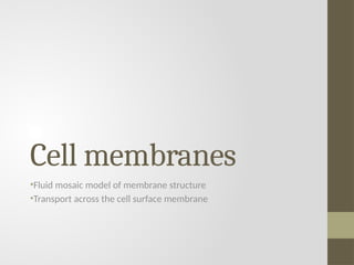
44.
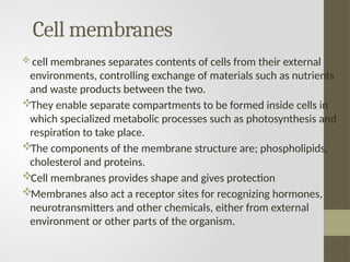
Cell membranes
cellmembranes separates contents of cells from their external
environments, controlling exchange of materials such as nutrients
and waste products between the two.
They enable separate compartments to be formed inside cells in
which specialized metabolic processes such as photosynthesis and
respiration to take place.
The components of the membrane structure are; phospholipids,
cholesterol and proteins.
Cell membranes provides shape and gives protection
Membranes also act a receptor sites for recognizing hormones,
neurotransmitters and other chemicals, either from external
environment or other parts of the organism.
45.
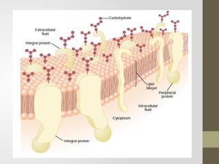
Cell membrane structureis composed
almost entirely of proteins and lipids in the
following ratios approximately 55 % proteins;
25 % phospholipids; 13 % cholesterol; 4 %
other lipids and carbohydrates, 3 %.
They mainly contain proteins and lipids
The drawing which follows shows the
contents of the cell membrane.
47.
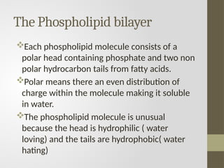
The Phospholipid bilayer
Eachphospholipid molecule consists of a
polar head containing phosphate and two non
polar hydrocarbon tails from fatty acids.
Polar means there an even distribution of
charge within the molecule making it soluble
in water.
The phospholipid molecule is unusual
because the head is hydrophilic ( water
loving) and the tails are hydrophobic( water
hating)
48.
Phospholipid continues
The nonpolar hydrophobic tail project out of the
water while the polar hydrophilic heads lie in the
surface of water
If the phospholipid is present in large enough
amounts more than cover the surface of the water,
or if it is shake up with water particles known as
micelles are formed in which hydrophobic tails
project inwards away from water.
The two layers of phospholipids hence referred to
as phospholipid bilayer
49.
The fluid modelof membrane structure
Singer and Nicolson developed the fluid mosaic model in 1972.
This model is the one in which protein molecules float about in
a phospholipid bilayer.
The structure of cell surface shows a fluid model because of
mosaic arrangement of proteins throughout its phospholipid
bilayer which is also capable of movement hence it is fluid in
nature.
The scattered protein molecules resemble a mosaic but since
the phospholipid bilayer is fluid, the proteins form a fluid
mosaic pattern.
Cholesterol is a main fluidity of the phospholipid bilayer
Make sure every EHT student should be able to draw a fluid mosaic model of membrane structure
50.
Transport across thecell surface
membrane
Transport across the membranes occur due to
the following reasons;
a) To obtain nutrients.
b) To excrete waste substances
c) To maintain the suitable PH and ionic
concentration
This happens by four mechanisms namely;
diffusion, osmosis, active transport and bulk
transport (endocytosis or exocytosis).
51.
Diffusion and facilitated
diffusion
Diffusionis the movement of molecules or ions from the
region of their high concentration to a region of their low
concentration down the diffusion gradient.
For example, oxygen diffuses from the lungs into the blood
while at the same time carbon dioxide diffuses in the opposite
direction.
Channel proteins that are found along the cell membrane
also play a role in transporting water soluble ions across the
membrane by the process of facilitated diffusion.
Facilitated diffusion involves the carrier proteins to facilitate
the transfer of the substance down the diffusion gradient.
52.
Osmosis
Osmosis is thepassage of water
molecules from the region of their
high concentration to a region of their
low concentration through a partially
permeable membrane.
It is best regarded as a form of
diffusion in which only water
molecules moves.
53.
Active transport
Active transportis a transport of substances
across cell membranes done against a
concentration gradient that is from a region of low
concentration to one of high concentration.
Energy is required to drive active transport and is
obtained from ATP, energy produced by
mitochondria.
This transport involves carrier proteins situated in
the cell surface membrane to carry the
compounds such as amino acids, across the
membrane.
54.
Bulk transport (endocytosisand
exocytosis )
endocytosis and exocytosis are active
process involving the bulk transport of
materials through membranes, either into
cells (endocytosis) or out of cells
(exocytosis).
Endocytosis
Endocytosis occurs by infolding or extension of
the cell surface membrane to form a vesicle or
vacuole. It is in the following types
55.
Endocytosis continuation
Phagocytosis (celleating) is when
material taken up by the cells is in
solid form.
Pinocytosis (cell drinking) is when
material taken up by the cells is in
liquid form.
56.
Exocytosis
It is thereverse process of endocytosis
This is when waste materials are removed
from cells, such as solids, undigested
remains from phagocytic vacuoles or
useful materials are secreted.