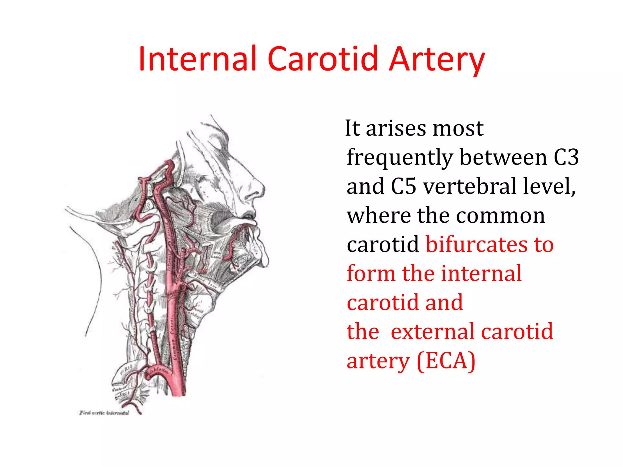
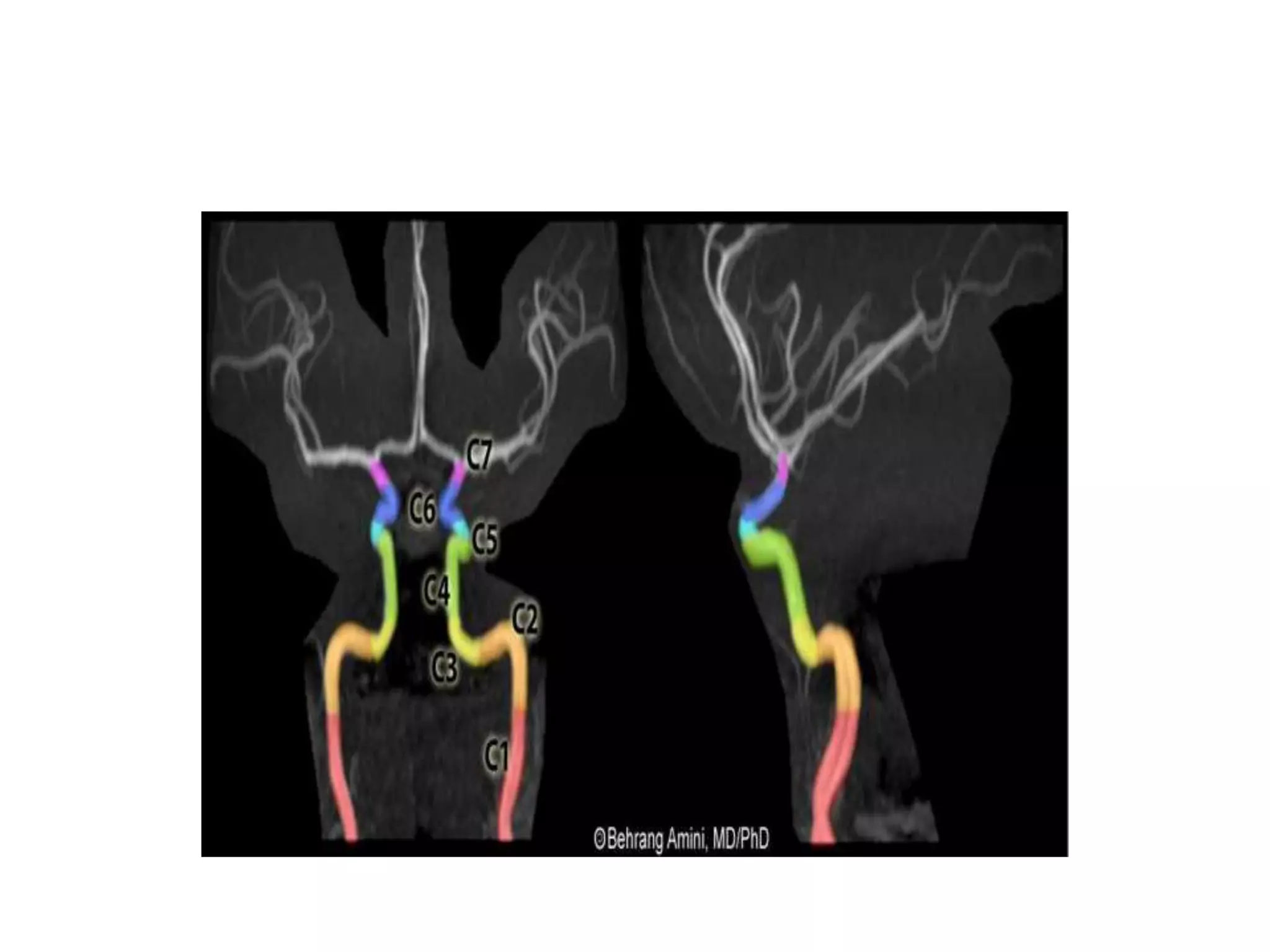
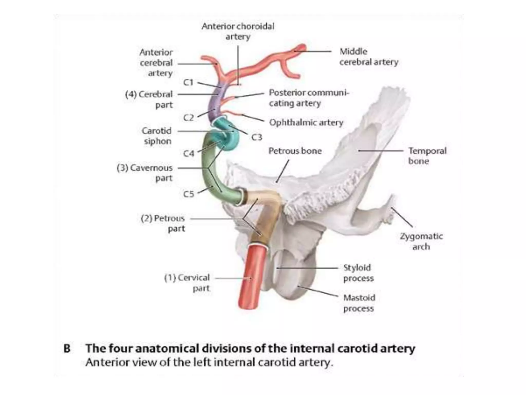
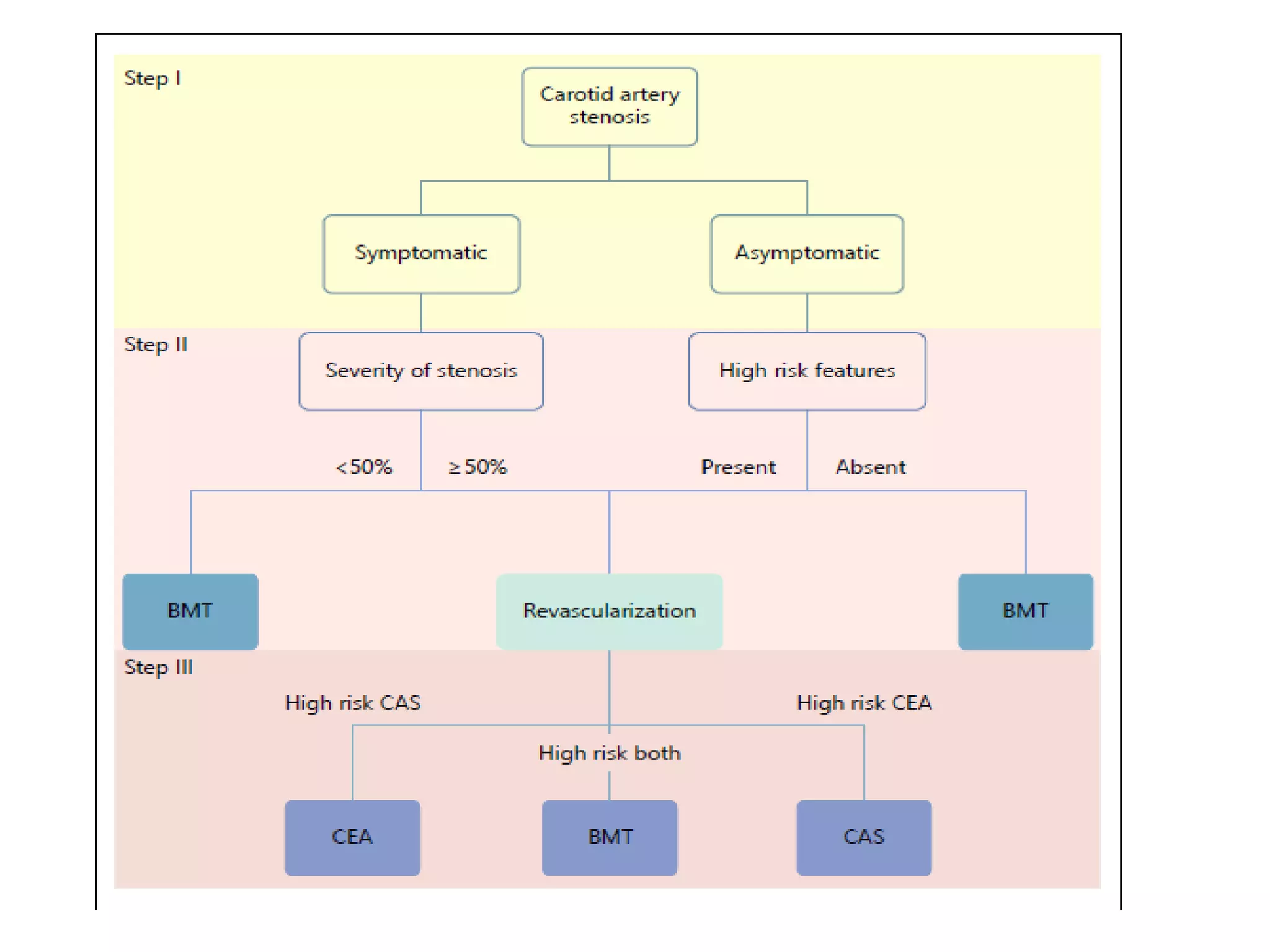
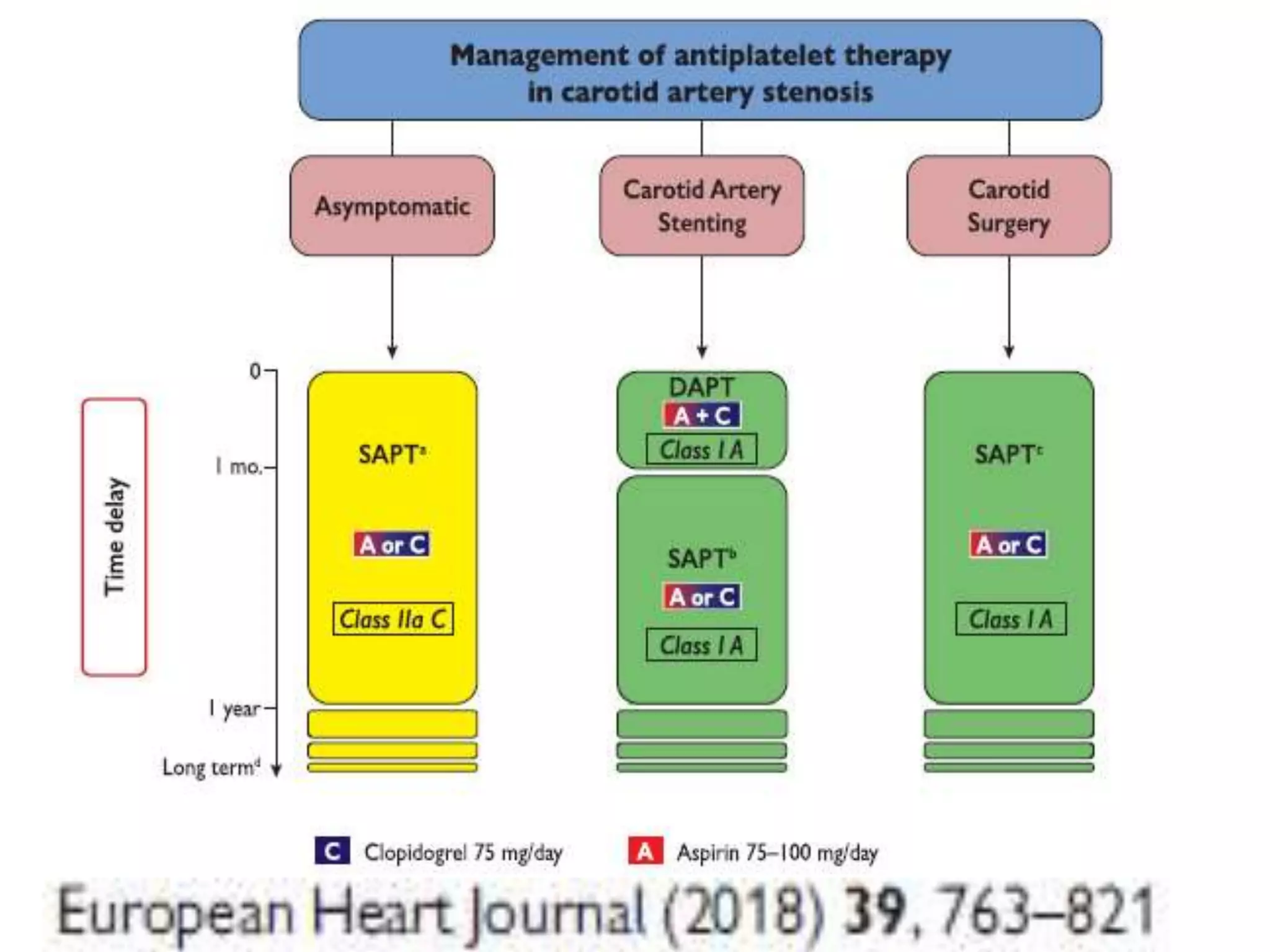
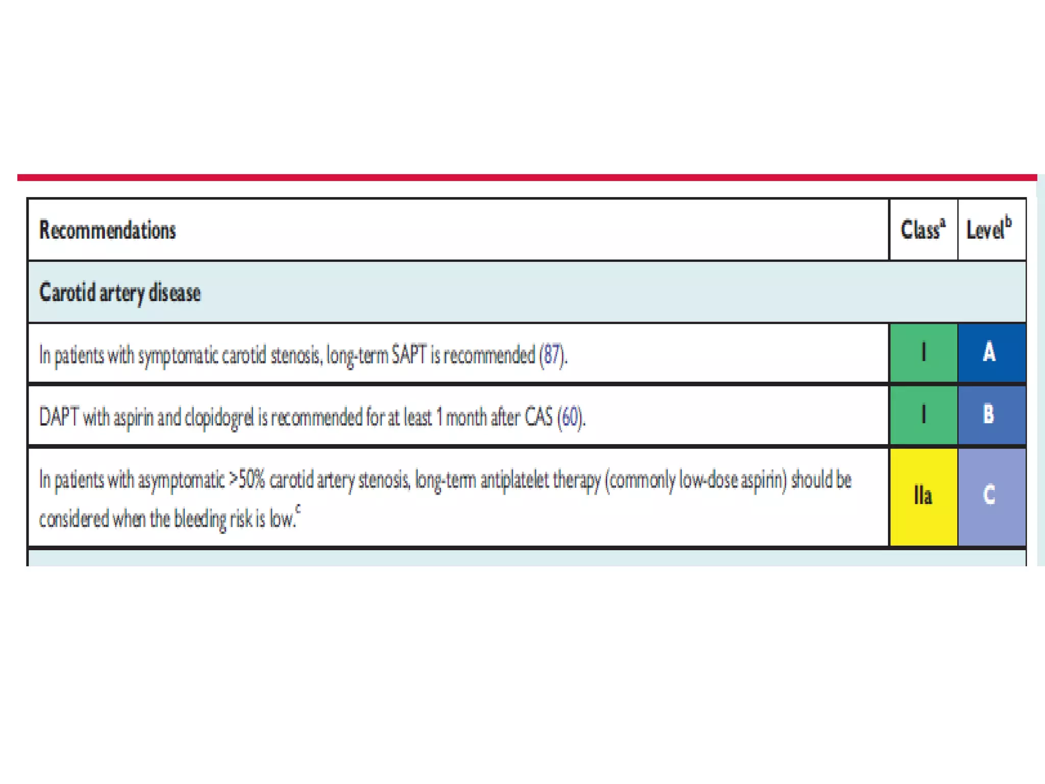
CAROTID ARTERY STENOSIS
Dr Pankaj S Rathi
Definition
Carotid artery stenosis refers to a ≥ 50%
stenosis of the extracranial internal carotid
artery (ICA), with stenosis severity
estimated using the North American
Symptomatic Carotid Endarterectomy Trial
(NASCET) method.
Carotid Artery Anatomy
Internal Carotid Artery
It arises most
frequently between C3
and C5 vertebral level,
where the common
carotid bifurcates to
form the internal
carotid and
the external carotid
artery (ECA)
Anatomical variations and segments of
ICA
• Variations in level of
bifurcation
– left higher 50%
– right higher 22%
– same height 28%
• Variations in origin
C1/2: 0.3%
C2/3: 3.7%
C3/4: 34.2%
C4/5: 48.1%
C5/6: 13%
C6/7: 0.15%
ICA Segments
Bouthillier Classification
• cervical segment
• petrous (horizontal)
segment
• lacerum segment
• cavernous segment
• clinoid segment
• ophthalmic (supraclinoid)
segment
• communicating (terminal)
segment
Indian figures of Ischemic stroke
• Prevalence rate : 545 per 100,000
• Mortality rate in stroke is 7.5 per 1000
• Carotid stenosis is an established risk factor of
ischemic stroke
• Atherosclerosis of internal carotid artery (ICA) is a
major risk factors for stroke
• Causative factor in approximately 30% of all
ischemic strokes
Ann Indian Acad Neurology 2015 Oct-Dec; 18(4): 412–414
Neurology India : 2017, Vol 65 Issue 2 Page : 279-285
Severity of The Problem in India
• Asymptomatic carotid artery stenosis (ACAS) of
more than 50% has a 2–6% annual risk of stroke
• 5.2% of asymptomatic individuals > 40 years of
age harbor significant extracranial carotid artery
disease
Neurology India : 2017, Vol 65 Issue 2 Page : 279-285
Prevalence and features of carotid
atherosclerosis
Neurology India : 2017, Vol 65 Issue 2 Page : 279-285
Natural History of Atherosclerotic
Carotid Artery Disease
• Extracranial atherosclerotic disease accounts for up to
15% to 20% of all ischemic strokes
• Clear correlation between the degree of stenosis and the
risk of stroke in the NASCET study
After 18 months of medical therapy without
revascularization
Risk of stroke rates
19% in those with 70% to 79% initial stenosis,
28% in those with 80% to 89% stenosis
33% in the 90% to 99% stenosis
Risk diminished with near-occlusion
Circulation. 2011;124:e54-e130
Characterization of Atherosclerotic Lesions
in the Extracranial Carotid Arteries and
stroke
Plaque morphology
Ulceration, echolucency, intraplaque hemorrhage,
and high lipid content
Hypoechoic plaques
Subcortical and cortical cerebral infarcts of
suspected embolic origin
Hyperechoic plaques
Diffuse white matter infarcts of presumed
hemodynamic origin
Methods of Diagnosing Carotid Artery
Stenosis
• Carotid Duplex Doppler
• CT Angiography
• MR Angiography
• DSA
Indications for carotid duplex
sonography
• Cervical bruit in an asymptomatic patient
• Follow-up of known stenosis (50%) in asymptomatic
individuals
• Vascular assessment in a patient with multiple risk
factors for atherosclerosis
• Stroke risk assessment in a patient with CAD or PAD
• Amaurosis fugax
• Hemispheric TIA
• Stroke in a candidate for carotid revascularization
• Follow-up after a carotid revascularization procedure
• Intraoperative assessment during CEA or stenting
Grading Of ICA Stenosis
Method of Grading
Asymptomatic & Symptomatic Carotid
Artery Disease
• Asymptomatic carotid stenosis
If patients lack a recent history (6 months) of ipsilateral carotid
ischemic stroke or transient ischemic attack .
• Symptomatic carotid stenosis
Stenosis in the internal carotid artery, either intracranial or
extracranial, leading to symptoms of amaurosis fugax, transient
ischemic attacks, or ischemic stroke ipsilateral to the lesion
• Severe stenosis (70–99%) :
Highest risk for recurrent stroke or TIA
Stroke. 2017;48:00-00
Curr Treat Options Cardio Med (2017) 19: 62
Indications for Screening Carotid
Artery
Our Patient
• Mr. X is a 74 year old man
• On a statin for elevated cholesterol and ASA for
primary prevention
• Has attended 3-4 health check up over past 7
years to request carotid artery screening
• Was told of 50% stenosis
• Worried about risk for stroke
• Would consider procedure to open artery if
would reduce risk of stroke
Past Medical and Surgical History
• Elevated cholesterol
• Low back pain
• Cervical spondylosis
• GERD
• BPH
• S/p shoulder surgery
Social and Family History
• Retired medical officer
• Travels twice yearly from Indore to Jabalpur
with wife
• Helps to care for 4 grandchildren
• No cigarettes
• Social alcohol use, 2-3 glasses whisky/week
• Family history: no cardiovascular disease or
stroke
Medication History
• Simvastatin 20 mg qd
• Aspirin 81 mg qd
• Omeprazole 20 mg qd
• Tamsulosin 0.8 mg qhs
Health Examination
• Well appearing
• Bp 124/82, HR 72, weight 72, BMI 26.7
• Chest - clear
• Cardiac – RRR no murmurs or S4
• Extremities - normal
Would you screen this patient for
carotid artery stenosis?
Evaluation of Asymptomatic Patients
at Risk of Extracranial Carotid Artery
Disease
Recommendations for Duplex Ultrasonography
to Evaluate Asymptomatic Patients With
Known or Suspected Carotid Stenosis
Circulation. 2011;124:e54-e130
Duplex ultrasonography to detect
hemodynamically significant carotid stenosis may
be considered in asymptomatic patients with
symptomatic PAD, coronary artery disease (CAD),
or atherosclerotic aortic aneurysm
Circulation. 2011;124:e54-e130
I IIaIIb III
Duplex ultrasonography might be considered to detect carotid stenosis in
asymptomatic patients without clinical evidence of atherosclerosis who
have 2 or more of the following risk factors: hypertension, hyperlipidemia,
tobacco smoking, a family history in a first degree relative of
atherosclerosis manifested before age 60 years, or a family history of
ischemic stroke. However, it is unclear whether establishing a diagnosis of
ECVD would justify actions that affect clinical outcomes.
Circulation. 2011;124:e54-e130
I IIa IIb III
In a metaanalysis, DUS, MRA and CTA were
equivalent for detecting significant carotid
stenosis
Accurate, practical and cost-effective assessment of carotid stenosis in
the UK. Health Technol Assess 2006;10:iii–iv, ix–x, 1 - 182.
Decision making in management of
carotid artery stenosis
Asymptomatic Carotid Stenosis:
What is Medical Intervention,
and Is It Effective?
Best Medical Therapy in Carotid
Stenosis
Best medical therapy (BMT) includes CV risk factor
management, including best pharmacological therapy, as
well as nonpharmacological measures such as smoking
cessation, healthy diet, weight loss and regular physical
exercise.
Pharmacological component
Antihypertensive
Lipid-lowering
Antithrombotic drugs
Optimal glucose level control
Eur Heart J 2007;28:2375–2414
Moving targets
Carotid stenting has evolved.
Outcomes are improving and the
procedure is maturing. Randomized
trials (including CREST) now
supported by large post-market
surveillance outcomes
Medical therapy has also evolved with
evidence suggesting the risk of stroke in
asymptomatic patients today may be
much lower than even 10 years ago
ASA
Early
1900s
Late
1900s
Today
ASA
HTN RX
1800s
ASA
HTN RX (ACEI,
beta blockers)
DM Control,
Lipid Rx
ASA
HTN RX
Tight DM Control
High dose statins
ACE and ARB
Annual TIA and stroke in asymptomatic
control arms of selected randomized trials
0%
2%
4%
6%
8%
10%
12%
14%
16%
18%
20%
Ipsilateral stroke/ TIA
Effect of Antiplatelet Therapy in
Patients with TIA or Stroke
Antiplatelet Trialists’ Collaboration. BMJ 2002;324:71-86
287 Studies: 135,000 Patients in Comparisons of
Antiplatelet Rx vs Control
Use of ramipril in preventing stroke:
double blind randomised trial
BMJ VOLUME 324 23 MARCH 2002 bmj.com
Stroke 2009;40
• 1007 patients with carotid stenosis
(not requiring revascularization) at baseline
– 3271 patients had no carotid stenosis at baseline
• All patients had stroke/TIA within 6 months of
randomization
– Randomized to Atorvastatin 80 mg/d vs Placebo
• No known CHD
• LDL Cholesterol between 100-190 mg/dL
Of those patients with carotid artery stenosis at
baseline…
• Atorvastatin lowered any stroke risk by 33%
• Atorvastatin lowered any CHD event by 43%
• Later carotid revascularization was reduced by
56%!
The Carotid “Prescription”
• ASA 81 mg/d
– No role for dual antiplatelet therapy for stroke
“prevention”
• Antihypertensive Therapy
– ACE Inhibitor
– Angiotensin Receptor Antagonist
• Lipid Lowering Therapy
– LDL-Cholesterol <100 mg/dL
• Tobacco Cessation
• Glycemic Control (HbA1C <7.0%)
Asymptomatic Carotid stenosis
with High Risk Factors
Asymptomatic Carotid Stenosis with
High Risk Factors
When Invasive Procedure ?
Treatment for Asymptomatic Carotid
Artery Stenosis with high risk factors
Revascularization
 Carotid endarterectomy
(CEA)
 Carotid artery stenting
(CAS)
Carotid endarterectomy (CEA)
Carotid artery stenting (CAS)
Endovascular techniques
Carotid Artery Stenting
Less invasive
Low risk of cranial nerve
injury,
Wound complications
Neck haematoma
Carotid Endarterectomy
CAS offers advantages
‘hostile neck’ (previous
radiation, recurrent
stenosis), contralateral
recurrent laryngeal nerve
palsy or in the case of
challenging surgical access
[very high ICA lesions,
proximal common carotid
artery (CCA) lesion
CREST (Carotid Revascularization Endarterectomy versus Stenting Trial),
SAPPHIRE (Stenting and Angioplasty with Protection in Patients at High Risk
for Endarterectomy) ACT trial ( Asymptomatic Carotid Trail )
ACST 10-year Follow-up Results Provide Most Rigorous Comparison Of revascularization
And BMT Lancet. 2010 Sep 25; 376(9746): 1074–1084
Symptomatic Carotid Artery Disease
High Risk Features for CEA & CAS
Failure of Medical Therapy in
Symptomatic Carotid Artery Disease
• 50–99% stenosis if
treated medically
• Increasing age (>75
years)
• Symptoms within 14 days
& Male sex
• Hemispheric symptoms
• Cortical stroke
• Medical comorbidities
• Irregular stenosis
• Increasing stenosis
severity
• Contralateral occlusion
Timing of CEA
P.M. Rothwell et al. Stroke. 2004;35:2855-2861
Copyright © American Heart Association, Inc. All rights reserved.
Timing of CEA
CEA within 14 days ( 50–69% )
ARR for stroke at 5 years ( 14.8% NNT = 7)
ARR (3.3% NNT = 30)when delay was 2–4 weeks
ARR (2.5% NNT 40 ) when the delay was 4–12weeks
Beyond 12weeks, no strokes were prevented by CEA.
CEA within 14 days ( 70–99%)
ARR for stroke at 5 years was 23.0% (NNT = 4)
ARR (15.9% NNT 6 ) where delays were 2–4weeks
ARR (7.9% NNT 13 ) for delays of 4–12weeks
ARR (7.4 % NNT 14 ) at 5 yrs when done beyond
12weeks
Endovascular therapy vs. open surgery
• The risk of ‘any stroke’ and ‘death/ stroke’ was 50% higher
following CAS, primarily because CAS was associated with
a significantly higher rate of minor stroke
• CAS was associated with higher periprocedural
death/stroke, especially in patients >70 years of age, but
with significantly lower risks for MI, cranial nerve injury
and haematoma
Conclusion
• CEA and CAS should largely be used for symptomatic
patients
• Most asymptomatic patients should get best medical
treatment ( BMT ) & neither CAS or CAS ( except for small
% )
• Except in extraordinary circumstances, carotid
revascularization by either CEA or CAS is not
recommended when atherosclerosis narrows the lumen by
less than 50%
• Carotid revascularization is not recommended for patients
with chronic total occlusion of the targeted carotid artery
• Carotid revascularization is not recommended for patients
with severe disability caused by cerebral infarction that
precludes preservation of useful function
THANK YOU

Carotid Artery Stenosis Dr Pankaj Rathi DM Traine Shri Aurobindo Medical College Indore

  • 1.
  • 2.
    Definition Carotid artery stenosisrefers to a ≥ 50% stenosis of the extracranial internal carotid artery (ICA), with stenosis severity estimated using the North American Symptomatic Carotid Endarterectomy Trial (NASCET) method.
  • 3.
  • 4.
    Internal Carotid Artery Itarises most frequently between C3 and C5 vertebral level, where the common carotid bifurcates to form the internal carotid and the external carotid artery (ECA)
  • 5.
    Anatomical variations andsegments of ICA • Variations in level of bifurcation – left higher 50% – right higher 22% – same height 28% • Variations in origin C1/2: 0.3% C2/3: 3.7% C3/4: 34.2% C4/5: 48.1% C5/6: 13% C6/7: 0.15% ICA Segments Bouthillier Classification • cervical segment • petrous (horizontal) segment • lacerum segment • cavernous segment • clinoid segment • ophthalmic (supraclinoid) segment • communicating (terminal) segment
  • 8.
    Indian figures ofIschemic stroke • Prevalence rate : 545 per 100,000 • Mortality rate in stroke is 7.5 per 1000 • Carotid stenosis is an established risk factor of ischemic stroke • Atherosclerosis of internal carotid artery (ICA) is a major risk factors for stroke • Causative factor in approximately 30% of all ischemic strokes Ann Indian Acad Neurology 2015 Oct-Dec; 18(4): 412–414 Neurology India : 2017, Vol 65 Issue 2 Page : 279-285
  • 9.
    Severity of TheProblem in India • Asymptomatic carotid artery stenosis (ACAS) of more than 50% has a 2–6% annual risk of stroke • 5.2% of asymptomatic individuals > 40 years of age harbor significant extracranial carotid artery disease Neurology India : 2017, Vol 65 Issue 2 Page : 279-285
  • 10.
    Prevalence and featuresof carotid atherosclerosis Neurology India : 2017, Vol 65 Issue 2 Page : 279-285
  • 11.
    Natural History ofAtherosclerotic Carotid Artery Disease • Extracranial atherosclerotic disease accounts for up to 15% to 20% of all ischemic strokes • Clear correlation between the degree of stenosis and the risk of stroke in the NASCET study After 18 months of medical therapy without revascularization Risk of stroke rates 19% in those with 70% to 79% initial stenosis, 28% in those with 80% to 89% stenosis 33% in the 90% to 99% stenosis Risk diminished with near-occlusion Circulation. 2011;124:e54-e130
  • 13.
    Characterization of AtheroscleroticLesions in the Extracranial Carotid Arteries and stroke Plaque morphology Ulceration, echolucency, intraplaque hemorrhage, and high lipid content Hypoechoic plaques Subcortical and cortical cerebral infarcts of suspected embolic origin Hyperechoic plaques Diffuse white matter infarcts of presumed hemodynamic origin
  • 14.
    Methods of DiagnosingCarotid Artery Stenosis • Carotid Duplex Doppler • CT Angiography • MR Angiography • DSA
  • 15.
    Indications for carotidduplex sonography • Cervical bruit in an asymptomatic patient • Follow-up of known stenosis (50%) in asymptomatic individuals • Vascular assessment in a patient with multiple risk factors for atherosclerosis • Stroke risk assessment in a patient with CAD or PAD • Amaurosis fugax • Hemispheric TIA • Stroke in a candidate for carotid revascularization • Follow-up after a carotid revascularization procedure • Intraoperative assessment during CEA or stenting
  • 16.
  • 17.
  • 18.
    Asymptomatic & SymptomaticCarotid Artery Disease • Asymptomatic carotid stenosis If patients lack a recent history (6 months) of ipsilateral carotid ischemic stroke or transient ischemic attack . • Symptomatic carotid stenosis Stenosis in the internal carotid artery, either intracranial or extracranial, leading to symptoms of amaurosis fugax, transient ischemic attacks, or ischemic stroke ipsilateral to the lesion • Severe stenosis (70–99%) : Highest risk for recurrent stroke or TIA Stroke. 2017;48:00-00 Curr Treat Options Cardio Med (2017) 19: 62
  • 19.
  • 21.
    Our Patient • Mr.X is a 74 year old man • On a statin for elevated cholesterol and ASA for primary prevention • Has attended 3-4 health check up over past 7 years to request carotid artery screening • Was told of 50% stenosis • Worried about risk for stroke • Would consider procedure to open artery if would reduce risk of stroke
  • 22.
    Past Medical andSurgical History • Elevated cholesterol • Low back pain • Cervical spondylosis • GERD • BPH • S/p shoulder surgery
  • 23.
    Social and FamilyHistory • Retired medical officer • Travels twice yearly from Indore to Jabalpur with wife • Helps to care for 4 grandchildren • No cigarettes • Social alcohol use, 2-3 glasses whisky/week • Family history: no cardiovascular disease or stroke
  • 24.
    Medication History • Simvastatin20 mg qd • Aspirin 81 mg qd • Omeprazole 20 mg qd • Tamsulosin 0.8 mg qhs
  • 25.
    Health Examination • Wellappearing • Bp 124/82, HR 72, weight 72, BMI 26.7 • Chest - clear • Cardiac – RRR no murmurs or S4 • Extremities - normal
  • 26.
    Would you screenthis patient for carotid artery stenosis?
  • 27.
    Evaluation of AsymptomaticPatients at Risk of Extracranial Carotid Artery Disease Recommendations for Duplex Ultrasonography to Evaluate Asymptomatic Patients With Known or Suspected Carotid Stenosis Circulation. 2011;124:e54-e130
  • 29.
    Duplex ultrasonography todetect hemodynamically significant carotid stenosis may be considered in asymptomatic patients with symptomatic PAD, coronary artery disease (CAD), or atherosclerotic aortic aneurysm Circulation. 2011;124:e54-e130 I IIaIIb III
  • 30.
    Duplex ultrasonography mightbe considered to detect carotid stenosis in asymptomatic patients without clinical evidence of atherosclerosis who have 2 or more of the following risk factors: hypertension, hyperlipidemia, tobacco smoking, a family history in a first degree relative of atherosclerosis manifested before age 60 years, or a family history of ischemic stroke. However, it is unclear whether establishing a diagnosis of ECVD would justify actions that affect clinical outcomes. Circulation. 2011;124:e54-e130 I IIa IIb III
  • 32.
    In a metaanalysis,DUS, MRA and CTA were equivalent for detecting significant carotid stenosis Accurate, practical and cost-effective assessment of carotid stenosis in the UK. Health Technol Assess 2006;10:iii–iv, ix–x, 1 - 182.
  • 33.
    Decision making inmanagement of carotid artery stenosis
  • 35.
    Asymptomatic Carotid Stenosis: Whatis Medical Intervention, and Is It Effective?
  • 36.
    Best Medical Therapyin Carotid Stenosis Best medical therapy (BMT) includes CV risk factor management, including best pharmacological therapy, as well as nonpharmacological measures such as smoking cessation, healthy diet, weight loss and regular physical exercise. Pharmacological component Antihypertensive Lipid-lowering Antithrombotic drugs Optimal glucose level control Eur Heart J 2007;28:2375–2414
  • 37.
    Moving targets Carotid stentinghas evolved. Outcomes are improving and the procedure is maturing. Randomized trials (including CREST) now supported by large post-market surveillance outcomes Medical therapy has also evolved with evidence suggesting the risk of stroke in asymptomatic patients today may be much lower than even 10 years ago
  • 38.
    ASA Early 1900s Late 1900s Today ASA HTN RX 1800s ASA HTN RX(ACEI, beta blockers) DM Control, Lipid Rx ASA HTN RX Tight DM Control High dose statins ACE and ARB
  • 39.
    Annual TIA andstroke in asymptomatic control arms of selected randomized trials 0% 2% 4% 6% 8% 10% 12% 14% 16% 18% 20% Ipsilateral stroke/ TIA Ipsilateral stroke/ TIA
  • 40.
    Effect of AntiplateletTherapy in Patients with TIA or Stroke Antiplatelet Trialists’ Collaboration. BMJ 2002;324:71-86 287 Studies: 135,000 Patients in Comparisons of Antiplatelet Rx vs Control
  • 43.
    Use of ramiprilin preventing stroke: double blind randomised trial BMJ VOLUME 324 23 MARCH 2002 bmj.com
  • 44.
    Stroke 2009;40 • 1007patients with carotid stenosis (not requiring revascularization) at baseline – 3271 patients had no carotid stenosis at baseline • All patients had stroke/TIA within 6 months of randomization – Randomized to Atorvastatin 80 mg/d vs Placebo • No known CHD • LDL Cholesterol between 100-190 mg/dL
  • 45.
    Of those patientswith carotid artery stenosis at baseline… • Atorvastatin lowered any stroke risk by 33% • Atorvastatin lowered any CHD event by 43% • Later carotid revascularization was reduced by 56%!
  • 46.
    The Carotid “Prescription” •ASA 81 mg/d – No role for dual antiplatelet therapy for stroke “prevention” • Antihypertensive Therapy – ACE Inhibitor – Angiotensin Receptor Antagonist • Lipid Lowering Therapy – LDL-Cholesterol <100 mg/dL • Tobacco Cessation • Glycemic Control (HbA1C <7.0%)
  • 48.
  • 50.
    Asymptomatic Carotid Stenosiswith High Risk Factors
  • 51.
  • 52.
    Treatment for AsymptomaticCarotid Artery Stenosis with high risk factors Revascularization  Carotid endarterectomy (CEA)  Carotid artery stenting (CAS)
  • 53.
  • 54.
  • 55.
    Endovascular techniques Carotid ArteryStenting Less invasive Low risk of cranial nerve injury, Wound complications Neck haematoma Carotid Endarterectomy CAS offers advantages ‘hostile neck’ (previous radiation, recurrent stenosis), contralateral recurrent laryngeal nerve palsy or in the case of challenging surgical access [very high ICA lesions, proximal common carotid artery (CCA) lesion
  • 57.
    CREST (Carotid RevascularizationEndarterectomy versus Stenting Trial), SAPPHIRE (Stenting and Angioplasty with Protection in Patients at High Risk for Endarterectomy) ACT trial ( Asymptomatic Carotid Trail )
  • 60.
    ACST 10-year Follow-upResults Provide Most Rigorous Comparison Of revascularization And BMT Lancet. 2010 Sep 25; 376(9746): 1074–1084
  • 61.
  • 62.
  • 63.
    High Risk Featuresfor CEA & CAS
  • 67.
    Failure of MedicalTherapy in Symptomatic Carotid Artery Disease • 50–99% stenosis if treated medically • Increasing age (>75 years) • Symptoms within 14 days & Male sex • Hemispheric symptoms • Cortical stroke • Medical comorbidities • Irregular stenosis • Increasing stenosis severity • Contralateral occlusion
  • 68.
  • 69.
    P.M. Rothwell etal. Stroke. 2004;35:2855-2861 Copyright © American Heart Association, Inc. All rights reserved.
  • 70.
    Timing of CEA CEAwithin 14 days ( 50–69% ) ARR for stroke at 5 years ( 14.8% NNT = 7) ARR (3.3% NNT = 30)when delay was 2–4 weeks ARR (2.5% NNT 40 ) when the delay was 4–12weeks Beyond 12weeks, no strokes were prevented by CEA. CEA within 14 days ( 70–99%) ARR for stroke at 5 years was 23.0% (NNT = 4) ARR (15.9% NNT 6 ) where delays were 2–4weeks ARR (7.9% NNT 13 ) for delays of 4–12weeks ARR (7.4 % NNT 14 ) at 5 yrs when done beyond 12weeks
  • 71.
    Endovascular therapy vs.open surgery • The risk of ‘any stroke’ and ‘death/ stroke’ was 50% higher following CAS, primarily because CAS was associated with a significantly higher rate of minor stroke • CAS was associated with higher periprocedural death/stroke, especially in patients >70 years of age, but with significantly lower risks for MI, cranial nerve injury and haematoma
  • 72.
    Conclusion • CEA andCAS should largely be used for symptomatic patients • Most asymptomatic patients should get best medical treatment ( BMT ) & neither CAS or CAS ( except for small % ) • Except in extraordinary circumstances, carotid revascularization by either CEA or CAS is not recommended when atherosclerosis narrows the lumen by less than 50% • Carotid revascularization is not recommended for patients with chronic total occlusion of the targeted carotid artery • Carotid revascularization is not recommended for patients with severe disability caused by cerebral infarction that precludes preservation of useful function
  • 73.