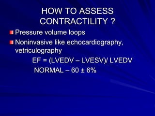
Cardiovascular physiology
Dr. Shyam Dhake
Dr. Sadashiv Swain
www.anaesthesia.co.in
anaesthesia.co.in@gmail.com
Cardiac output
DEFINATION
 Cardiac output : vol of blood pumped by heart
per minute. It is measure of ventricular systolic
function.
C.O = S V × H R
 Stroke volume: vol of blood pumped per
contraction
 Cardiac index : C I = C O / BSA
normal value 2.5 to 4.2 l / min / m2
DETERMINANTS OF C .O
Intrinsic factors
Heart rate
Contractility
Extrinsic factors
Pre load
After load
Heart rate
No of beats per minute
C .O directly proportional to HR
HR is intrinsic function of SA node
HR is modified by autonomic, humoral,
local factors
Enhanced vagal activity decrease HR
Enhanced sympathetic activity increase
HR
Contractility
Intrinsic ability of myocardium to pump in
absence of changes in preload and after
load
Factors modifying contractility are
exercise, adrenergic stimulation, changes
in Ph, temperature, drugs, ischemia
anoxia.
Frank starling relationship
Relation between sarcomere length and
myocardial force
States that if cardiac muscle is stretched it
develops greater contractile tension
Increase in venous return increases
contractility and CO
Clinical application is relation between
LVEDV and SV
Frank straling relationship
Length
Tension
(= preload)
HOW TO ASSESS
CONTRACTILITY ?
Pressure volume loops
Noninvasive like echocardiography,
vetriculography
EF = (LVEDV – LVESV)/ LVEDV
NORMAL – 60 ± 6%
PRELOAD
Defined as ventricular load at the end of
diastole before contraction has started
In clinical practice PCWP or CVP are used
to estimate preload
Determinants of preload
Venous return
Blood volume
Heart rate
Atrial contraction
AFTERLOAD
Defined as systolic load on LV after
contraction has began
Aortic compliance is determinant of
afterload e.g. AS or chronic hypertension
both impede ventricular ejection
Measurement of afterload DONE BY
echocardiography
systolic BP or SVR
AFTERLOAD
Wall stress: Laplace law states that wall
stress is product of pressure and radius
divided by wall thickness
wall stress= P × R/ 2H
RV load depends on PVR.
CARDIAC WORK
External work( stroke work) is work done
to eject blood under pressure. stroke
work= SV×P
Internal work is work done to change
shape of heart for ejection. Wall stress
directly proportional to internal work
Both internal work and external work
consume oxygen
Wall motion abnormalities
Valvular dysfunction
Methods to measure CO
Fick principal
Thermodilution
Dye dilution
Ultrasonography
Thoracic bioimpedance
Pressure volume loop
Anatomy and physiology of
coronary circulation
Rt coronary artery
- arises from anterior aortic sinus
- supply RA, RV, inferior wall of LV,
(60% ) SA node, (80%) AV node
Posterior descending artery
- 80% branch of RCA (rt dominant
circulation)
- 20% branch of LCA ( lt dominant
circulation)
- supplies interventricular septum and
inferior wall
ARTERIAL SUPPLY
Left coronary artery
arises from posterior aortic sinus
supply LA, LV, most of
interventricular septum
Left anterior descending
septum and anterior wall
Left circumflex
lateral wall
Venous drianage
Coronary sinus
great cardiac vein
middle cardiac vein
small cardiac vein
oblique vein
Anterior cardiac vein
Venae cordae minimae
VENOUS DRIANAGE
Determinants of coronary perfusion
Coronary perfusion is intermittent
compared to continous in other organs
CPP = Aortic diastolic pressure –
LVEDP
LV is perfused entirely during diastole
RV is perfused during both systole &
diastole
Autoregulation of coronary blood
flow
Coronary blood flow = 250 ml/min at rest
Myocardium regulates its blood supply
between 50 to 170 mmhg
Metabolic control
Neurohumoral control
When blood pressure decreases

Blood flow decreases

Vascular smooth muscle relaxation

Blood flow increases
Metabolic control
When blood flow decreases

Metabolites accumulate

Vasodilatation occurs

Blood flow increases
Myocardial oxygen balance
Myocardium extracts 65% 02 in arterial
blood compared to 25% in most other
tissues
Cannot compensates for reduction in
blood flow by extracting more 02 from Hb
Any increase in demand must be met by
an increase in coronary blood flow
Myocardial 02 supply & demand
Supply
HR
coronary perfusion pressure
arterial 02 content
coronary vessel diameter
Myocardial 02 supply & demand
Demand
basal requirement
HR
wall tension
contractility
Systemic circulation
Arteries (wind kessel vessels)
Arterioles (resistance vessels)
Capillaries
Veins ( capacitance vessels)
Normal distribution of blood volume
Heart 7%
Pulmonary circulation 9%
Systemic circulation
Arteries 15%
Capillaries 5%
Veins 64%
Autoregulation
Defination
Ability of organ to maintain constant blood
flow over wide range of perfusion pressure
Mechanism
metabolic
myogenic
Arterial blood pressure
Mean arterial pressure
MAP = DP + PP/3
Control of arterial blood pressure
Immediate control
Intermediate control
Long term control
Immediate control
Minute to minute control of BP
central sensors
Peripheral baroreceptor( stretch receptors)
aortic
carotid
Chemoreceptor
Intermediate control
After few minutes of sustained decrease in
BP
Renin angiotensin aldosteron system
ANP
Altered capillary permiability
Renin angiotensin aldosterone
system
Atrial Natriuretic Peptide
Produced by the atria of the heart.
Stretch of atria stimulates production of
ANP.
– Antagonistic to aldosterone and angiotensin
II.
– Promotes Na+ and H20 excretion in the urine
by the kidney.
– Promotes vasodilation.
Long term control
After hours of sustained change in BP
Sodium and water retension
Cardiac reflexes
Baroreceptor reflex
Chemoreceptor reflex
Bainbridge reflex
Bezold jarish reflex
Valsalva maneuver
Cushings reflex
Occulocardiac reflex
Baroreceptor reflex
↑ BP

↑ BR in carotid sinus & aortic arch

Sinus nerve & Aortic nerve

IX & X nerve

N. solitarius

↑ vagal tone

↓ HR
Chemoreceptor reflex
↓pO2 ↑ pCO2 & ↓pH

↑ CR in carotid body & aortic arch

Sinus nerve & Aortic nerve

IX & X nerve

↑ Respiratory centre

↑ ventilatory drive
Bainbridge reflex
Venous engorgement of atria & great veins

Stimulation of stretch receptors

X nerve

CVS center medulla

↓ Vagal tone

↑ HR
Bezold jarish reflex
Ischemia

Receptors in LV

X nerve

Reflex bradycardia, Hypotension &
coronary artery dilation
Valsalva maneuver
Forced expiration against closed glottis
↑ Intrathoracic pressure → ↑CVP → ↓ V.R
→ ↓ CO &BP → sensed by BR → ↑ HR &
contractility
When glottis opens
↑ VR → ↑ contractility → ↑ BP →sensed
by BR → ↓ HR & BP
Cushings reflex
↑ Intracranial pressure

Cerebral ischemia

↑ VMC

↑SNS - ↑BP

↑BR

↑CIC

↑Vagal tone

reflex bradycardia ↓ HR
Occulocardiac reflex
Pressure on eye

long & short ciliary nvs

ciliary ganglion

gasserion ganglia

↑ PNS → BRADYCARDIA
Thank you
www.anaesthesia.co.in
anaesthesia.co.in@gmail.com

Cardiovascular Physiology.ppt

  • 1.
    Cardiovascular physiology Dr. ShyamDhake Dr. Sadashiv Swain www.anaesthesia.co.in anaesthesia.co.in@gmail.com
  • 2.
    Cardiac output DEFINATION  Cardiacoutput : vol of blood pumped by heart per minute. It is measure of ventricular systolic function. C.O = S V × H R  Stroke volume: vol of blood pumped per contraction  Cardiac index : C I = C O / BSA normal value 2.5 to 4.2 l / min / m2
  • 3.
    DETERMINANTS OF C.O Intrinsic factors Heart rate Contractility Extrinsic factors Pre load After load
  • 4.
    Heart rate No ofbeats per minute C .O directly proportional to HR HR is intrinsic function of SA node HR is modified by autonomic, humoral, local factors Enhanced vagal activity decrease HR Enhanced sympathetic activity increase HR
  • 5.
    Contractility Intrinsic ability ofmyocardium to pump in absence of changes in preload and after load Factors modifying contractility are exercise, adrenergic stimulation, changes in Ph, temperature, drugs, ischemia anoxia.
  • 6.
    Frank starling relationship Relationbetween sarcomere length and myocardial force States that if cardiac muscle is stretched it develops greater contractile tension Increase in venous return increases contractility and CO Clinical application is relation between LVEDV and SV
  • 7.
  • 8.
    HOW TO ASSESS CONTRACTILITY? Pressure volume loops Noninvasive like echocardiography, vetriculography EF = (LVEDV – LVESV)/ LVEDV NORMAL – 60 ± 6%
  • 9.
    PRELOAD Defined as ventricularload at the end of diastole before contraction has started In clinical practice PCWP or CVP are used to estimate preload
  • 10.
    Determinants of preload Venousreturn Blood volume Heart rate Atrial contraction
  • 11.
    AFTERLOAD Defined as systolicload on LV after contraction has began Aortic compliance is determinant of afterload e.g. AS or chronic hypertension both impede ventricular ejection Measurement of afterload DONE BY echocardiography systolic BP or SVR
  • 12.
    AFTERLOAD Wall stress: Laplacelaw states that wall stress is product of pressure and radius divided by wall thickness wall stress= P × R/ 2H RV load depends on PVR.
  • 13.
    CARDIAC WORK External work(stroke work) is work done to eject blood under pressure. stroke work= SV×P Internal work is work done to change shape of heart for ejection. Wall stress directly proportional to internal work Both internal work and external work consume oxygen
  • 14.
  • 15.
    Methods to measureCO Fick principal Thermodilution Dye dilution Ultrasonography Thoracic bioimpedance
  • 16.
  • 17.
    Anatomy and physiologyof coronary circulation Rt coronary artery - arises from anterior aortic sinus - supply RA, RV, inferior wall of LV, (60% ) SA node, (80%) AV node Posterior descending artery - 80% branch of RCA (rt dominant circulation) - 20% branch of LCA ( lt dominant circulation) - supplies interventricular septum and inferior wall
  • 18.
  • 19.
    Left coronary artery arisesfrom posterior aortic sinus supply LA, LV, most of interventricular septum Left anterior descending septum and anterior wall Left circumflex lateral wall
  • 20.
    Venous drianage Coronary sinus greatcardiac vein middle cardiac vein small cardiac vein oblique vein Anterior cardiac vein Venae cordae minimae
  • 21.
  • 22.
    Determinants of coronaryperfusion Coronary perfusion is intermittent compared to continous in other organs CPP = Aortic diastolic pressure – LVEDP LV is perfused entirely during diastole RV is perfused during both systole & diastole
  • 24.
    Autoregulation of coronaryblood flow Coronary blood flow = 250 ml/min at rest Myocardium regulates its blood supply between 50 to 170 mmhg Metabolic control Neurohumoral control
  • 25.
    Neurohumoral control When bloodpressure decreases  Blood flow decreases  Vascular smooth muscle relaxation  Blood flow increases
  • 26.
    Metabolic control When bloodflow decreases  Metabolites accumulate  Vasodilatation occurs  Blood flow increases
  • 27.
    Myocardial oxygen balance Myocardiumextracts 65% 02 in arterial blood compared to 25% in most other tissues Cannot compensates for reduction in blood flow by extracting more 02 from Hb Any increase in demand must be met by an increase in coronary blood flow
  • 28.
    Myocardial 02 supply& demand Supply HR coronary perfusion pressure arterial 02 content coronary vessel diameter
  • 29.
    Myocardial 02 supply& demand Demand basal requirement HR wall tension contractility
  • 30.
    Systemic circulation Arteries (windkessel vessels) Arterioles (resistance vessels) Capillaries Veins ( capacitance vessels)
  • 31.
    Normal distribution ofblood volume Heart 7% Pulmonary circulation 9% Systemic circulation Arteries 15% Capillaries 5% Veins 64%
  • 32.
    Autoregulation Defination Ability of organto maintain constant blood flow over wide range of perfusion pressure Mechanism metabolic myogenic
  • 33.
    Arterial blood pressure Meanarterial pressure MAP = DP + PP/3
  • 34.
    Control of arterialblood pressure Immediate control Intermediate control Long term control
  • 35.
    Immediate control Minute tominute control of BP central sensors Peripheral baroreceptor( stretch receptors) aortic carotid Chemoreceptor
  • 36.
    Intermediate control After fewminutes of sustained decrease in BP Renin angiotensin aldosteron system ANP Altered capillary permiability
  • 37.
  • 38.
    Atrial Natriuretic Peptide Producedby the atria of the heart. Stretch of atria stimulates production of ANP. – Antagonistic to aldosterone and angiotensin II. – Promotes Na+ and H20 excretion in the urine by the kidney. – Promotes vasodilation.
  • 39.
    Long term control Afterhours of sustained change in BP Sodium and water retension
  • 40.
    Cardiac reflexes Baroreceptor reflex Chemoreceptorreflex Bainbridge reflex Bezold jarish reflex Valsalva maneuver Cushings reflex Occulocardiac reflex
  • 41.
    Baroreceptor reflex ↑ BP  ↑BR in carotid sinus & aortic arch  Sinus nerve & Aortic nerve  IX & X nerve  N. solitarius  ↑ vagal tone  ↓ HR
  • 42.
    Chemoreceptor reflex ↓pO2 ↑pCO2 & ↓pH  ↑ CR in carotid body & aortic arch  Sinus nerve & Aortic nerve  IX & X nerve  ↑ Respiratory centre  ↑ ventilatory drive
  • 43.
    Bainbridge reflex Venous engorgementof atria & great veins  Stimulation of stretch receptors  X nerve  CVS center medulla  ↓ Vagal tone  ↑ HR
  • 44.
    Bezold jarish reflex Ischemia  Receptorsin LV  X nerve  Reflex bradycardia, Hypotension & coronary artery dilation
  • 45.
    Valsalva maneuver Forced expirationagainst closed glottis ↑ Intrathoracic pressure → ↑CVP → ↓ V.R → ↓ CO &BP → sensed by BR → ↑ HR & contractility When glottis opens ↑ VR → ↑ contractility → ↑ BP →sensed by BR → ↓ HR & BP
  • 46.
    Cushings reflex ↑ Intracranialpressure  Cerebral ischemia  ↑ VMC  ↑SNS - ↑BP  ↑BR  ↑CIC  ↑Vagal tone  reflex bradycardia ↓ HR
  • 47.
    Occulocardiac reflex Pressure oneye  long & short ciliary nvs  ciliary ganglion  gasserion ganglia  ↑ PNS → BRADYCARDIA
  • 48.