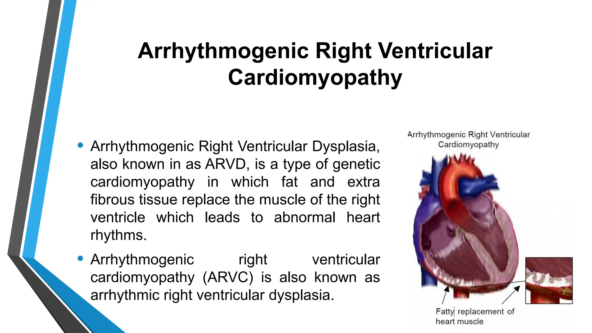
CARDIOMYOPATHIES
MODERATOR PRESENTER
DR SMITA DAS SANDHYA VERMA
ASSOCIATE PROFESSOR MSc NURSING STUDENT
CON,AIIMS CON, AIIMS
OBJECTIVES
1.To introduce the topic.
2.To define cardiomyopathies.
3.To brief anatomy and physiology of heart.
4.To explain classification of cardiomyopathy.
5.To describe different cardiomyopathy with their etiology,
pathophysiology ,clinical manifestation , diagnostic evaluation and its
management.
6.To explain about patient and caregiver teaching’
7.To enlist various preventive approach for cardiomyopathy in general.
CARDIOMYOPATHY
• Cardiomyopathy = Cardio (heart)+ Myo (muscle myocardium)+ pathy
(disease) is a disease of the heart muscle that makes it harder for the
heart to pump blood to the rest of the body.
• Cardiomyopathy (CMP) is a group of diseases that directly affect
myocardial structure or function. CMP can be classified as primary or
secondary.
• The World Health Organization (WHO) defines cardiomyopathies as
diseases of the myocardium associated with cardiac dysfunction.
DEFINITION
• The American Heart Association describes cardiomyopathies as
• “A heterogeneous group of diseases of the myocardium associated
with mechanical and/or electric dysfunction that usually (but not
invariably) exhibit inappropriate ventricular hypertrophy or dilatation
due to a variety of etiologies that are frequently genetic.
Cardiomyopathies are either confined to the heart or part of
generalised systemic disorders and often lead to cardiovascular death
or progressive heart failure-related disability” (Maron et al, 2006).
ANATOMY OF HEART
• The heart has three layers of tissue: the endocardium, myocardium,
and epicardium:
• Endocardium: The thin inner layer that lines the heart chambers and
valves. It is made of connective tissue and smooth muscle fibers.
• Myocardium: The thick middle layer of muscle that contracts and
relaxes to pump blood. The myocardium is especially thick in the left
ventricle, which pumps blood throughout the body.
• Epicardium :Also known as the visceral pericardium, this layer
protects the other heart layers and produces pericardial fluid.
• The heart is also surrounded by a sac called the pericardium, which is
made of two thin layers of tissue. The space between the layers, called
the pericardial cavity, contains pericardial fluid that reduces friction
between the heart and surrounding tissues.
PHYSIOLOGY OF HEART
Cardiac myocytes
• Cardiac muscle cells, or cardiomyocytes, are
striated, branched, and contain many
mitochondria. They have a single nucleus,
surrounded by a cell membrane called the
sarcolemma.
Function
• Cardiac muscle is responsible for pumping
blood, regulating blood flow, and removing
waste products from the body. Cardiac muscle
is under involuntary control. The action potential
of cardiac muscle is activated by changes in the
permeability of the plasma membrane to
potassium, sodium, and calcium ions.
TYPES OF CARDIOMYOPATHY
• Primary CMP refers to those conditions in which the
etiology of the disease is idiopathic. The heart muscle in
this case is the only part of the heart involved, and other
heart structures are unaffected.
• In secondary CMP the cause of the myocardial disease
is known and is secondary to another disease process
TYPES OF CARDIOMYOPATHY
 Dilated cardiomyopathy
 Ischemic cardiomyopathy
 Arrhythmogenic right ventricular dysplasia
 Restrictive Cardiomyopathy
 Takotsubo cardiomyopathy(Broken heart)
 Chemotherapy induced cardiomyopathy
 Alcohol Induced cardiomyopathy
 Peripartum cardiomyopathy
DILATED CARDIOMYOPATHY
• In Dilated Cardiomyopathy, the heart
muscle stretches and becomes thin
that causes dilation and enlargement
of ventricles (mainly left).
• Dilated CMP is characterized by a
diffuse inflammation and rapid
degeneration of heart fibers.
• The heart muscles become too weak
to pump blood efficiently.
• DCM is the most common
cardiomyopathy at 60% of the total
cases.
ETIOLOGY
• Cardiotoxic agents: alcohol, cocaine, doxorubicin
• Coronary artery disease • Genetic (autosomal
dominant)
• Hypertension • Metabolic disorders
• Muscular dystrophy • Myocarditis
• Pregnancy • Valve disease
• Aortic stenosis
Pathophysiology
Exposure to risk factors
Increase in myocardial mass & reduction in
ventricular wall thickness
Globular shape of heart & ventricular chamber
dilation
The heart muscles become too weak to pump blood
efficiently.
Signs of heart failure
Clinical Manifestations : Symptoms
• Develop acutely after an infection or slowly over time
• Decreased exercise capacity
• Fatigue
• Dyspnea at rest
• Paroxysmal nocturnal dyspnea
• Orthopnea
• As the disease progresses, the patient may experience dry cough,
palpitations, abdominal bloating, nausea, vomiting, and anorexia.
• Clot formation and embolization
Clinical Manifestations : Signs
• Abnormal S3 and/or S4
• Dysrhythmias
• Heart murmurs
• Pulmonary crackles
• Edema
• Weak peripheral pulses
• Pallor
• Hepatomegaly
• JVD
Diagnostic studies
.
HISTORY
PHYSICAL
EXAMINATIO
N
ECG CHEST X-RAY
ECHOCARDI
OGRAPHY
BNP MUGA SCAN BIOPSY
Management
Drug Therapy
• Nitrates (e.g., nitroglycerin)
• diuretics (e.g., furosemide) decrease preload,
• ACE inhibitors (eg., captopril) reduce afterload.
• ß-Blockers (e.g., metoprolol) and aldosterone antagonists (eg.,
spironolactone) control the neurohormonal stimulation that occurs in HF.
• Dysrhythmias are treated with antidysrhythmic.
• Anticoagulation therapy
• The use of statins (e.g., atorvastatin)
• Patients are often hospitalized for infusions of dobutamine or milrinone
followed by aggressive diuresis.
Nondrug therapies:
A) Devices to correct arrhythmias
1.Pacemakers
• A pacemaker is a small battery-operated device that helps the heart
beat in a regular rhythm.
• Pacemakers have three parts: a generator, wires (leads) and sensors
(electrodes). Some newer pacemakers are wireless.
• It produces electrical impulses to help control abnormal heartbeats. It's
implanted under the skin through a small incision
• Single-lead pacemakers use one lead usually placed in the right
ventricle (the lower right chamber of your heart).
• Dual-lead pacemakers use one lead in the right atrium and one lead in
the right ventricle.
2.
Implantable cardioverter defibr
illators (ICDs)
.
• An implantable cardioverter defibrillator (ICD) is a
medical device that’s surgically placed under the skin
on chest.
• The battery-powered device constantly tracks heart
rate and rhythm. Its pulse generator delivers an
electric shock when needed to correct arrhythmia.
The leads carry the shock to heart.
• ICDs are used to:
Correct arrythmia,
Prevent sudden cardiac arrest
B) Devices to improve blood flow
1.Cardiac resynchronization therapy (CRT).
• Cardiac resynchronization therapy, also known as CRT or biventricular
pacing, is a procedure for implanting a special type of pacemaker.
• A biventricular pacemaker helps most people who have this
procedure get both of their heart ventricles contracting at the same
time., a
• CRT, or biventricular pacemaker, has three lead:- One lead goes to
each of ventricles. The third goes to right atrium.
• 2.
Left ventricular assist devi
ce (LVAD)
.
• A left ventricular assist device
(LVAD) is a mechanical pump that
providers implant in people who
have heart failure. The device
helps the lower left chamber (left
ventricle) of heart pump blood out
of the ventricle to aorta and the
rest of body. Because it helps left
ventricle, it’s a left ventricular
assist device.
Bridge to transplant (BTT)
• Bridge to transplant (BTT) helps to
survive until patient can receive a
donor’s heart. The LVAD assists
heart and allows to have a better
quality of life and fewer symptoms. A
surgeon removes the device at the
time of transplant.
• The amount of time receive support
from an LVAD until heart
transplantation varies and depends
on medical condition, blood type
and body size.
Destination therapy (DT)
• Destination therapy (DT) is for people with heart failure who aren’t
candidates for heart transplant surgery. considered this only for
people after failure of like medications, lifestyle changes and invasive
procedures procedures.
• A left ventricular assist device supports heart’s function and improve
quality of life for the rest of life.
Heart Transplantation
• DCM is a leading reason for cardiac transplantation.
• A heart transplant, or a cardiac transplant, is a surgical transplant
procedure performed on patients with end-stage heart failure when
other medical or surgical treatments have failed.
Nursing management
1. Control blood pressure.
2. Maintain sinus heart rhythm.
3. Teach the recommended diet.
4. Assist the patient in meal planning.
5. Improve blood flow.
6. Manage chronic conditions.
7. Prevent blood clot formation.
8. Discuss other surgical options.
9. Refer the patient to cardiac rehab.
Hypertrophic Cardiomyopathy
Hypertrophic cardiomyopathy (HCM, or HOCM )
is a condition in which muscle tissues of
the heart become thickened without an obvious
cause. The parts of the heart most commonly
affected are the interventricular septum and
the ventricles This results in the heart being
less able to pump blood effectively and also
may cause electrical conduction problems.
Hypertrophic CMP is the most common cause of
SCD in otherwise healthy young people.
Types of Hypertrophic Cardiomyopathy
• There are two main types of HCM:
• Obstructive: The most common type, hypertrophic obstructive
cardiomyopathy (HOCM) or asymmetric septal hypertrophy means the
wall (septum) between the left ventricle and right ventricle thickens.
This can block (obstruct) blood flow from the left ventricle to the aorta
(the body's main artery).
• Nonobstructive: In nonobstructive HCM, the heart muscle thickens but
doesn't block blood flow.
• It is more common in men than in women. It is usually diagnosed in
young adults and, most often, in active, athletic persons.
Etiology
Genetics : There is a link between genetic
mutations in MYH7 and MYBPC3 (found in
about 50% of cases of HOCMP).
Hypertension,
Diabetes
Thyroid conditions
Pathophysiology
Massive
ventricular
hypertrophy
Rapid, forceful
contraction of the
left ventricle
Impaired
relaxation
(diastole)
Obstruction to
aortic outflow
(not present in all
patients).
(LVOTO)
Clinical Manifestations
• May be asymptomatic
• Exertional dyspnea
• Fatigue
• Angina
• Syncope
• Fatigue
• Common dysrhythmias include supraventricular
tachycardia, atrial fibrillation, ventricular tachycardia,
and ventricular fibrillation.
• Any of these dysrhythmias may lead to syncope or
SCD
Diagnostic Studies
History
Physical
Examinatio
n
ECG
ECHO
Management
Drug Therapy
Goals of care are to improve ventricular filling by reducing ventricular
contractility and relieving left ventricular outflow obstruction.
• ß-blockers (e.g., metoprolol)
• Calcium channel blockers (e.g., verapamil).
• Digitalis is considered if needed to treat atrial fibrillation.
• For patients at risk for SCD, a cardioverter-defibrillator is needed .
• Positive inotropes and chronotropes should be avoided as they
worsen LVOTO.
• Nitrates decrease preload so must be used with caution as that will
also worsen LVOTO.
Surgical Management
Septal myectomy: interventricular
septum (the wall between the left and
right ventricles) to relieve the left
ventricular outflow tract (LVOT)
obstruction. It involves cutting into the
thickened septal muscle and removing
some of the ventricular muscle.
It directly targets the bulky tissue causing
the obstruction and is the most common
surgery for patients with symptomatic
obstructive hypertrophic cardiomyopathy
Surgical Management
Ventriculomyotomy
It involves making incisions in the
hypertrophied muscle of the ventricle to
reduce tension and relieve obstruction.
It is a more conservative approach where
the muscle is cut to release tension but
without removing tissue.
Incisions are made into the thickened
ventricular muscle, typically without excising
any muscle, in an attempt to relieve the
LVOT obstruction.
Interventional procedure :
Alcohol-induced, percutaneous transluminal
septal myocardial ablation
• This procedure consists of injecting alcohol
into the first septal artery branching off the left
anterior descending artery.
• This causes ischemia and septal wall
infarction. Ablation of the septal wall
decreases the obstruction to flow, and the
patient's symptoms decrease.
• The procedure improves HF symptoms and
exercise capacity 3 months after ablation.
Interventional procedure:
RFA catheter ablation
Radiofrequency ablation (RA) is a minimally
invasive procedure that can be used). It's an
alternative to surgical myectomy or alcohol
septal ablation.
Ablation is done through a procedure called
cardiac catheterisation that passes flexible
tubes, or catheters, through blood vessels until
they reach heart.
A machine will send either radiofrequency (RF)
waves, extremely cold temperatures, or laser
light through the catheter tip to create a scar on
the heart. The scar prevents abnormal electrical
Nursing interventions for hypertrophic CMP
• Teach patient avoid strenuous activity and dehydration.
• Any activity that causes an increase in systemic vascular resistance
(thus increasing the obstruction to forward flow) is dangerous and
should be avoided.
• Rest and elevation of the feet to improve venous return to the heart
can manage chest pain in these patients.
• Vasodilators such as nitroglycerin may worsen the chest pain by
decreasing venous return and further increasing obstruction of blood
flow from the heart.
Restrictive Cardiomyopathy
• The heart's ventricles become rigid, stiff because
abnormal tissue, such as scar tissue, replaces the
normal heart muscle. As a result, the ventricles
can't relax normally and fill with blood, and the
atria become enlarged.
• It It is a disease of the myocardium that impairs
diastolic filling and stretch, Systolic function
remains unchanged.
• With restrictive CMP, the ventricles are resistant to
filling and therefore demand high diastolic filling
pressures to maintain CO
Etiology
• Amyloidosis
• Endomyocardial fibrosis
• Neoplastic tumor
• Post-radiation therapy
• Sarcoidosis
• Ventricular thrombus
Pathophysiology
An increase in
stiffness of the
ventricular walls,
Causes impaired
diastolic filling of the
ventricle and heart
failure
The ventricles are
resistant to filling and
therefore
Demand high
diastolic filling
pressures to maintain
CO
Increase RA & LA
pressure leads to
systemic venous
congestion &
pulmonary
congestion
Symptoms of heart
failure
Clinical Manifestations.
• Fatigue
• Exercise intolerance
• Dyspnea.
• Peripheral edema
• Hepatomegaly,
• Ascites
• Anasarca.
• Angina
• Orthopnea
• Syncope
• Palpitations
• Weight gain
• JVD
Diagnostic Studies
History
Physical
Examination
ECG
ECHO
Endomyocardi
al Biopsy
Medical Management
• Currently, there is no specific treatment for
restrictive CMP.
• Supportive managements includes
diuretics, corticosteroids, or anticoagulants,
depending on the etiology and
manifestations.
• Cardiac pacing may improve symptoms
• Treatment includes conventional therapy
for HF and dysrhythmias.
• Heart transplant may also be a
consideration.
Nursing management
• Assess heart rate, rhythm, and pulses.
• Monitor blood pressure.
• Assess and monitor urine output.
• Explain regarding importance of treatment compliance.
• Monitor for early signs of heart failure.
• Pace activities and rest.
Arrhythmogenic Right Ventricular
Cardiomyopathy
• Arrhythmogenic Right Ventricular Dysplasia,
also known in as ARVD, is a type of genetic
cardiomyopathy in which fat and extra
fibrous tissue replace the muscle of the right
ventricle which leads to abnormal heart
rhythms.
• Arrhythmogenic right ventricular
cardiomyopathy (ARVC) is also known as
arrhythmic right ventricular dysplasia.
Etiology
• Familial disease is common in ARVC,
typically with autosomal dominant
inheritance and incomplete penetrance.
A recessive form has also been
identified.
• Several genes and gene loci are
associated with ARVC.
Pathophysiology
The fibrofatty mechanism of this
disease leads to the progressive loss
of myocytes. ARVC may also involve
the left ventricle.
Structural and functional
abnormalities of the right ventricle
lead to arrhythmias and progressive
right ventricular failure.
Diagnostic Studies
History
Physical
Examination
ECG
ECHO
Endomyocardi
al Biopsy
Medical management
• ICD
• Antiarrhythmic therapy
• Neurohormonal blockade with ACE
inhibitors
• In individuals progressing to overt heart
failure, management involves the same
principles for the treatment of other forms
of cardiomyopathy.
• Heart transplantation is indicated for
patients overt cardiac failure.
Nursing Management
1. Encourage the patient to
rest.
2. Provide supplemental
oxygen.
3. Administer medications as
indicated.
4. Prepare for ICD insertion.
Takotsubo cardiomyopathy
• TCM is a transient heart syndrome that mimics acute coronary
syndrome.
• Patients often have chest pain, ST segment elevation on ECG, and
elevated cardiac biomarkers consistent with an MI. However, when the
patient undergoes heart catheterization, there is no significant
coronary artery disease.
• TCM is an acute, stress-related syndrome that is more common in
postmenopausal women. Most patients recover rapidly without
lasting symptoms.
NURSING DIAGNOSIS
• 1. Decreased Cardiac Output related to altered contractility of the heart,
structural changes, or valvular dysfunction as evidenced by fatigue,
dyspnea, reduced peripheral pulses, hypotension, edema, and cool
extremities.
• 2. Activity Intolerance related to decreased oxygen delivery, fatigue, or
decreased cardiac output evidenced by Shortness of breath, fatigue upon
exertion, and reports of feeling exhausted after minimal activities.
• 3. Impaired Gas Exchange related to fluid accumulation in the lungs
(pulmonary edema) secondary to heart failure evidenced by crackles on
lung auscultation, dyspnea, decreased oxygen saturation, and cyanosis.
NURSING DIAGNOSIS
• 4. Excess Fluid Volume related to compromised heart function leading
to fluid retention as evidenced by peripheral edema, weight gain,
jugular vein distension, and ascites.
• 5. Acute Pain related to myocardial ischemia or inflammation of the
heart muscle as evidenced by chest pain, changes in heart rate, and
elevated blood pressure during episodes of pain.
• 6. Ineffective Tissue Perfusion (Peripheral) related to decreased
cardiac output leading to reduced blood flow to tissues as evidenced
by cool, pale extremities, delayed capillary refill, and diminished
pulses.
Patient & Caregiver Teaching
1. Take all drugs as prescribed and regularly follow up with HCP.
2. Follow a low-sodium diet (if ordered) and read all product labels for sodium
content.
4. Regular blood pressure chek up.
3. Achieve and maintain a reasonable weight and avoid large meals.
5. Avoid alcohol, caffeine, diet pills, and over-the-counter cold medicines that may
contain stimulants.
6. Balance activity and rest periods.
7. Avoid heavy lifting or vigorous isometric exercises.
8. Use stress management techniques: relaxation breathing, guided imagery.
9. Report any signs of heart failure to HCP, including weight gain, edema,
dyspnea.
10. Encourage caregivers to learn CPR because of the potential for cardiac
Prevention for cardiomyopathies
• Lifestyle changes
• Healthy diet and physical activity
• Choose foods and beverages that are low in added sugar and
salt
• Avoid drinking alcohol
• Manage healthy weight
• Quit smoking and tobacco usage.
• Manage stress
• Managing underlying conditions such as hypertension, sleep
apnea or diabetes
• Scheduling regular checkups with a healthcare provider
• Taking all medications as prescribed
CONCLUSION
Cardiomyopathy is a serious condition characterized by the
weakening or abnormal functioning of the heart muscle, leading
to impaired cardiac function. It can result in complications like
heart failure, arrhythmias, and even sudden cardiac arrest if not
managed appropriately. While cardiomyopathy can be caused
by genetic factors, infections, chronic conditions like
hypertension or diabetes, and lifestyle factors, early diagnosis
and tailored treatment are crucial for improving outcomes.
• a
cardiomyopathies, types and nursing management.pptx

cardiomyopathies, types and nursing management.pptx

  • 1.
    CARDIOMYOPATHIES MODERATOR PRESENTER DR SMITADAS SANDHYA VERMA ASSOCIATE PROFESSOR MSc NURSING STUDENT CON,AIIMS CON, AIIMS
  • 2.
    OBJECTIVES 1.To introduce thetopic. 2.To define cardiomyopathies. 3.To brief anatomy and physiology of heart. 4.To explain classification of cardiomyopathy. 5.To describe different cardiomyopathy with their etiology, pathophysiology ,clinical manifestation , diagnostic evaluation and its management. 6.To explain about patient and caregiver teaching’ 7.To enlist various preventive approach for cardiomyopathy in general.
  • 3.
    CARDIOMYOPATHY • Cardiomyopathy =Cardio (heart)+ Myo (muscle myocardium)+ pathy (disease) is a disease of the heart muscle that makes it harder for the heart to pump blood to the rest of the body. • Cardiomyopathy (CMP) is a group of diseases that directly affect myocardial structure or function. CMP can be classified as primary or secondary. • The World Health Organization (WHO) defines cardiomyopathies as diseases of the myocardium associated with cardiac dysfunction.
  • 4.
    DEFINITION • The AmericanHeart Association describes cardiomyopathies as • “A heterogeneous group of diseases of the myocardium associated with mechanical and/or electric dysfunction that usually (but not invariably) exhibit inappropriate ventricular hypertrophy or dilatation due to a variety of etiologies that are frequently genetic. Cardiomyopathies are either confined to the heart or part of generalised systemic disorders and often lead to cardiovascular death or progressive heart failure-related disability” (Maron et al, 2006).
  • 5.
    ANATOMY OF HEART •The heart has three layers of tissue: the endocardium, myocardium, and epicardium: • Endocardium: The thin inner layer that lines the heart chambers and valves. It is made of connective tissue and smooth muscle fibers. • Myocardium: The thick middle layer of muscle that contracts and relaxes to pump blood. The myocardium is especially thick in the left ventricle, which pumps blood throughout the body. • Epicardium :Also known as the visceral pericardium, this layer protects the other heart layers and produces pericardial fluid. • The heart is also surrounded by a sac called the pericardium, which is made of two thin layers of tissue. The space between the layers, called the pericardial cavity, contains pericardial fluid that reduces friction between the heart and surrounding tissues.
  • 7.
    PHYSIOLOGY OF HEART Cardiacmyocytes • Cardiac muscle cells, or cardiomyocytes, are striated, branched, and contain many mitochondria. They have a single nucleus, surrounded by a cell membrane called the sarcolemma. Function • Cardiac muscle is responsible for pumping blood, regulating blood flow, and removing waste products from the body. Cardiac muscle is under involuntary control. The action potential of cardiac muscle is activated by changes in the permeability of the plasma membrane to potassium, sodium, and calcium ions.
  • 8.
    TYPES OF CARDIOMYOPATHY •Primary CMP refers to those conditions in which the etiology of the disease is idiopathic. The heart muscle in this case is the only part of the heart involved, and other heart structures are unaffected. • In secondary CMP the cause of the myocardial disease is known and is secondary to another disease process
  • 9.
    TYPES OF CARDIOMYOPATHY Dilated cardiomyopathy  Ischemic cardiomyopathy  Arrhythmogenic right ventricular dysplasia  Restrictive Cardiomyopathy  Takotsubo cardiomyopathy(Broken heart)  Chemotherapy induced cardiomyopathy  Alcohol Induced cardiomyopathy  Peripartum cardiomyopathy
  • 10.
    DILATED CARDIOMYOPATHY • InDilated Cardiomyopathy, the heart muscle stretches and becomes thin that causes dilation and enlargement of ventricles (mainly left). • Dilated CMP is characterized by a diffuse inflammation and rapid degeneration of heart fibers. • The heart muscles become too weak to pump blood efficiently. • DCM is the most common cardiomyopathy at 60% of the total cases.
  • 11.
    ETIOLOGY • Cardiotoxic agents:alcohol, cocaine, doxorubicin • Coronary artery disease • Genetic (autosomal dominant) • Hypertension • Metabolic disorders • Muscular dystrophy • Myocarditis • Pregnancy • Valve disease • Aortic stenosis
  • 12.
    Pathophysiology Exposure to riskfactors Increase in myocardial mass & reduction in ventricular wall thickness Globular shape of heart & ventricular chamber dilation The heart muscles become too weak to pump blood efficiently. Signs of heart failure
  • 13.
    Clinical Manifestations :Symptoms • Develop acutely after an infection or slowly over time • Decreased exercise capacity • Fatigue • Dyspnea at rest • Paroxysmal nocturnal dyspnea • Orthopnea • As the disease progresses, the patient may experience dry cough, palpitations, abdominal bloating, nausea, vomiting, and anorexia. • Clot formation and embolization
  • 14.
    Clinical Manifestations :Signs • Abnormal S3 and/or S4 • Dysrhythmias • Heart murmurs • Pulmonary crackles • Edema • Weak peripheral pulses • Pallor • Hepatomegaly • JVD
  • 15.
    Diagnostic studies . HISTORY PHYSICAL EXAMINATIO N ECG CHESTX-RAY ECHOCARDI OGRAPHY BNP MUGA SCAN BIOPSY
  • 16.
    Management Drug Therapy • Nitrates(e.g., nitroglycerin) • diuretics (e.g., furosemide) decrease preload, • ACE inhibitors (eg., captopril) reduce afterload. • ß-Blockers (e.g., metoprolol) and aldosterone antagonists (eg., spironolactone) control the neurohormonal stimulation that occurs in HF. • Dysrhythmias are treated with antidysrhythmic. • Anticoagulation therapy • The use of statins (e.g., atorvastatin) • Patients are often hospitalized for infusions of dobutamine or milrinone followed by aggressive diuresis.
  • 17.
    Nondrug therapies: A) Devicesto correct arrhythmias 1.Pacemakers • A pacemaker is a small battery-operated device that helps the heart beat in a regular rhythm. • Pacemakers have three parts: a generator, wires (leads) and sensors (electrodes). Some newer pacemakers are wireless. • It produces electrical impulses to help control abnormal heartbeats. It's implanted under the skin through a small incision • Single-lead pacemakers use one lead usually placed in the right ventricle (the lower right chamber of your heart). • Dual-lead pacemakers use one lead in the right atrium and one lead in the right ventricle.
  • 18.
    2. Implantable cardioverter defibr illators(ICDs) . • An implantable cardioverter defibrillator (ICD) is a medical device that’s surgically placed under the skin on chest. • The battery-powered device constantly tracks heart rate and rhythm. Its pulse generator delivers an electric shock when needed to correct arrhythmia. The leads carry the shock to heart. • ICDs are used to: Correct arrythmia, Prevent sudden cardiac arrest
  • 19.
    B) Devices toimprove blood flow 1.Cardiac resynchronization therapy (CRT). • Cardiac resynchronization therapy, also known as CRT or biventricular pacing, is a procedure for implanting a special type of pacemaker. • A biventricular pacemaker helps most people who have this procedure get both of their heart ventricles contracting at the same time., a • CRT, or biventricular pacemaker, has three lead:- One lead goes to each of ventricles. The third goes to right atrium.
  • 20.
    • 2. Left ventricularassist devi ce (LVAD) . • A left ventricular assist device (LVAD) is a mechanical pump that providers implant in people who have heart failure. The device helps the lower left chamber (left ventricle) of heart pump blood out of the ventricle to aorta and the rest of body. Because it helps left ventricle, it’s a left ventricular assist device.
  • 21.
    Bridge to transplant(BTT) • Bridge to transplant (BTT) helps to survive until patient can receive a donor’s heart. The LVAD assists heart and allows to have a better quality of life and fewer symptoms. A surgeon removes the device at the time of transplant. • The amount of time receive support from an LVAD until heart transplantation varies and depends on medical condition, blood type and body size.
  • 22.
    Destination therapy (DT) •Destination therapy (DT) is for people with heart failure who aren’t candidates for heart transplant surgery. considered this only for people after failure of like medications, lifestyle changes and invasive procedures procedures. • A left ventricular assist device supports heart’s function and improve quality of life for the rest of life.
  • 23.
    Heart Transplantation • DCMis a leading reason for cardiac transplantation. • A heart transplant, or a cardiac transplant, is a surgical transplant procedure performed on patients with end-stage heart failure when other medical or surgical treatments have failed.
  • 24.
    Nursing management 1. Controlblood pressure. 2. Maintain sinus heart rhythm. 3. Teach the recommended diet. 4. Assist the patient in meal planning. 5. Improve blood flow. 6. Manage chronic conditions. 7. Prevent blood clot formation. 8. Discuss other surgical options. 9. Refer the patient to cardiac rehab.
  • 25.
    Hypertrophic Cardiomyopathy Hypertrophic cardiomyopathy(HCM, or HOCM ) is a condition in which muscle tissues of the heart become thickened without an obvious cause. The parts of the heart most commonly affected are the interventricular septum and the ventricles This results in the heart being less able to pump blood effectively and also may cause electrical conduction problems. Hypertrophic CMP is the most common cause of SCD in otherwise healthy young people.
  • 26.
    Types of HypertrophicCardiomyopathy • There are two main types of HCM: • Obstructive: The most common type, hypertrophic obstructive cardiomyopathy (HOCM) or asymmetric septal hypertrophy means the wall (septum) between the left ventricle and right ventricle thickens. This can block (obstruct) blood flow from the left ventricle to the aorta (the body's main artery). • Nonobstructive: In nonobstructive HCM, the heart muscle thickens but doesn't block blood flow. • It is more common in men than in women. It is usually diagnosed in young adults and, most often, in active, athletic persons.
  • 27.
    Etiology Genetics : Thereis a link between genetic mutations in MYH7 and MYBPC3 (found in about 50% of cases of HOCMP). Hypertension, Diabetes Thyroid conditions
  • 28.
    Pathophysiology Massive ventricular hypertrophy Rapid, forceful contraction ofthe left ventricle Impaired relaxation (diastole) Obstruction to aortic outflow (not present in all patients). (LVOTO)
  • 29.
    Clinical Manifestations • Maybe asymptomatic • Exertional dyspnea • Fatigue • Angina • Syncope • Fatigue • Common dysrhythmias include supraventricular tachycardia, atrial fibrillation, ventricular tachycardia, and ventricular fibrillation. • Any of these dysrhythmias may lead to syncope or SCD
  • 30.
  • 31.
    Management Drug Therapy Goals ofcare are to improve ventricular filling by reducing ventricular contractility and relieving left ventricular outflow obstruction. • ß-blockers (e.g., metoprolol) • Calcium channel blockers (e.g., verapamil). • Digitalis is considered if needed to treat atrial fibrillation. • For patients at risk for SCD, a cardioverter-defibrillator is needed . • Positive inotropes and chronotropes should be avoided as they worsen LVOTO. • Nitrates decrease preload so must be used with caution as that will also worsen LVOTO.
  • 32.
    Surgical Management Septal myectomy:interventricular septum (the wall between the left and right ventricles) to relieve the left ventricular outflow tract (LVOT) obstruction. It involves cutting into the thickened septal muscle and removing some of the ventricular muscle. It directly targets the bulky tissue causing the obstruction and is the most common surgery for patients with symptomatic obstructive hypertrophic cardiomyopathy
  • 33.
    Surgical Management Ventriculomyotomy It involvesmaking incisions in the hypertrophied muscle of the ventricle to reduce tension and relieve obstruction. It is a more conservative approach where the muscle is cut to release tension but without removing tissue. Incisions are made into the thickened ventricular muscle, typically without excising any muscle, in an attempt to relieve the LVOT obstruction.
  • 34.
    Interventional procedure : Alcohol-induced,percutaneous transluminal septal myocardial ablation • This procedure consists of injecting alcohol into the first septal artery branching off the left anterior descending artery. • This causes ischemia and septal wall infarction. Ablation of the septal wall decreases the obstruction to flow, and the patient's symptoms decrease. • The procedure improves HF symptoms and exercise capacity 3 months after ablation.
  • 35.
    Interventional procedure: RFA catheterablation Radiofrequency ablation (RA) is a minimally invasive procedure that can be used). It's an alternative to surgical myectomy or alcohol septal ablation. Ablation is done through a procedure called cardiac catheterisation that passes flexible tubes, or catheters, through blood vessels until they reach heart. A machine will send either radiofrequency (RF) waves, extremely cold temperatures, or laser light through the catheter tip to create a scar on the heart. The scar prevents abnormal electrical
  • 36.
    Nursing interventions forhypertrophic CMP • Teach patient avoid strenuous activity and dehydration. • Any activity that causes an increase in systemic vascular resistance (thus increasing the obstruction to forward flow) is dangerous and should be avoided. • Rest and elevation of the feet to improve venous return to the heart can manage chest pain in these patients. • Vasodilators such as nitroglycerin may worsen the chest pain by decreasing venous return and further increasing obstruction of blood flow from the heart.
  • 37.
    Restrictive Cardiomyopathy • Theheart's ventricles become rigid, stiff because abnormal tissue, such as scar tissue, replaces the normal heart muscle. As a result, the ventricles can't relax normally and fill with blood, and the atria become enlarged. • It It is a disease of the myocardium that impairs diastolic filling and stretch, Systolic function remains unchanged. • With restrictive CMP, the ventricles are resistant to filling and therefore demand high diastolic filling pressures to maintain CO
  • 38.
    Etiology • Amyloidosis • Endomyocardialfibrosis • Neoplastic tumor • Post-radiation therapy • Sarcoidosis • Ventricular thrombus
  • 39.
    Pathophysiology An increase in stiffnessof the ventricular walls, Causes impaired diastolic filling of the ventricle and heart failure The ventricles are resistant to filling and therefore Demand high diastolic filling pressures to maintain CO Increase RA & LA pressure leads to systemic venous congestion & pulmonary congestion Symptoms of heart failure
  • 40.
    Clinical Manifestations. • Fatigue •Exercise intolerance • Dyspnea. • Peripheral edema • Hepatomegaly, • Ascites • Anasarca. • Angina • Orthopnea • Syncope • Palpitations • Weight gain • JVD
  • 41.
  • 42.
    Medical Management • Currently,there is no specific treatment for restrictive CMP. • Supportive managements includes diuretics, corticosteroids, or anticoagulants, depending on the etiology and manifestations. • Cardiac pacing may improve symptoms • Treatment includes conventional therapy for HF and dysrhythmias. • Heart transplant may also be a consideration.
  • 43.
    Nursing management • Assessheart rate, rhythm, and pulses. • Monitor blood pressure. • Assess and monitor urine output. • Explain regarding importance of treatment compliance. • Monitor for early signs of heart failure. • Pace activities and rest.
  • 44.
    Arrhythmogenic Right Ventricular Cardiomyopathy •Arrhythmogenic Right Ventricular Dysplasia, also known in as ARVD, is a type of genetic cardiomyopathy in which fat and extra fibrous tissue replace the muscle of the right ventricle which leads to abnormal heart rhythms. • Arrhythmogenic right ventricular cardiomyopathy (ARVC) is also known as arrhythmic right ventricular dysplasia.
  • 45.
    Etiology • Familial diseaseis common in ARVC, typically with autosomal dominant inheritance and incomplete penetrance. A recessive form has also been identified. • Several genes and gene loci are associated with ARVC.
  • 46.
    Pathophysiology The fibrofatty mechanismof this disease leads to the progressive loss of myocytes. ARVC may also involve the left ventricle. Structural and functional abnormalities of the right ventricle lead to arrhythmias and progressive right ventricular failure.
  • 47.
  • 48.
    Medical management • ICD •Antiarrhythmic therapy • Neurohormonal blockade with ACE inhibitors • In individuals progressing to overt heart failure, management involves the same principles for the treatment of other forms of cardiomyopathy. • Heart transplantation is indicated for patients overt cardiac failure.
  • 49.
    Nursing Management 1. Encouragethe patient to rest. 2. Provide supplemental oxygen. 3. Administer medications as indicated. 4. Prepare for ICD insertion.
  • 50.
    Takotsubo cardiomyopathy • TCMis a transient heart syndrome that mimics acute coronary syndrome. • Patients often have chest pain, ST segment elevation on ECG, and elevated cardiac biomarkers consistent with an MI. However, when the patient undergoes heart catheterization, there is no significant coronary artery disease. • TCM is an acute, stress-related syndrome that is more common in postmenopausal women. Most patients recover rapidly without lasting symptoms.
  • 51.
    NURSING DIAGNOSIS • 1.Decreased Cardiac Output related to altered contractility of the heart, structural changes, or valvular dysfunction as evidenced by fatigue, dyspnea, reduced peripheral pulses, hypotension, edema, and cool extremities. • 2. Activity Intolerance related to decreased oxygen delivery, fatigue, or decreased cardiac output evidenced by Shortness of breath, fatigue upon exertion, and reports of feeling exhausted after minimal activities. • 3. Impaired Gas Exchange related to fluid accumulation in the lungs (pulmonary edema) secondary to heart failure evidenced by crackles on lung auscultation, dyspnea, decreased oxygen saturation, and cyanosis.
  • 52.
    NURSING DIAGNOSIS • 4.Excess Fluid Volume related to compromised heart function leading to fluid retention as evidenced by peripheral edema, weight gain, jugular vein distension, and ascites. • 5. Acute Pain related to myocardial ischemia or inflammation of the heart muscle as evidenced by chest pain, changes in heart rate, and elevated blood pressure during episodes of pain. • 6. Ineffective Tissue Perfusion (Peripheral) related to decreased cardiac output leading to reduced blood flow to tissues as evidenced by cool, pale extremities, delayed capillary refill, and diminished pulses.
  • 53.
    Patient & CaregiverTeaching 1. Take all drugs as prescribed and regularly follow up with HCP. 2. Follow a low-sodium diet (if ordered) and read all product labels for sodium content. 4. Regular blood pressure chek up. 3. Achieve and maintain a reasonable weight and avoid large meals. 5. Avoid alcohol, caffeine, diet pills, and over-the-counter cold medicines that may contain stimulants. 6. Balance activity and rest periods. 7. Avoid heavy lifting or vigorous isometric exercises. 8. Use stress management techniques: relaxation breathing, guided imagery. 9. Report any signs of heart failure to HCP, including weight gain, edema, dyspnea. 10. Encourage caregivers to learn CPR because of the potential for cardiac
  • 54.
    Prevention for cardiomyopathies •Lifestyle changes • Healthy diet and physical activity • Choose foods and beverages that are low in added sugar and salt • Avoid drinking alcohol • Manage healthy weight • Quit smoking and tobacco usage. • Manage stress • Managing underlying conditions such as hypertension, sleep apnea or diabetes • Scheduling regular checkups with a healthcare provider • Taking all medications as prescribed
  • 55.
    CONCLUSION Cardiomyopathy is aserious condition characterized by the weakening or abnormal functioning of the heart muscle, leading to impaired cardiac function. It can result in complications like heart failure, arrhythmias, and even sudden cardiac arrest if not managed appropriately. While cardiomyopathy can be caused by genetic factors, infections, chronic conditions like hypertension or diabetes, and lifestyle factors, early diagnosis and tailored treatment are crucial for improving outcomes.
  • 56.