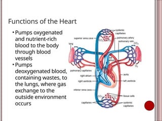
Functions of theHeart
• Pumps oxygenated
and nutrient-rich
blood to the body
through blood
vessels
• Pumps
deoxygenated blood,
containing wastes, to
the lungs, where gas
exchange to the
outside environment
occurs
3.
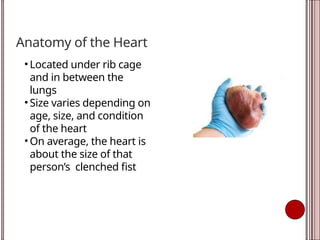
Anatomy of theHeart
• Located under rib cage
and in between the
lungs
• Size varies depending on
age, size, and condition
of the heart
• On average, the heart is
about the size of that
person’s clenched fist
4.
Anatomical relationship
• Anteriorly:The heart is positioned behind the body of the
sternum (breastbone) and the adjoining costal cartilages
(rib cartilages).
• Posteriorly: It lies in front of the thoracic vertebrae
(specifically T5-T8 when lying down). The esophagus,
descending thoracic aorta, and other veins are also
located behind it.
• Inferiorly: The heart rests on the central tendon of the
diaphragm, the muscle that separates the chest from the
abdomen
5.
Anatomical relationship
• Laterally:It is situated between the right and left lungs,
which are enclosed by the pleura (a membrane). The left
lung has a slight indentation called the "cardiac notch" to
accommodate the heart's position, which is tilted slightly
to the left.
• Superiorly (Base): The broader upper part (base) is the
attachment point for several large blood vessels, including
the superior and inferior venae cavae, the aorta, and the
pulmonary trunk.
• Inferiorly (Apex): The pointed lower tip (apex) is formed by
the left ventricle and points downward, forward, and to
the left, typically located at the level of the fifth left
intercostal space, medial to the midclavicular line.
6.
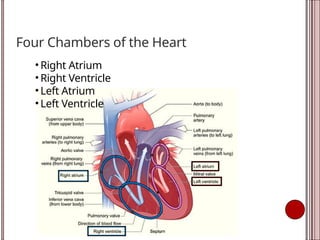
Four Chambers ofthe Heart
• Right Atrium
• Right Ventricle
• Left Atrium
• Left Ventricle
7.
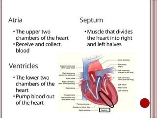
Atria
• The uppertwo
chambers of the heart
• Receive and collect
blood
Ventricles
• The lower two
chambers of the
heart
• Pump blood out
of the heart
Septum
• Muscle that divides
the heart into right
and left halves
8.
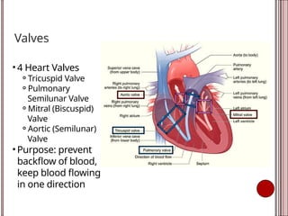
Valves
• 4 HeartValves
⚬Tricuspid Valve
⚬Pulmonary
Semilunar Valve
⚬Mitral (Biscuspid)
Valve
⚬Aortic (Semilunar)
Valve
• Purpose: prevent
backflow of blood,
keep blood flowing
in one direction
9.
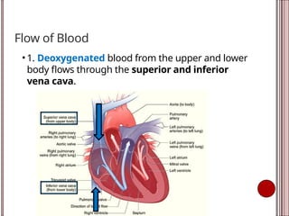
Flow of Blood
•1. Deoxygenated blood from the upper and lower
body flows through the superior and inferior
vena cava.
10.
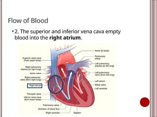
Flow of Blood
•2. The superior and inferior vena cava empty
blood into the right atrium.
11.
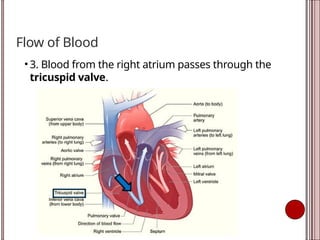
Flow of Blood
•3. Blood from the right atrium passes through the
tricuspid valve.
12.
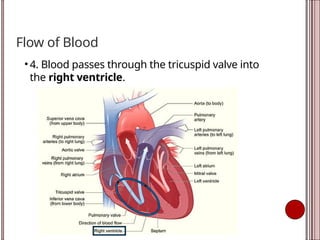
Flow of Blood
•4. Blood passes through the tricuspid valve into
the right ventricle.
13.
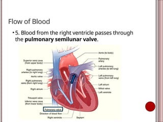
Flow of Blood
•5. Blood from the right ventricle passes through
the pulmonary semilunar valve.
14.
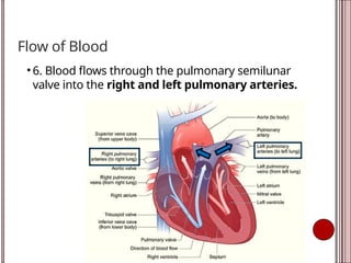
Flow of Blood
•6. Blood flows through the pulmonary semilunar
valve into the right and left pulmonary arteries.
15.
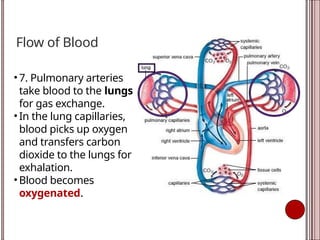
Flow of Blood
•7. Pulmonary arteries
take blood to the lungs
for gas exchange.
• In the lung capillaries,
blood picks up oxygen
and transfers carbon
dioxide to the lungs for
exhalation.
• Blood becomes
oxygenated.
16.
Flow of Blood
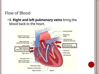
•8. Right and left pulmonary veins bring the
blood back to the heart.
17.
Flow of Blood
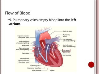
•9. Pulmonary veins empty blood into the left
atrium.
18.
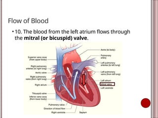
Flow of Blood
•10. The blood from the left atrium flows through
the mitral (or bicuspid) valve.
19.
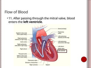
Flow of Blood
•11. After passing through the mitral valve, blood
enters the left ventricle.
20.
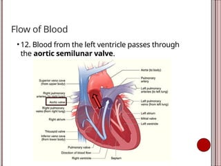
Flow of Blood
•12. Blood from the left ventricle passes through
the aortic semilunar valve.
21.
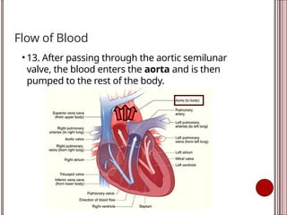
Flow of Blood
•13. After passing through the aortic semilunar
valve, the blood enters the aorta and is then
pumped to the rest of the body.
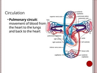
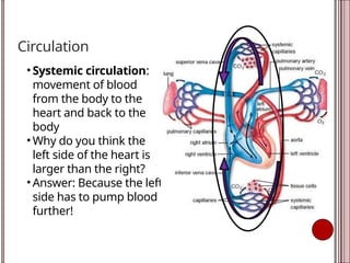
Circulation
• Systemic circulation:
movementof blood
from the body to the
heart and back to the
body
• Why do you think the
left side of the heart is
larger than the right?
• Answer: Because the left
side has to pump blood
further!
24.
Contraction
• Systole -contract
⚬Atrial Systole: when the atria contract and pump blood
into the ventricles
⚬Ventricular Systole: when the ventricles contract and
pump blood out of the heart to the lungs or body
• Diastole - relax
⚬When the atria and ventricles relax and start to fill with
blood
25.
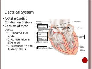
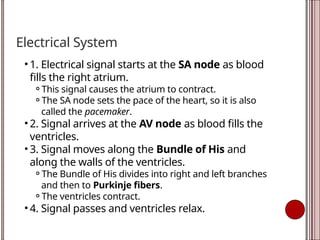
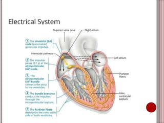
Electrical System
• AKAthe Cardiac
Conduction System
• Consists of three
parts:
⚬1. Sinoatrial (SA)
node
⚬2. Atrioventricular
(AV) node
⚬3. Bundle of His and
Purkinje fibers
26.
Electrical System
• 1.Electrical signal starts at the SA node as blood
fills the right atrium.
⚬This signal causes the atrium to contract.
⚬The SA node sets the pace of the heart, so it is also
called the pacemaker.
• 2. Signal arrives at the AV node as blood fills the
ventricles.
• 3. Signal moves along the Bundle of His and
along the walls of the ventricles.
⚬The Bundle of His divides into right and left branches
and then to Purkinje fibers.
⚬The ventricles contract.
• 4. Signal passes and ventricles relax.
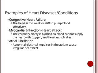
Examples of HeartDiseases/Conditions
• Congestive Heart Failure
⚬The heart is too weak or stiff to pump blood
effectively.
• Myocardial Infarction (Heart attack!)
⚬The coronary artery is blocked so blood cannot supply
the heart with oxygen, and heart muscle dies.
• Atrial Fibrillation
⚬Abnormal electrical impulses in the atrium cause
irregular heart beat.
#23 See if students can answer this question about why the left side of the heart is larger. The left side of the heart is responsible for pumping blood to the body (from head to toe). This covers a much larger distance than to just the lungs, which is what the right side is responsible for.
23