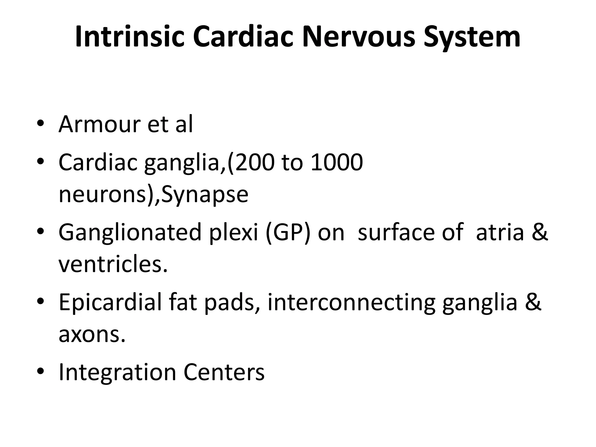
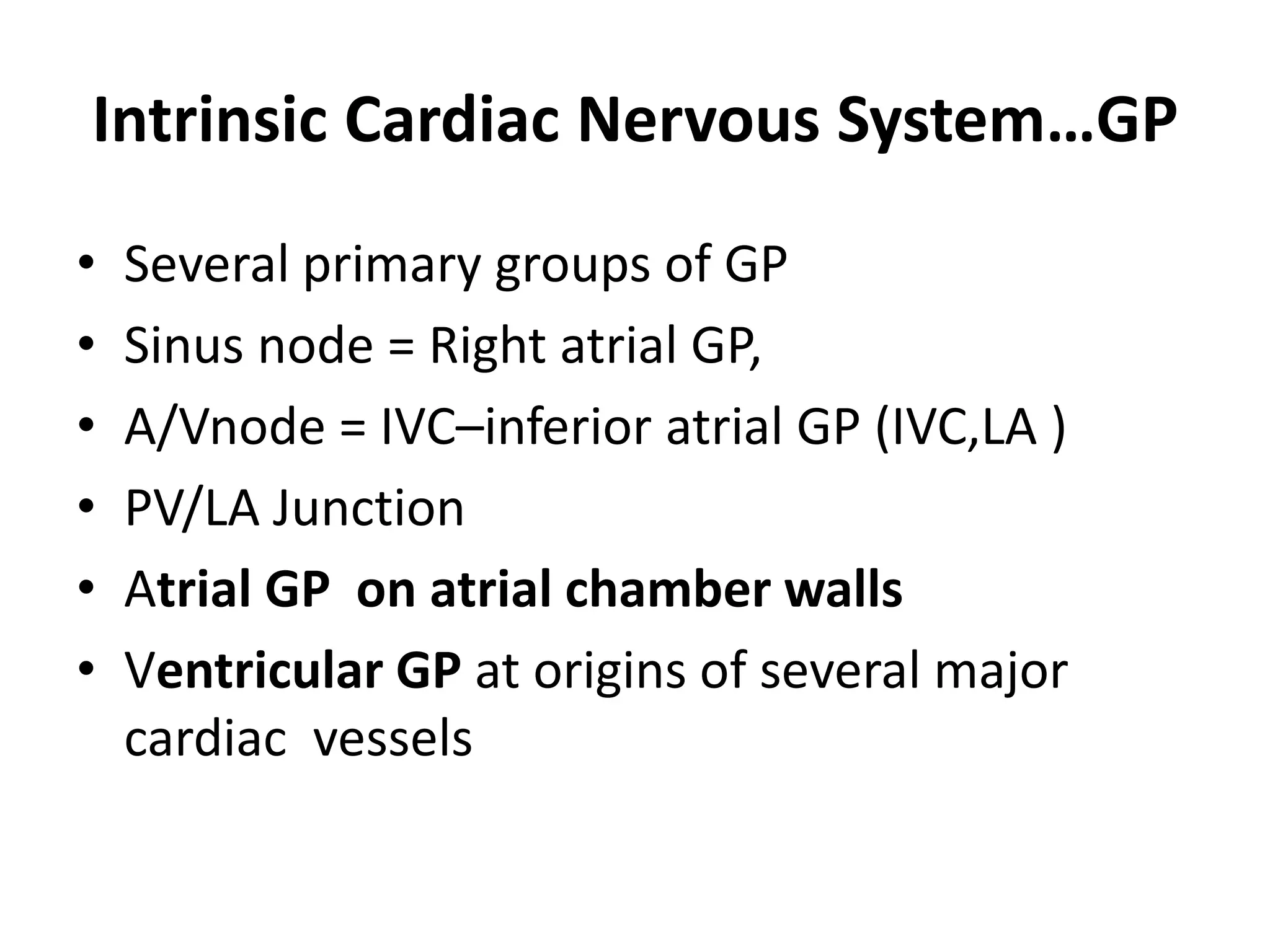
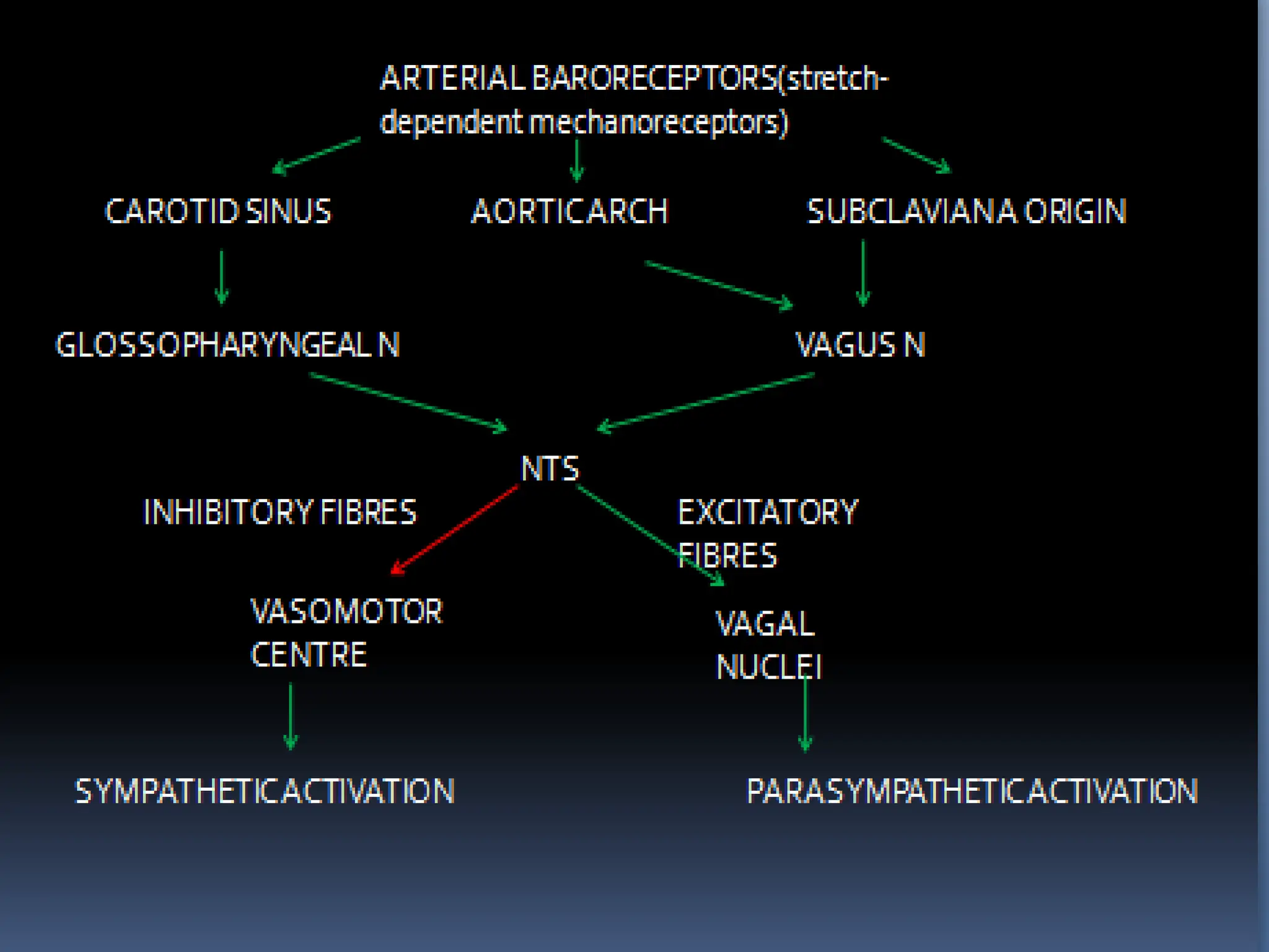
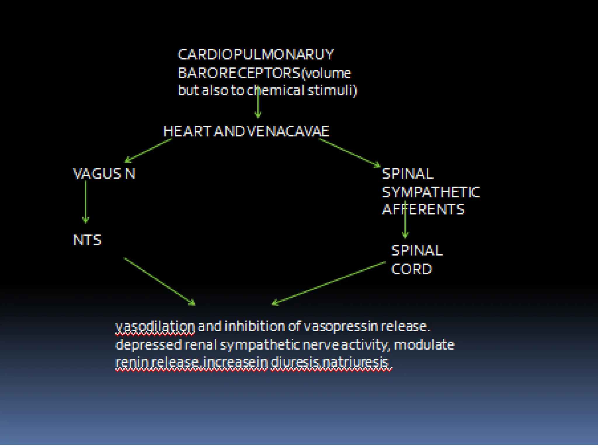
CARDIAC AUTONOMIC SYSTEM
CLINICAL SIGNIFICANCE
LONG SEMINAR
BROAD HEADINGS
• ANATOMY & PHYSIOLOGY
• ANS EVALUATION & ORTHOSTATIC
INTOLERANCE
• ANS & ARRYTHMIAS
• ANS & HTN
• ANS & HF
INTRODUCTION
• 1628, William Harvey hinted at link B/W
brain & heart when he wrote, “For every
affection of mind that is attended with
either pain or pleasure, hope or fear, is the
cause of an agitation whose influence
extends to heart.”
INTRODUCTION
• Numerous anatomic & physiological studies
of cardiac ANS investigated this link & found it
to be very complex.
• Autonomic activation alters not only heart
rate, conduction, & hemodynamics, but also
cellular & subcellular properties of individual
myocytes.
• ANATOMY & PHYSIOLOGY
Normal Autonomic Innervation of the
Heart
Extrinsic & Intrinsic
• Extrinsic comprises fibers mediate
connections B/W heart & nervous system,
whereas
• Intrinsic consists of autonomic nerve fibers
once they enter pericardial sac.
Extrinsic Cardiac Nervous System
Sympathetic & parasympathetic
components.
• Sympathetic fibers derived -- autonomic
ganglia along cervical & thoracic spinal cord.
Superior cervical ganglia, C1–3
Stellate (cervicothoracic) ganglia C7–8 to T1–2
Thoracic ganglia (as low as T7 thoracic ganglion).
These ganglia house the cell bodies of most postganglionic sympathetic neuronswhose
axons form the superior, middle, and inferior cardiac nerves and terminate on the surface
of the heart.
The parasympathetic
innervation
• Origin nucleus ambigus (medulla).
• Parasympathetic preganglionic fibers are
carried almost entirely within vagus nerve &
divided into superior, middle, & inferior
branches .
• Most vagal nerve fibers converge at fat pad
B/W SVC & Ao (3RD Fat Pad) en route to
sinus & AV nodes.
Cardiac Sympathetic &
parasympathetic nerves
• SN fibers located subepicardially & travel along major
coronary arteries representing predominant
autonomic component in ventricles.
• Parasympathetic fibers run with vagus nerve
subendocardially & are mainly present in atrial & less
abundantly in ventricle .
• Ventricular sympathetic innervation characterized
gradient base to apex
• Some degree of sidedness ,Rt Sympathetic, Rt Vagus
SN>AV Node & Vice versa
Intrinsic Cardiac Nervous System
• Armour et al
• Cardiac ganglia,(200 to 1000
neurons),Synapse
• Ganglionated plexi (GP) on surface of atria &
ventricles.
• Epicardial fat pads, interconnecting ganglia &
axons.
• Integration Centers
Intrinsic Cardiac Nervous System…GP
• Several primary groups of GP
• Sinus node = Right atrial GP,
• A/Vnode = IVC–inferior atrial GP (IVC,LA )
• PV/LA Junction
• Atrial GP on atrial chamber walls
• Ventricular GP at origins of several major
cardiac vessels
OVERVIEW OF
NEURAL CIRCULATORY CONTROL
• Interpaly B/W Sympathetic/parasympathetic
• Sympathetic activity is inhibited and
parasympathetic activity predominates-
normally
– Baroreflex
– Heart rate modulation
– Chemoreflexes
HEART RATE MODULTION
• Predominantly sympathetic activation
• Mainly via arterial baroreceptors
• Intrinsic sinus nodal rate is 95 to 110 beats/min
• Acetylcholine ↑ conductance of K across cell
membrane,inhibits hyperpolarization-activated
pacemaker current
• Epinephrine & norepinephrine->cAMP-mediated
phosphorylation of membrane proteins -
>increase in inward ca current
>accelerated slow diastolic depolarization
CHEMOREFLEXES
• Modulators of sympathetic activation
• Peripheral (carotid bodies) -hypoxemia
• Central (brainstem) hypercapnia
• ↓chemoreflex stretch of pulm afferents &
with activation of baroreflex, (Peripheral)
• Sleep apnea - sympathetic vasoconstrictor
response to hypoxia is potentiated
• Hypertension-increased ventilatory response
to hypoxemia
DIVING REFLEX
• Prolonged apnea->simultaneous increased
parasympathetic drive to the heart and
increased sympathetic drive to the vasculature
Anatomy and Physiology of Renal SNS..
Autonomic centers in medulla oblongata and midbrain
Receive and integrate afferent signals from end organ sensors and
baroreceptors, hypothalamus, cortex, limbic system
efferent signals to sympathetic pre-ganglionic neurons in
intermediolateral column of spinal cord
Anatomy and Physiology of Renal SNS..
Fibers from neurons in intermediolateral column (T10 -L2) extend
via splanchnic nerves to post-ganglionic neurons located in pre-
vertebral ganglia
Post-ganglionic neurons reach kidney via adventitia of renal arteries
Efferent (exclusively noradrenergic) sympathetic fibers supply renal
tubular cells, JG apparatus and vasculature
Nerves arise from T10-L2
Arborize around artery
(primarily lie within adventitia)
Renal Nerve Anatomy..
Vessel
Lumen
Media
Adventitia
Renal
Nerves
22
22
Renal Sympathetic Activation:
Efferent Nerves of Kidney  Recipient of Sympathetic Signals
Renal Efferent
Nerves
↑ Renin Release  RAAS activation
↑ Sodium Retention
↓ Renal Blood Flow
23
Stimulation of efferent sympathetic fibers..
Activation of adluminal basolateral Na/K ATPase  Na/water
retention (alpha 1)
Renin secretion via JG (beta 1)
Vasoconstriction of renal arterioles (alpha 1)
Graded response depending on intensity of sympathetic signal (stimulation of renin
secretion first affected f/b tubular sodium reabsorption and renal vascular tone)
Anatomy and Physiology of Renal SNS..
Afferent sympathetic fibers – cell bodies located in dorsal root
Ganglia  neurons of posterior gray column of ipsilateral spinal
cord  autonomic centers in CNS as well as to contralateral kidney
Afferent fiber endings are found in all parts of the kidney;
Richest network located in the renal pelvis
Signals are transmitted by 2 families of receptors….
Mechanosensitive receptors
relay information regarding hydrostatic renal pelvic pressure, renal
arterial and venous pressure
Chemosensitive receptors
activated by renal ischemia and changes in chemical milieu of
renal interstitium
Hypertrophy
Arrhythmia
Oxygen Consumption
Vasoconstriction
Atherosclerosis
Insulin
Resistance
Renal Sympathetic Activation: Afferent Nerves
Kidney as Origin of Central Sympathetic Drive
Renal Afferent
Nerves
↑ Renin Release  RAAS activation
↑ Sodium Retention
↓ Renal Blood Flow
Sleep
Disturbances
27
Kidney -- effect on the overall sympathetic tone
EVALUATION OF AUTONOMIC SYSTEM
• Orthostatics
• Valsalva Maneuver
• Baroreflex sensitivity
• Heart rate variability
• Heart Rate Recovery
• Tilt-Table Testing
• Direct nerve recordings using implanted
radiotransmitters.
• Microneurography & NE Spill over(SNS)
ORTHOSTATICS
• Orthostatic Hypotension :↓of > 20 mm in SBP
or >10 mm Hg in DBP after rising to a standing
position from a supine position once symptoms
develop or after 3 minutes-
• 700 mL of blood drained from thorax on standing
• Significant decrease in BP without a
corresponding rise in heart rate - abnormal
autonomic innervation to heart, chronotropic
incompetence or drug therapy, such as beta
blockade.
Valsalva Maneuver
• Continuous ECG monitoring
• 12 seconds at 40 mm Hg
• Fastest heart rate during maneuver is divided
by slowest heart rate immediately afterward-
>1.4
BAROREFLEX SENSITIVITY
• Measure of parasympathetic input to sinus node
• Reflex increase in R-R interval in response to an
increase in BP
• Intravenous injection of a bolus of phenylephrine
• Linear relationship b/w increase in RR & increase
in Systolic pressure
• Slope of RR interval vs arterial BP
• Steep in healty individuals but flattens when
abnormal.
• FINAPRES (from finger arterial pressure) device
Heart rate variability
• Oscillation of heart rate & R-R intervals
• Time domain & frequency domain analysis
• Time domain graph of value shows how much
signal varies over time
• Frequency domain graph shows how much of
signal lies within given frequency bands over
a range of requencies
• Phase domain measurement & other
nonlinear dynamic methods
Time domain analysis
• SDNN, the standard deviation of NN intervals-
over a 24-hour period.
• SDANN, standard deviation of the average NN
intervals calculated over short periods, usually 5
minutes
• RMSSD, the square root of the mean squared
difference of successive NNs.
• NN50, the number of pairs of successive NNs that
differ by more than 50 ms.
• pNN50, the proportion of NN50 divided by total
number of NNs.
Frequency-domain methods
• Discrete fourier transformation
• HF-0.15-0.4Hz->vagal activity-respiration
• LF-0.04-0.15Hz->vagal and sympathetic
activity – delay in baroreceptor reflex loop
• VLF- 0.0033-0.04Hz->physical activity
• ULF-0-0.0033Hz->diurnal variation
Utility of HRV
• Predictor of
– post infarction mortality
– diabetic neuropathy
– ?allograft rejection after cardiac transplantation
Heart Rate Recovery
• Rate at which the heart rate returns to baseline,
measured over 1st min after exercise
• HRR =HR (peak) –HR (1min later)
• Normal- >12bpm(upright),>18bpm(supine)
• Delayed recovery-> decreased vagal activity
• Prognostic value independent of peak exercise
level,betablocker use,severity of coronary
disease,LV function,chronotropic
incompetence,presence of exercise induced
angina or ischaemic ECG changes
Tilt-Table Testing
• 30 to 45 minutes after a 20-minute horizontal
pre-tilt stabilization phase, at angle B/W 60 &
80 degrees
• Induction of reflex hypotension
(Vasodepressor) or Bradycardia
(Cardioinhibitory) with reproduction of
syncope --diagnostic
• Isoprenaline 1-3 ug /min (HR 25%),NTG 300-
400 ug spray , specifity 90% absence of drugs
ORTHOSTATIC INTOLERANCE
• Postural orthostatic tachycardia syndrome
(POTS)
• Neurally mediated syncope
• Chronic Fatigue Syndrome
• Baroreflex Failure
• Carotid Sinus Hypersensitivity
Postural orthostatic tachycardia
syndrome (POTS)
• F:M = 5 : 1 20 to 40 years of age
• Diagnostic criteria
• ▪ Orthostatic tachycardia greater than 30 beats/min,
usually to 120 beats/min or higher
▪ Transient systolic blood pressure decrease of more
than 20 mm Hg, with recovery within the first minute
of tilt
▪ Standing plasma norepinephrine level higher than
600 pg/mL
▪ Severe orthostatic symptoms
Postural orthostatic tachycardia
syndrome (POTS) Rx
• Increasing intravascular volume ,
• Compression garments,
• Beta blockers,midodrine & fludrocortisone,
• Exercise training and improved physical
conditioning
Neurally mediated syncope
Bezold–Jarisch reflex
• ↓ed venous return to heart-↓ed Ventricular
preload --↓ed CO & BP –sensed by
Baroreceptors --↑Catecholamine levels –
Vigrously contracting volume depleted
Ventricle –Mechanoreceptors/C fibres –Dorsal
Vagal Nucleus of medulla ---paradoxical
withdrawal of peripheral sympathetic tone &
↑ed vagal tone ---Vasodilation & bradycardia -
--Syncope .
Neurally mediated syncope…Rx
• Increasing fluid & salt intake
• Beta blockers-inhibition of mechanoreceptor
activation
• Fludrocortisone-expands central fluid volume
• Vasoconstrictors and selective serotonin reuptake
inhibitors-regulating sympathetic nervous system
activity
• Cardiac pacing- addresses the bradycardia,but
does not compensate for vasodepressor
component (II B –Highly symptomatic associated
with bradycardia)
Chronic Fatigue Syndrome
• Unexplaine fatigue >6 months ,unrelieved by
rest ,No clear cause
• Syncope - decrease in sympathetic outflow in
the absence of ventricular hypovolemia or
hypercontractility.
• Midorine or beta-blockers, exercise
conditioning program
• Fludrocortisone and salt loading not effective
Baroreflex Failure
• Sx, radiation therapy, & CVA
• Damage to afferent neuronal input (via vagus &
glossopharyngeal nerves) or from damage to
brainstem nuclei or interneurons
• Pheochromacytoma like pressor crisis
• Labile BP ,may rise to extremely high levels
• Prolonged & exaggerated responses to cold
pressor test
• Plasma & urinary norepinephrine levels may be
high, with plasma levels in 1000- to 3000-pg/mL
range
• Depressor response to a small dose of clonidine
• Treatment-nitroprusside ,clonidine,
methyldopa,low-dose benzodiazepines
Carotid Sinus Hypersensitivity
• Ventricular pause longer than 3 seconds
and/or a fall in systolic BP of more than
50 mm Hg with carotid sinus massage
• Carotid Sinus Syndrome = carotid Sinus
Hypersensitivity + Spontaneous Syncope
• ? Cardiac pacing
SLEEP
• Parasympathetic tone increases during non–
rapid eye movement (NREM) sleep
• REM sleep, predominant in the later hours of
sleep, just before waking-increase in
sympathetic outflow -tachycardia and cardiac
ischemia
• Predomionance of Cadiac events during early
waking hours after sleep.
ANS & Arrythmias
Autonomic Influences on Cardiac
Electrophysiology
• Interplay Between Sympa & Parasympathetic
Systems
• Accentuated antagonism : Enhanced negative
chronotropic effect of vagal stimulation in
presence of background sympathetic stimulation.
• Shen et al observed that chronic lt-sided cervical
VNS led to a significant reduction in sympathetic
nerve activity from LSG .
• Vagal antagnistic –Pre/Post Junctional –
Chronotropic effect/Ventricular Performance/Ca
handling, & Cardiac EP
Normal Autonomic Tone in Cardiac
Electrophysiology
• Sympathetic influences ,complex ,modulated
by myocardial function.
• In normal heart, sympathetic stimulation ↓
APD & transmural dispersion ofrepolarization.
• In pathological states,HF &LQTS , sympathetic
stimulation is a potent stimulus for
arrhythmias, ↑dispersion of repolarization or
by generation of afterdepolarizations
Normal Autonomic Tone in Cardiac
Electrophysiology
Differential effect of parasympathetic
• Parasympathetic stimulation proarrhythmic in
atria(↓ atrial ERP ,↑ spatial
electrophysiological heterogeneity, EAD) but
antiarrhythmic in ventricles (↑ action
potential duration & ERP),
• Sympathetic stimulation seems to be
proarrhythmic for both chambers
Abnormal Autonomic Tone in Cardiac
Arrhythmias…..
AF
• Coumel et al 1978 first reported that cardiac
autonomic activities might predispose patients to
develop paroxysmal atrial arrhythmias.
• Subsequent studies showed that onset of AF can
be associated with simultaneous discharge of
both limbs, leading to an imbalance between
these 2 arms of cardiac ANS.
• Extrinsic & intrinsic cardiac ANS.(11% Intrinsic
only…studies)
AF….
• Cryoablation of bilateral stellate ganglia & of
superior cardiac branches of lt vagus nerve
eliminated all episodes of paroxysmal AF,
consistent with causal relationship
Tan AY, Zhou S, Ogawa M, Song J, Chu M, Li H, Fishbein MC, Lin
SF,Chen LS, Chen PS. Neural mechanisms of paroxysmal atrial
fibrillation and paroxysmal atrial tachycardia in ambulatory
canines. Circulation. 2008
ANS & VT
• Experimentally, SNS stimulation ECG repolarization &
↓ of fibrillation threshold, facilitating initiation of VF.
↑ Presence of cardiac ischemia.
• Regional cellular and tissue remodeling
• Heterogeneity of SNS innervation.
• Nerve sprouting
Explanted hearts :Ventricular arrthmias ↑
Nerve growth factor / LSG/ electric stimulation of
LSG in animals MI resulted in sympathetic nerve
sprouting & ↑VF & SCD, suggesting a causal
relationship
MI causes the upregulation NGF - infarcted site,
BSG
ANS & Inherited Arrhythmia
Syndromes
• Normal individuals, sympathetic stimulation
shortens ventricular APD & QT .
• LQTS types 1 & 2, ↑ adrenergic tone
can prolong QT interval.
• LQT1 >LQT2>>LQT3
ANS & Brugada Syndrome
• Most episodes VF observed during periods of
↑ vagal tone(rest, during sleep)
• Kasanuki et al reported sudden ↑ of vagal
activity just before episodes of VF .
Idiopathic VF/J-Wave Syndrome
• J-waves – transmural voltage gradient of
myocardial cells in phase 1 of AP, which is created
by transient outward potassium currents (Ito).
• Bradycardia ↑ Ito & ↑ J-wave amplitude in
idiopathic VF.
• Isoproterenol infusion may eliminate J-waves &
↓VF.
• Sympathetic stimulation worsens virtually all
ventricular tachyarrhythmias, except in Brugada
, J-wave syndromes where it can prevent them.
Catecholaminergic Polymorphic
Ventricular Tachycardia(CPVT)
• ↑Sympathetic activity in young patients with
structurally normal hearts & ECG.
• Mutations in RYR2(60%)- Leaky Ca release
channels ,Excessive Ca release ,Ca Overload , DAD
, Ventricular arrythmias (Sympathetic
stimulation).
• β-Blockade either with β-blocker
pharmacotherapy or left-sided stellectomy
(which also interrupts α stimulation) .
• Flecainide to block the ryanodine receptor.
ANS & ARVD
• Arrhythmias precipitated by ↑ sympathetic
activity & ↓ by an antiarrhythmic drug
regimen with antiadrenergic properties.
• In ARVD ,Significant reduction of myocardial β-
adrenergic receptor density, which has been
theorized to be due to (downregulation)
increased firing of efferent sympathetic
nerves.
Autonomic Modulation for Treatment
of Arrhythmias Atrial Fibrillation
Neural Ablation
• PV ISOLATION
• GP ABLATION
• Scherlag et al showed that GP ablation in
addition to PVI increased ablation success
from 70% to 91% among patients with
paroxysmal or persistent AF after 1Yr of f/u
Atrial Fibrillation
Neural Ablation
Autonomic Modulation for Treatment
of Arrhythmias Atrial Fibrillation
Neural Ablation
• A recent randomized, multicenter clinical trial that
enrolled 242 patients compared efficacy of PVI, GP
ablation alone, & PVI followed by GP ablation after 2
years of f/u.
• Freedom from AF was achieved in 56%, 48%, & 74%,
respectively, after 2 ys of f/u.
• These results suggest that addition of GP ablation to
PVI confers a significantly higher success rate
compared with either PVI or GP alone in patients with
PAF. Autonomic denervation added to pulmonary vein isolation for
paroxysmal atrial fibrillation: a randomized clinical
trial. J Am Coll Cardiol. 2013;.
Atrial Fibrillation
Neural Ablation
• Ablation Of extrinsic cardiac nerves particularly LSG has
been shown to abolish episodes of atrial
tachyarrhythmias.(animals)
• To test antiarrhythmic property of renal denervation
directly, Pokushalov et al compared efficacy of
combined renal denervation with PVI to PVI alone in a
study enrolling 27 patients of paroxysmal or persistent
AF.
• They found that at 1-yr f/u, 69% of patients who
received both procedures were free of AF, compared
with 29% of those in PVI-only group.
• Both Afferent & Efferent nerves affected
Atrial Fibrillation
Neural Ablation
• However, despite the promising results, large
multicenter randomized trials are needed
before a widespread application of renal
denervation in treatment of AF may be
advised.
Atrial Fibrillation
Neural Stimulation
Low level cervical VNS
• Despite its profibrillatory effects , cervical VNS at
a stimulus strength 1 V below the threshold
needed to reduce heart rate- (LL-VNS)—can
lower intrinsic cardiac nerve activity &,
paradoxically, suppress electrically induced AF.
Spinal cord stimulation (SCS) .
Olgin et al demonstrated that SCS at T1-T2
enhances parasympathetic activity by slowing
sinus rate & prolong AV nodal conduction
Ventricular Tachyarrhythmias
Neural Ablation
• β-Blockers have been shown to ↓ incidence
of recurrentventriculararrhythmias,reinforcing
crucial role of SNS in pathogenesis of
ventricular arrhythmias, (cardiac ischemia).
• Jonnesco first performed left stellectomy in a
patient with angina pectoris complicated by
serious ventricular arrhythmias & succeeded
in terminating both angina & arrhythmias
Left cardiac sympathetic denervation (LCSD)
• Left cardiac sympathetic denervation (LCSD),
first described in 1971 has been a safe
&effective procedure
• Video-assisted thoracoscopic (VATS) LCSD
was first reported by Reardon 2000
• Largest experience in children with refractory
LQTS f/b CPVT, now indications expanding ,
being done in patients with structural heart
disease with refractory ventricular arrhythmias
Left cardiac sympathetic denervation (LCSD)
Thoracic Epidural Anesthesia
• Thoracic epidural anesthesia is a therapeutic
option that selectively targets nerve fibers that
innervate myocardium
• Involves application of LA directly onto
sympathetic chain which results in almost
immediate sympatholysis
• TEA is given via an epidural catheter placed at
T1-2 or T2-3 interspace via a paramedian
approach.
• Bupivacaine , 1ml bolus of 0.25% Bupivicane
followed by an infusion at a rate of 2ml/hour is
administered
• 75 year old male, post
CABG with ICMP
presented with 86 shocks
and 42 ATPs, ICD
battery depleted and
required external shocks
• CAG- no targets for
revascularisation
• Managed with TEA for
72 hours, VT ablation
was done after 48 hrs of
TEA
Bilateral cardiac sympathetic denervation (BCSD)
• When LCSD is ineffective in suppressing VAs,
adjunctive right cardiac sympathetic denervation may
be an option
• Ajijola et al et al published data of 6 patients(age 47-
75%, EF-15-40%) who had refractory ventricular
arrhythmias after VT ablation
• All these patients were taken for BCSD or RCSD was
done as adjunct to LCSD
Bilateral cardiac sympathetic denervation (BCSD)
• After BCSD, complete
response was observed in
66.7% of patients (4 of 6),
while partial response was
seen in 16.7% of patients (1 of
6) and no response in 16.7% (1
of 6).
• Implantable cardioverter-
defibrillator shocks and
antitachycardia pacing
decreased to no shocks or
episodes in 3 patients and
decreased by 50% in 1 patient
Renal Denervation
• Kidney receives a dense innervation of
sympathetic & sensory fibers & can be both a
target of sympathetic activity & a source of
signals that drive sympathetic tone.
• So targeting innervation of kidneys results in
reduction of overall sympathetic tone.
Catheter based Renal denervation-Technique
• No more than six radiofrequency ablation
lesions separated both longitudinally and
rotationally (a "spiral pattern", ) to be placed
per renal artery.
• The power can be started at 10 W and titrated
to a maximum 20 W
• Each lesion should be between 30-120
seconds in duration (no more than 120 seconds
per lesion.
Safety and Efficacy of Renal Denervation as a Novel
Treatment for Ventricular Tachycardia Storm in Patients
with Cardiomyopathy
Remo et al ,Heart rhythm 2014
• Largest case series to date using RDN as adjunctive therapy for
refractory VT in patients with underlying cardiomyopathy
• 4 patients with recurrent VT despite maximal antiarrhythmics
and prior ablations
• The number of VT episodes was decreased from 11.0 ± 4.2
[5.0-14.0] during the month prior to the ablation to 0.3 ± 0.1
[0.2-0.4] per month after the ablation. All VT episodes occurred
in the first four months after ablation (2.6 ± 1.5 months).
• RDN was well tolerated acutely and demonstrated no clinically
significant complications during 8.8 ± 2.6 [5.0-11.0] months
follow-up
Ongoing trials ..
REnal SympathetiC Denervation to sUpprEss
Ventricular Tachyarrhythmias (RESCUE-VT)
• Patients are randomised to ICD with or without
renal denervation
• REnal Sympathetic dEnervaTion as an a
Adjunct to Catheter-based VT Ablation
(RESET-VT
• Patients undergoing VT ablation will be
randomized to either VT ablation alone or VT
ablation + RSDN
Neural Stimulation
• More than 150 years ago, Einbrodt first
demonstrated cervical VNS increased
threshold of experimentally induced VF.
VNS helps by
• Directly antagonize sympathetic actions at
both pre- & postjunctional levels.
• Histological remodeling of LSG which ↓
sympathetic outflow to the heart.
• ↓ Heart rate ,systemic inflammation
• ↑ expression of connexin-43,
SPINAL CORD STIMULATION
• Issa et al observed that SCS stimulation
during transient myocardial ischemia can
reduce VT & VF events from 59% to 23% in
Animal studies .
• A multicenter, prospective clinical trial,
Determining the Feasibility of Spinal Cord
Neuromodulation for the Treatment of
Chronic Heart Failure (DEFEAT-HF), is
underway to evaluate effects of SCS in HF.
Inherited Arrhythmia Syndromes
LQT1 , CPVT
• β-blocker pharmacotherapy is the cornerstone
of medical therapy.
• However, some patients are either intolerant
or refractory to this therapy.
• LCSD has emerged to become a treatment
modality for such patients & has proved to be
a safe and effective treatment option.
• ANS & Heart failure
ACTIVATION OF SNS
• ↓ CO --↑SNS & Parasympathetic withdrawal
• Loss of Inhibitory input – Baro receptors
• Increased Excitatory reflexs—peripheral Chemo &
Muscle metaboreceptors
• Loss of HRV & ↑ed peripheral vascular resistance
• NE levels 2-3 times of that of normal.
• ADMIRE HF / MIBG Uptake
• Short term support & long term maladaptive
• RAAS Activation
ACTIVATION OF SNS
Heart failure
Sympathetic nervous system activation
in heart failure
• Evidences for activation of the sympathetic
nervous system
• Urine and plasma noradrenaline
Renal & Sympathetic nerves
Rate of removal low (Low CO)
• Decreased Myocardial B1 Receptors
Selectively down-regulated/ increased rates of
sympathetic nerve firing and transmitter release .
(b2 adrenoceptors extrajunctional)
• Cardiac noradrenaline spillover
Adverse effects of chronic sympathetic
activation
• Cohn et al., first demonstrated that prevailing plasma
concentration of noradrenaline was a predictor of HF
patient survival, survival being worse at the highest
plasma concentrations of transmitter
• Kaye DM, Lefkovits J, Jennings GL, Bergin P, Broughton
A, Esler MD. Adverse consequences of high
sympathetic nervous activity in the failing human
heart. J Am Coll Cardiology 1995
• Brunner-La Rocca HP, Esler MD, Jennings GL, Kaye DM.
Effect of cardiac sympathetic nervous activity on
mode of death in congestive heart failure. Eur Heart J
2001
Heart rate variability and baroreflex
sensitivity
• In patients with HF ,HRV & baroreflex
sensitivity markedly reduced, & their
reduction has been found to be a predictor of
arrhythmic mortality.
• When examined in conjunction with
depressed LVEF, also BRS contributes to risk
stratification.
Antagonism of sympathetic activation
in heart failure
Beta-adrenergic blockade
• Clear prolongation of survival, with
carvediolol, metoprolol, bisoprolol, and
nebivolol,
• Not a class effect
Central Inhibition of sympathetic outflow
• Moxinidine actually increased mortality
ANS & HYPERTENSION
SYMPATHETIC NERVOUS SYSTEM &
Hypertension
Sympathetic nervous system activation
in essential hypertension
ANS & HYPERTENSION
• Reduced arterial baroreflex sensitivity
• Impaired parasympathetic cardiac control.
• Increase in the activity of the sympathetic
nervous system to the heart.
• Systematic imbalance between
parasympathetic and sympathetic cardiac
modulation in hypertensive patients
Consequences of sympathetic nervous system
activation
in hypertension, beyond blood pressure elevation: left
ventricular hypertrophy, insulin resistance
• A Study demonstrated proportionality between
increases in LV mass (normalized for blood
pressure) & cardiac noradrenaline spillover in
patients with essential hypertension.
• Reduced skeletal muscle blood flow in essential
hypertension resulting from
sympatheticallymediated vasoconstriction is
probable primary cause of insulin resistance
Sympathetic nervous system activation
in secondary forms of hypertension
• In ESRD SNS is at a very high level, higher than
in essential HTN & >= HF .
• Renal transplantation restores renal function
but does not abolish HTN.
• Nephrectomy on other hand does reduce
blood pressure, through normalization of
sympathetic tone via removal of the
sympathetic excitatory influence of renal
afferents from diseased kidneys.
Sympathetic nervous system activation
in secondary forms of hypertension
• Less studied are renovascular hypertension &
pregnancy hypertension, where SNS is
activated, & primary aldosteronism &
Cushing’s , where SNS is suppressed.
• OSA - resistant hypertension associated with
increased sympathetic activity to cardiac and
vascular targets, ↑hypoxic & hypercapnic
chemoreflexes --↑SNA
Neurogenic essential hypertension:
research translation via anti-adrenergic
therapies
Non-pharmacological treatment
• Aerobic exercise training & calorie restriction,
inhibit the sympathetic nervous system.
• CPAP ventilation in OSA related resistant
hypertension prevents nocturnal obstruction
of upper airways, reduces sympathetic activity
& favour BP reduction.
Anti-adrenergic drugs
• Early anti-hypertensives commonly anti-
adrenergic,& potent, but fell out of favour
because of SE.
• Beta-blocking drugs (lipids,Insulin resistance)
• Alpha-adrenergic blocking drug(Postural
hypotension,Heart failure risk)
• Centrally acting sympathetic suppressants
A clinically meaningful measure, those achieving a SBP of 140 mm Hg, yielded a significant
difference betweenthe groups. The weight of the overall evidence suggests that over the
long-term, BAT can safely reduceSBP in patients with resistant hypertension
RENAL DENERVATION
Basis/evidence to consider renal denervation therapy..
Early studies -- activation of renal sympathetic outflow in essential
HTN (increased norepinephrine spillover from sympathetic nerves of kidneys to plasma)
Increased efferent sympathetic outflow to kidneys  elevation of
BP via release of renin  activation of RAS  increased
tubular sodium retention, reduced renal blood flow
Basis/evidence to consider renal denervation therapy..
Overactivity of SNS in patients with ESRD requiring dialysis, with
normalization after bilateral nephrectomy
Sympathetic overactivity persists in patients without nephrectomy
(Uremic toxins removed after transplantation)
Removal of diseased kidney  BP normalization
Catheter-based renal denervation..
Nerves are not imaged or mapped before treatment
Currently available.. Symplicity Catheter System (Medtronic)
6 F compatible single‐use disposable catheter, reusable RF generator
Standard interventional techniques via femoral artery, positioned in the renal artery under
fluoroscopic guidance
Procedure performed on both sides
4-6 sites ablated in a longitudinal and rotational manner in 2-minute
treatments at each site to cover full circumference
(delivery of relatively low‐power and precisely focused two minute bursts of RF
energy 8W or less endoluminally)
Mechanism by which renal sympathetic denervation
improves BP..
- Decreasing efferent sympathetic signaling to kidneys
- Reducing norepinephrine spillover
- Natriuresis
- Increasing renal blood flow
- Lowering plasma rennin activity
- Decreasing renal afferent signalling
- Decreasing central sympathetic activation
SIMPLICITY 3
CONCLUDING
• Cardiac ANS plays important role in various
physiological & pathological cardiovascular
conditions .
• Evaluation of ANS ranges from simple bed side
manouvers to procedures ,microneurography,NE
Spill over
• Translation of knowledge of pathophysiology has
lead to achievement of transition,
from“mechanisms to management,” in various
cardiovascular disorders.
• Further research is warranted concerning
modulation of ANS in treatment of various
disorders
•THANK YOU
CARDIAC AUTONOMIC SYSTEM  CLINICAL SIGNIFICANCE.pptx

CARDIAC AUTONOMIC SYSTEM CLINICAL SIGNIFICANCE.pptx

  • 1.
    CARDIAC AUTONOMIC SYSTEM CLINICALSIGNIFICANCE LONG SEMINAR
  • 2.
    BROAD HEADINGS • ANATOMY& PHYSIOLOGY • ANS EVALUATION & ORTHOSTATIC INTOLERANCE • ANS & ARRYTHMIAS • ANS & HTN • ANS & HF
  • 3.
    INTRODUCTION • 1628, WilliamHarvey hinted at link B/W brain & heart when he wrote, “For every affection of mind that is attended with either pain or pleasure, hope or fear, is the cause of an agitation whose influence extends to heart.”
  • 4.
    INTRODUCTION • Numerous anatomic& physiological studies of cardiac ANS investigated this link & found it to be very complex. • Autonomic activation alters not only heart rate, conduction, & hemodynamics, but also cellular & subcellular properties of individual myocytes.
  • 5.
    • ANATOMY &PHYSIOLOGY
  • 6.
    Normal Autonomic Innervationof the Heart Extrinsic & Intrinsic • Extrinsic comprises fibers mediate connections B/W heart & nervous system, whereas • Intrinsic consists of autonomic nerve fibers once they enter pericardial sac.
  • 7.
    Extrinsic Cardiac NervousSystem Sympathetic & parasympathetic components. • Sympathetic fibers derived -- autonomic ganglia along cervical & thoracic spinal cord. Superior cervical ganglia, C1–3 Stellate (cervicothoracic) ganglia C7–8 to T1–2 Thoracic ganglia (as low as T7 thoracic ganglion).
  • 8.
    These ganglia housethe cell bodies of most postganglionic sympathetic neuronswhose axons form the superior, middle, and inferior cardiac nerves and terminate on the surface of the heart.
  • 9.
    The parasympathetic innervation • Originnucleus ambigus (medulla). • Parasympathetic preganglionic fibers are carried almost entirely within vagus nerve & divided into superior, middle, & inferior branches . • Most vagal nerve fibers converge at fat pad B/W SVC & Ao (3RD Fat Pad) en route to sinus & AV nodes.
  • 10.
    Cardiac Sympathetic & parasympatheticnerves • SN fibers located subepicardially & travel along major coronary arteries representing predominant autonomic component in ventricles. • Parasympathetic fibers run with vagus nerve subendocardially & are mainly present in atrial & less abundantly in ventricle . • Ventricular sympathetic innervation characterized gradient base to apex • Some degree of sidedness ,Rt Sympathetic, Rt Vagus SN>AV Node & Vice versa
  • 11.
    Intrinsic Cardiac NervousSystem • Armour et al • Cardiac ganglia,(200 to 1000 neurons),Synapse • Ganglionated plexi (GP) on surface of atria & ventricles. • Epicardial fat pads, interconnecting ganglia & axons. • Integration Centers
  • 12.
    Intrinsic Cardiac NervousSystem…GP • Several primary groups of GP • Sinus node = Right atrial GP, • A/Vnode = IVC–inferior atrial GP (IVC,LA ) • PV/LA Junction • Atrial GP on atrial chamber walls • Ventricular GP at origins of several major cardiac vessels
  • 13.
    OVERVIEW OF NEURAL CIRCULATORYCONTROL • Interpaly B/W Sympathetic/parasympathetic • Sympathetic activity is inhibited and parasympathetic activity predominates- normally – Baroreflex – Heart rate modulation – Chemoreflexes
  • 16.
    HEART RATE MODULTION •Predominantly sympathetic activation • Mainly via arterial baroreceptors • Intrinsic sinus nodal rate is 95 to 110 beats/min • Acetylcholine ↑ conductance of K across cell membrane,inhibits hyperpolarization-activated pacemaker current • Epinephrine & norepinephrine->cAMP-mediated phosphorylation of membrane proteins - >increase in inward ca current >accelerated slow diastolic depolarization
  • 17.
    CHEMOREFLEXES • Modulators ofsympathetic activation • Peripheral (carotid bodies) -hypoxemia • Central (brainstem) hypercapnia • ↓chemoreflex stretch of pulm afferents & with activation of baroreflex, (Peripheral) • Sleep apnea - sympathetic vasoconstrictor response to hypoxia is potentiated • Hypertension-increased ventilatory response to hypoxemia
  • 18.
    DIVING REFLEX • Prolongedapnea->simultaneous increased parasympathetic drive to the heart and increased sympathetic drive to the vasculature
  • 19.
    Anatomy and Physiologyof Renal SNS.. Autonomic centers in medulla oblongata and midbrain Receive and integrate afferent signals from end organ sensors and baroreceptors, hypothalamus, cortex, limbic system efferent signals to sympathetic pre-ganglionic neurons in intermediolateral column of spinal cord
  • 21.
    Anatomy and Physiologyof Renal SNS.. Fibers from neurons in intermediolateral column (T10 -L2) extend via splanchnic nerves to post-ganglionic neurons located in pre- vertebral ganglia Post-ganglionic neurons reach kidney via adventitia of renal arteries Efferent (exclusively noradrenergic) sympathetic fibers supply renal tubular cells, JG apparatus and vasculature
  • 22.
    Nerves arise fromT10-L2 Arborize around artery (primarily lie within adventitia) Renal Nerve Anatomy.. Vessel Lumen Media Adventitia Renal Nerves 22 22
  • 23.
    Renal Sympathetic Activation: EfferentNerves of Kidney  Recipient of Sympathetic Signals Renal Efferent Nerves ↑ Renin Release  RAAS activation ↑ Sodium Retention ↓ Renal Blood Flow 23
  • 24.
    Stimulation of efferentsympathetic fibers.. Activation of adluminal basolateral Na/K ATPase  Na/water retention (alpha 1) Renin secretion via JG (beta 1) Vasoconstriction of renal arterioles (alpha 1) Graded response depending on intensity of sympathetic signal (stimulation of renin secretion first affected f/b tubular sodium reabsorption and renal vascular tone)
  • 25.
    Anatomy and Physiologyof Renal SNS.. Afferent sympathetic fibers – cell bodies located in dorsal root Ganglia  neurons of posterior gray column of ipsilateral spinal cord  autonomic centers in CNS as well as to contralateral kidney Afferent fiber endings are found in all parts of the kidney; Richest network located in the renal pelvis
  • 26.
    Signals are transmittedby 2 families of receptors…. Mechanosensitive receptors relay information regarding hydrostatic renal pelvic pressure, renal arterial and venous pressure Chemosensitive receptors activated by renal ischemia and changes in chemical milieu of renal interstitium
  • 27.
    Hypertrophy Arrhythmia Oxygen Consumption Vasoconstriction Atherosclerosis Insulin Resistance Renal SympatheticActivation: Afferent Nerves Kidney as Origin of Central Sympathetic Drive Renal Afferent Nerves ↑ Renin Release  RAAS activation ↑ Sodium Retention ↓ Renal Blood Flow Sleep Disturbances 27 Kidney -- effect on the overall sympathetic tone
  • 28.
  • 29.
    EVALUATION OF AUTONOMICSYSTEM • Orthostatics • Valsalva Maneuver • Baroreflex sensitivity • Heart rate variability • Heart Rate Recovery • Tilt-Table Testing • Direct nerve recordings using implanted radiotransmitters. • Microneurography & NE Spill over(SNS)
  • 30.
    ORTHOSTATICS • Orthostatic Hypotension:↓of > 20 mm in SBP or >10 mm Hg in DBP after rising to a standing position from a supine position once symptoms develop or after 3 minutes- • 700 mL of blood drained from thorax on standing • Significant decrease in BP without a corresponding rise in heart rate - abnormal autonomic innervation to heart, chronotropic incompetence or drug therapy, such as beta blockade.
  • 31.
    Valsalva Maneuver • ContinuousECG monitoring • 12 seconds at 40 mm Hg • Fastest heart rate during maneuver is divided by slowest heart rate immediately afterward- >1.4
  • 32.
    BAROREFLEX SENSITIVITY • Measureof parasympathetic input to sinus node • Reflex increase in R-R interval in response to an increase in BP • Intravenous injection of a bolus of phenylephrine • Linear relationship b/w increase in RR & increase in Systolic pressure • Slope of RR interval vs arterial BP • Steep in healty individuals but flattens when abnormal. • FINAPRES (from finger arterial pressure) device
  • 33.
    Heart rate variability •Oscillation of heart rate & R-R intervals • Time domain & frequency domain analysis • Time domain graph of value shows how much signal varies over time • Frequency domain graph shows how much of signal lies within given frequency bands over a range of requencies • Phase domain measurement & other nonlinear dynamic methods
  • 34.
    Time domain analysis •SDNN, the standard deviation of NN intervals- over a 24-hour period. • SDANN, standard deviation of the average NN intervals calculated over short periods, usually 5 minutes • RMSSD, the square root of the mean squared difference of successive NNs. • NN50, the number of pairs of successive NNs that differ by more than 50 ms. • pNN50, the proportion of NN50 divided by total number of NNs.
  • 36.
    Frequency-domain methods • Discretefourier transformation • HF-0.15-0.4Hz->vagal activity-respiration • LF-0.04-0.15Hz->vagal and sympathetic activity – delay in baroreceptor reflex loop • VLF- 0.0033-0.04Hz->physical activity • ULF-0-0.0033Hz->diurnal variation
  • 37.
    Utility of HRV •Predictor of – post infarction mortality – diabetic neuropathy – ?allograft rejection after cardiac transplantation
  • 38.
    Heart Rate Recovery •Rate at which the heart rate returns to baseline, measured over 1st min after exercise • HRR =HR (peak) –HR (1min later) • Normal- >12bpm(upright),>18bpm(supine) • Delayed recovery-> decreased vagal activity • Prognostic value independent of peak exercise level,betablocker use,severity of coronary disease,LV function,chronotropic incompetence,presence of exercise induced angina or ischaemic ECG changes
  • 39.
    Tilt-Table Testing • 30to 45 minutes after a 20-minute horizontal pre-tilt stabilization phase, at angle B/W 60 & 80 degrees • Induction of reflex hypotension (Vasodepressor) or Bradycardia (Cardioinhibitory) with reproduction of syncope --diagnostic • Isoprenaline 1-3 ug /min (HR 25%),NTG 300- 400 ug spray , specifity 90% absence of drugs
  • 40.
    ORTHOSTATIC INTOLERANCE • Posturalorthostatic tachycardia syndrome (POTS) • Neurally mediated syncope • Chronic Fatigue Syndrome • Baroreflex Failure • Carotid Sinus Hypersensitivity
  • 41.
    Postural orthostatic tachycardia syndrome(POTS) • F:M = 5 : 1 20 to 40 years of age • Diagnostic criteria • ▪ Orthostatic tachycardia greater than 30 beats/min, usually to 120 beats/min or higher ▪ Transient systolic blood pressure decrease of more than 20 mm Hg, with recovery within the first minute of tilt ▪ Standing plasma norepinephrine level higher than 600 pg/mL ▪ Severe orthostatic symptoms
  • 42.
    Postural orthostatic tachycardia syndrome(POTS) Rx • Increasing intravascular volume , • Compression garments, • Beta blockers,midodrine & fludrocortisone, • Exercise training and improved physical conditioning
  • 43.
    Neurally mediated syncope Bezold–Jarischreflex • ↓ed venous return to heart-↓ed Ventricular preload --↓ed CO & BP –sensed by Baroreceptors --↑Catecholamine levels – Vigrously contracting volume depleted Ventricle –Mechanoreceptors/C fibres –Dorsal Vagal Nucleus of medulla ---paradoxical withdrawal of peripheral sympathetic tone & ↑ed vagal tone ---Vasodilation & bradycardia - --Syncope .
  • 44.
    Neurally mediated syncope…Rx •Increasing fluid & salt intake • Beta blockers-inhibition of mechanoreceptor activation • Fludrocortisone-expands central fluid volume • Vasoconstrictors and selective serotonin reuptake inhibitors-regulating sympathetic nervous system activity • Cardiac pacing- addresses the bradycardia,but does not compensate for vasodepressor component (II B –Highly symptomatic associated with bradycardia)
  • 45.
    Chronic Fatigue Syndrome •Unexplaine fatigue >6 months ,unrelieved by rest ,No clear cause • Syncope - decrease in sympathetic outflow in the absence of ventricular hypovolemia or hypercontractility. • Midorine or beta-blockers, exercise conditioning program • Fludrocortisone and salt loading not effective
  • 46.
    Baroreflex Failure • Sx,radiation therapy, & CVA • Damage to afferent neuronal input (via vagus & glossopharyngeal nerves) or from damage to brainstem nuclei or interneurons • Pheochromacytoma like pressor crisis • Labile BP ,may rise to extremely high levels • Prolonged & exaggerated responses to cold pressor test • Plasma & urinary norepinephrine levels may be high, with plasma levels in 1000- to 3000-pg/mL range • Depressor response to a small dose of clonidine • Treatment-nitroprusside ,clonidine, methyldopa,low-dose benzodiazepines
  • 47.
    Carotid Sinus Hypersensitivity •Ventricular pause longer than 3 seconds and/or a fall in systolic BP of more than 50 mm Hg with carotid sinus massage • Carotid Sinus Syndrome = carotid Sinus Hypersensitivity + Spontaneous Syncope • ? Cardiac pacing
  • 48.
    SLEEP • Parasympathetic toneincreases during non– rapid eye movement (NREM) sleep • REM sleep, predominant in the later hours of sleep, just before waking-increase in sympathetic outflow -tachycardia and cardiac ischemia • Predomionance of Cadiac events during early waking hours after sleep.
  • 49.
  • 50.
    Autonomic Influences onCardiac Electrophysiology • Interplay Between Sympa & Parasympathetic Systems • Accentuated antagonism : Enhanced negative chronotropic effect of vagal stimulation in presence of background sympathetic stimulation. • Shen et al observed that chronic lt-sided cervical VNS led to a significant reduction in sympathetic nerve activity from LSG . • Vagal antagnistic –Pre/Post Junctional – Chronotropic effect/Ventricular Performance/Ca handling, & Cardiac EP
  • 51.
    Normal Autonomic Tonein Cardiac Electrophysiology • Sympathetic influences ,complex ,modulated by myocardial function. • In normal heart, sympathetic stimulation ↓ APD & transmural dispersion ofrepolarization. • In pathological states,HF &LQTS , sympathetic stimulation is a potent stimulus for arrhythmias, ↑dispersion of repolarization or by generation of afterdepolarizations
  • 52.
    Normal Autonomic Tonein Cardiac Electrophysiology Differential effect of parasympathetic • Parasympathetic stimulation proarrhythmic in atria(↓ atrial ERP ,↑ spatial electrophysiological heterogeneity, EAD) but antiarrhythmic in ventricles (↑ action potential duration & ERP), • Sympathetic stimulation seems to be proarrhythmic for both chambers
  • 53.
    Abnormal Autonomic Tonein Cardiac Arrhythmias….. AF • Coumel et al 1978 first reported that cardiac autonomic activities might predispose patients to develop paroxysmal atrial arrhythmias. • Subsequent studies showed that onset of AF can be associated with simultaneous discharge of both limbs, leading to an imbalance between these 2 arms of cardiac ANS. • Extrinsic & intrinsic cardiac ANS.(11% Intrinsic only…studies)
  • 55.
    AF…. • Cryoablation ofbilateral stellate ganglia & of superior cardiac branches of lt vagus nerve eliminated all episodes of paroxysmal AF, consistent with causal relationship Tan AY, Zhou S, Ogawa M, Song J, Chu M, Li H, Fishbein MC, Lin SF,Chen LS, Chen PS. Neural mechanisms of paroxysmal atrial fibrillation and paroxysmal atrial tachycardia in ambulatory canines. Circulation. 2008
  • 56.
    ANS & VT •Experimentally, SNS stimulation ECG repolarization & ↓ of fibrillation threshold, facilitating initiation of VF. ↑ Presence of cardiac ischemia. • Regional cellular and tissue remodeling • Heterogeneity of SNS innervation. • Nerve sprouting Explanted hearts :Ventricular arrthmias ↑ Nerve growth factor / LSG/ electric stimulation of LSG in animals MI resulted in sympathetic nerve sprouting & ↑VF & SCD, suggesting a causal relationship MI causes the upregulation NGF - infarcted site, BSG
  • 59.
    ANS & InheritedArrhythmia Syndromes • Normal individuals, sympathetic stimulation shortens ventricular APD & QT . • LQTS types 1 & 2, ↑ adrenergic tone can prolong QT interval. • LQT1 >LQT2>>LQT3
  • 60.
    ANS & BrugadaSyndrome • Most episodes VF observed during periods of ↑ vagal tone(rest, during sleep) • Kasanuki et al reported sudden ↑ of vagal activity just before episodes of VF .
  • 61.
    Idiopathic VF/J-Wave Syndrome •J-waves – transmural voltage gradient of myocardial cells in phase 1 of AP, which is created by transient outward potassium currents (Ito). • Bradycardia ↑ Ito & ↑ J-wave amplitude in idiopathic VF. • Isoproterenol infusion may eliminate J-waves & ↓VF. • Sympathetic stimulation worsens virtually all ventricular tachyarrhythmias, except in Brugada , J-wave syndromes where it can prevent them.
  • 62.
    Catecholaminergic Polymorphic Ventricular Tachycardia(CPVT) •↑Sympathetic activity in young patients with structurally normal hearts & ECG. • Mutations in RYR2(60%)- Leaky Ca release channels ,Excessive Ca release ,Ca Overload , DAD , Ventricular arrythmias (Sympathetic stimulation). • β-Blockade either with β-blocker pharmacotherapy or left-sided stellectomy (which also interrupts α stimulation) . • Flecainide to block the ryanodine receptor.
  • 63.
    ANS & ARVD •Arrhythmias precipitated by ↑ sympathetic activity & ↓ by an antiarrhythmic drug regimen with antiadrenergic properties. • In ARVD ,Significant reduction of myocardial β- adrenergic receptor density, which has been theorized to be due to (downregulation) increased firing of efferent sympathetic nerves.
  • 64.
    Autonomic Modulation forTreatment of Arrhythmias Atrial Fibrillation Neural Ablation • PV ISOLATION • GP ABLATION • Scherlag et al showed that GP ablation in addition to PVI increased ablation success from 70% to 91% among patients with paroxysmal or persistent AF after 1Yr of f/u
  • 65.
  • 66.
    Autonomic Modulation forTreatment of Arrhythmias Atrial Fibrillation Neural Ablation • A recent randomized, multicenter clinical trial that enrolled 242 patients compared efficacy of PVI, GP ablation alone, & PVI followed by GP ablation after 2 years of f/u. • Freedom from AF was achieved in 56%, 48%, & 74%, respectively, after 2 ys of f/u. • These results suggest that addition of GP ablation to PVI confers a significantly higher success rate compared with either PVI or GP alone in patients with PAF. Autonomic denervation added to pulmonary vein isolation for paroxysmal atrial fibrillation: a randomized clinical trial. J Am Coll Cardiol. 2013;.
  • 67.
    Atrial Fibrillation Neural Ablation •Ablation Of extrinsic cardiac nerves particularly LSG has been shown to abolish episodes of atrial tachyarrhythmias.(animals) • To test antiarrhythmic property of renal denervation directly, Pokushalov et al compared efficacy of combined renal denervation with PVI to PVI alone in a study enrolling 27 patients of paroxysmal or persistent AF. • They found that at 1-yr f/u, 69% of patients who received both procedures were free of AF, compared with 29% of those in PVI-only group. • Both Afferent & Efferent nerves affected
  • 68.
    Atrial Fibrillation Neural Ablation •However, despite the promising results, large multicenter randomized trials are needed before a widespread application of renal denervation in treatment of AF may be advised.
  • 69.
    Atrial Fibrillation Neural Stimulation Lowlevel cervical VNS • Despite its profibrillatory effects , cervical VNS at a stimulus strength 1 V below the threshold needed to reduce heart rate- (LL-VNS)—can lower intrinsic cardiac nerve activity &, paradoxically, suppress electrically induced AF. Spinal cord stimulation (SCS) . Olgin et al demonstrated that SCS at T1-T2 enhances parasympathetic activity by slowing sinus rate & prolong AV nodal conduction
  • 70.
    Ventricular Tachyarrhythmias Neural Ablation •β-Blockers have been shown to ↓ incidence of recurrentventriculararrhythmias,reinforcing crucial role of SNS in pathogenesis of ventricular arrhythmias, (cardiac ischemia). • Jonnesco first performed left stellectomy in a patient with angina pectoris complicated by serious ventricular arrhythmias & succeeded in terminating both angina & arrhythmias
  • 71.
    Left cardiac sympatheticdenervation (LCSD) • Left cardiac sympathetic denervation (LCSD), first described in 1971 has been a safe &effective procedure • Video-assisted thoracoscopic (VATS) LCSD was first reported by Reardon 2000 • Largest experience in children with refractory LQTS f/b CPVT, now indications expanding , being done in patients with structural heart disease with refractory ventricular arrhythmias
  • 72.
    Left cardiac sympatheticdenervation (LCSD)
  • 74.
    Thoracic Epidural Anesthesia •Thoracic epidural anesthesia is a therapeutic option that selectively targets nerve fibers that innervate myocardium • Involves application of LA directly onto sympathetic chain which results in almost immediate sympatholysis • TEA is given via an epidural catheter placed at T1-2 or T2-3 interspace via a paramedian approach. • Bupivacaine , 1ml bolus of 0.25% Bupivicane followed by an infusion at a rate of 2ml/hour is administered
  • 75.
    • 75 yearold male, post CABG with ICMP presented with 86 shocks and 42 ATPs, ICD battery depleted and required external shocks • CAG- no targets for revascularisation • Managed with TEA for 72 hours, VT ablation was done after 48 hrs of TEA
  • 77.
    Bilateral cardiac sympatheticdenervation (BCSD) • When LCSD is ineffective in suppressing VAs, adjunctive right cardiac sympathetic denervation may be an option • Ajijola et al et al published data of 6 patients(age 47- 75%, EF-15-40%) who had refractory ventricular arrhythmias after VT ablation • All these patients were taken for BCSD or RCSD was done as adjunct to LCSD
  • 78.
    Bilateral cardiac sympatheticdenervation (BCSD) • After BCSD, complete response was observed in 66.7% of patients (4 of 6), while partial response was seen in 16.7% of patients (1 of 6) and no response in 16.7% (1 of 6). • Implantable cardioverter- defibrillator shocks and antitachycardia pacing decreased to no shocks or episodes in 3 patients and decreased by 50% in 1 patient
  • 79.
    Renal Denervation • Kidneyreceives a dense innervation of sympathetic & sensory fibers & can be both a target of sympathetic activity & a source of signals that drive sympathetic tone. • So targeting innervation of kidneys results in reduction of overall sympathetic tone.
  • 80.
    Catheter based Renaldenervation-Technique • No more than six radiofrequency ablation lesions separated both longitudinally and rotationally (a "spiral pattern", ) to be placed per renal artery. • The power can be started at 10 W and titrated to a maximum 20 W • Each lesion should be between 30-120 seconds in duration (no more than 120 seconds per lesion.
  • 83.
    Safety and Efficacyof Renal Denervation as a Novel Treatment for Ventricular Tachycardia Storm in Patients with Cardiomyopathy Remo et al ,Heart rhythm 2014 • Largest case series to date using RDN as adjunctive therapy for refractory VT in patients with underlying cardiomyopathy • 4 patients with recurrent VT despite maximal antiarrhythmics and prior ablations • The number of VT episodes was decreased from 11.0 ± 4.2 [5.0-14.0] during the month prior to the ablation to 0.3 ± 0.1 [0.2-0.4] per month after the ablation. All VT episodes occurred in the first four months after ablation (2.6 ± 1.5 months). • RDN was well tolerated acutely and demonstrated no clinically significant complications during 8.8 ± 2.6 [5.0-11.0] months follow-up
  • 84.
    Ongoing trials .. REnalSympathetiC Denervation to sUpprEss Ventricular Tachyarrhythmias (RESCUE-VT) • Patients are randomised to ICD with or without renal denervation • REnal Sympathetic dEnervaTion as an a Adjunct to Catheter-based VT Ablation (RESET-VT • Patients undergoing VT ablation will be randomized to either VT ablation alone or VT ablation + RSDN
  • 85.
    Neural Stimulation • Morethan 150 years ago, Einbrodt first demonstrated cervical VNS increased threshold of experimentally induced VF. VNS helps by • Directly antagonize sympathetic actions at both pre- & postjunctional levels. • Histological remodeling of LSG which ↓ sympathetic outflow to the heart. • ↓ Heart rate ,systemic inflammation • ↑ expression of connexin-43,
  • 86.
    SPINAL CORD STIMULATION •Issa et al observed that SCS stimulation during transient myocardial ischemia can reduce VT & VF events from 59% to 23% in Animal studies . • A multicenter, prospective clinical trial, Determining the Feasibility of Spinal Cord Neuromodulation for the Treatment of Chronic Heart Failure (DEFEAT-HF), is underway to evaluate effects of SCS in HF.
  • 87.
    Inherited Arrhythmia Syndromes LQT1, CPVT • β-blocker pharmacotherapy is the cornerstone of medical therapy. • However, some patients are either intolerant or refractory to this therapy. • LCSD has emerged to become a treatment modality for such patients & has proved to be a safe and effective treatment option.
  • 88.
    • ANS &Heart failure
  • 89.
    ACTIVATION OF SNS •↓ CO --↑SNS & Parasympathetic withdrawal • Loss of Inhibitory input – Baro receptors • Increased Excitatory reflexs—peripheral Chemo & Muscle metaboreceptors • Loss of HRV & ↑ed peripheral vascular resistance • NE levels 2-3 times of that of normal. • ADMIRE HF / MIBG Uptake • Short term support & long term maladaptive • RAAS Activation
  • 90.
  • 91.
    Heart failure Sympathetic nervoussystem activation in heart failure • Evidences for activation of the sympathetic nervous system • Urine and plasma noradrenaline Renal & Sympathetic nerves Rate of removal low (Low CO) • Decreased Myocardial B1 Receptors Selectively down-regulated/ increased rates of sympathetic nerve firing and transmitter release . (b2 adrenoceptors extrajunctional) • Cardiac noradrenaline spillover
  • 92.
    Adverse effects ofchronic sympathetic activation • Cohn et al., first demonstrated that prevailing plasma concentration of noradrenaline was a predictor of HF patient survival, survival being worse at the highest plasma concentrations of transmitter • Kaye DM, Lefkovits J, Jennings GL, Bergin P, Broughton A, Esler MD. Adverse consequences of high sympathetic nervous activity in the failing human heart. J Am Coll Cardiology 1995 • Brunner-La Rocca HP, Esler MD, Jennings GL, Kaye DM. Effect of cardiac sympathetic nervous activity on mode of death in congestive heart failure. Eur Heart J 2001
  • 93.
    Heart rate variabilityand baroreflex sensitivity • In patients with HF ,HRV & baroreflex sensitivity markedly reduced, & their reduction has been found to be a predictor of arrhythmic mortality. • When examined in conjunction with depressed LVEF, also BRS contributes to risk stratification.
  • 94.
    Antagonism of sympatheticactivation in heart failure Beta-adrenergic blockade • Clear prolongation of survival, with carvediolol, metoprolol, bisoprolol, and nebivolol, • Not a class effect Central Inhibition of sympathetic outflow • Moxinidine actually increased mortality
  • 95.
  • 96.
  • 97.
    Sympathetic nervous systemactivation in essential hypertension
  • 98.
    ANS & HYPERTENSION •Reduced arterial baroreflex sensitivity • Impaired parasympathetic cardiac control. • Increase in the activity of the sympathetic nervous system to the heart. • Systematic imbalance between parasympathetic and sympathetic cardiac modulation in hypertensive patients
  • 99.
    Consequences of sympatheticnervous system activation in hypertension, beyond blood pressure elevation: left ventricular hypertrophy, insulin resistance • A Study demonstrated proportionality between increases in LV mass (normalized for blood pressure) & cardiac noradrenaline spillover in patients with essential hypertension. • Reduced skeletal muscle blood flow in essential hypertension resulting from sympatheticallymediated vasoconstriction is probable primary cause of insulin resistance
  • 100.
    Sympathetic nervous systemactivation in secondary forms of hypertension • In ESRD SNS is at a very high level, higher than in essential HTN & >= HF . • Renal transplantation restores renal function but does not abolish HTN. • Nephrectomy on other hand does reduce blood pressure, through normalization of sympathetic tone via removal of the sympathetic excitatory influence of renal afferents from diseased kidneys.
  • 101.
    Sympathetic nervous systemactivation in secondary forms of hypertension • Less studied are renovascular hypertension & pregnancy hypertension, where SNS is activated, & primary aldosteronism & Cushing’s , where SNS is suppressed. • OSA - resistant hypertension associated with increased sympathetic activity to cardiac and vascular targets, ↑hypoxic & hypercapnic chemoreflexes --↑SNA
  • 102.
    Neurogenic essential hypertension: researchtranslation via anti-adrenergic therapies Non-pharmacological treatment • Aerobic exercise training & calorie restriction, inhibit the sympathetic nervous system. • CPAP ventilation in OSA related resistant hypertension prevents nocturnal obstruction of upper airways, reduces sympathetic activity & favour BP reduction.
  • 103.
    Anti-adrenergic drugs • Earlyanti-hypertensives commonly anti- adrenergic,& potent, but fell out of favour because of SE. • Beta-blocking drugs (lipids,Insulin resistance) • Alpha-adrenergic blocking drug(Postural hypotension,Heart failure risk) • Centrally acting sympathetic suppressants
  • 105.
    A clinically meaningfulmeasure, those achieving a SBP of 140 mm Hg, yielded a significant difference betweenthe groups. The weight of the overall evidence suggests that over the long-term, BAT can safely reduceSBP in patients with resistant hypertension
  • 106.
    RENAL DENERVATION Basis/evidence toconsider renal denervation therapy.. Early studies -- activation of renal sympathetic outflow in essential HTN (increased norepinephrine spillover from sympathetic nerves of kidneys to plasma) Increased efferent sympathetic outflow to kidneys  elevation of BP via release of renin  activation of RAS  increased tubular sodium retention, reduced renal blood flow
  • 107.
    Basis/evidence to considerrenal denervation therapy.. Overactivity of SNS in patients with ESRD requiring dialysis, with normalization after bilateral nephrectomy Sympathetic overactivity persists in patients without nephrectomy (Uremic toxins removed after transplantation) Removal of diseased kidney  BP normalization
  • 108.
    Catheter-based renal denervation.. Nervesare not imaged or mapped before treatment Currently available.. Symplicity Catheter System (Medtronic) 6 F compatible single‐use disposable catheter, reusable RF generator Standard interventional techniques via femoral artery, positioned in the renal artery under fluoroscopic guidance Procedure performed on both sides 4-6 sites ablated in a longitudinal and rotational manner in 2-minute treatments at each site to cover full circumference (delivery of relatively low‐power and precisely focused two minute bursts of RF energy 8W or less endoluminally)
  • 111.
    Mechanism by whichrenal sympathetic denervation improves BP.. - Decreasing efferent sympathetic signaling to kidneys - Reducing norepinephrine spillover - Natriuresis - Increasing renal blood flow - Lowering plasma rennin activity - Decreasing renal afferent signalling - Decreasing central sympathetic activation
  • 114.
  • 115.
    CONCLUDING • Cardiac ANSplays important role in various physiological & pathological cardiovascular conditions . • Evaluation of ANS ranges from simple bed side manouvers to procedures ,microneurography,NE Spill over • Translation of knowledge of pathophysiology has lead to achievement of transition, from“mechanisms to management,” in various cardiovascular disorders. • Further research is warranted concerning modulation of ANS in treatment of various disorders
  • 116.