SlideShare a Scribd company logo
1 of 144
BIOPROGRESSI
VE THERAPY
www.indiandentalacademy.comwww.indiandentalacademy.com
Introduction
The Management Umbrella
Principles of Bioprogressive Therapy
Visual Treatment Objective
The Use of Superimposition Areas
Orthopedics in Bioprogressive Therapy
The Utility and Sectional Arches
Bioprogressive Mixed Dentition Treatment
Mechanics Sequence for Extraction Cases
Mechanics Sequence for Class II Division 1 Cases
Mechanics Sequence for Class II Division 2 Cases
Finishing Procedures and Retentionwww.indiandentalacademy.comwww.indiandentalacademy.com
Bioprogressive Therapy was originated by Drs. Robert Ricketts
and Ruel Bench who combined contemporary edgewise mechanics
with solid diagnostic principles and an innovative approach to
sectional mechanics.
Bioprogressive Therapy accepts as its mission the treatment of the
total face rather than the narrower objective of the teeth or the
occlusion. Although the teeth and the occlusion are of critical
importance in achieving the broader goal of treating and improving
the face, orthodontic therapies must be designed to be applied
appropriately to specific facial types, muscular patterns, and
functional needs of individuals.
The relationship of the jaws to each other, with the resulting
convexity or concavity of the profile, suggests the orthopedic
alteration that will be required to achieve the desired result. The
progressive unfolding of these arches, in conjunction with the
purposeful alterations resulting from orthodontic therapy, combine
to produce the desired outcomes as they relate to aesthetic effect,
occlusal and respiratory function.
www.indiandentalacademy.comwww.indiandentalacademy.com
Basic to an understanding of these potential changes is the
dynamics of growth and function under normal relationships
with an appreciation for a range of variation from the normal
as applied to the individual with his specific needs and
potential.
Dr. Ricketts' orthodontic philosophy and therapy involves a
broad concept of total treatment, rather than a sequence of
technical and mechanical steps. Referred to as Bio-
Progressive Therapy, it takes advantage of biological
progressions including growth, development, and function,
and directs them in a fashion that normalizes function and
enhances aesthetic effect.
www.indiandentalacademy.comwww.indiandentalacademy.com
THE MANAGEMENT UMBRELLA CONCEPT
The management umbrella comprises of the following
 Planning
 Organizing
 Leading
Controlling
Allen management system
JCO 1977; 11(9): 616-627
www.indiandentalacademy.comwww.indiandentalacademy.com
JCO 1977; 11(9): 616-627
www.indiandentalacademy.comwww.indiandentalacademy.com
JCO 1977; 11(9): 616-627
www.indiandentalacademy.comwww.indiandentalacademy.com
JCO 1977; 11(9): 616-627
www.indiandentalacademy.comwww.indiandentalacademy.com
JCO 1977; 11(9): 616-627
www.indiandentalacademy.comwww.indiandentalacademy.com
JCO 1977; 11(9): 616-627
www.indiandentalacademy.comwww.indiandentalacademy.com
PRINCIPLES OF BIOPROGRESSIVE THERAPY
1.The use of a systems approach to diagnosis and
treatment by the application of the visual
treatment objective in planning treatment,
evaluating anchorage and monitoring results.
2. Torque control throughout treatment.
3. Muscular and cortical bone anchorage.
4. Movement of all teeth in any direction with the
proper application of pressure.
5. Orthopedic alteration.
JCO 1977; 11(10): 661-682
www.indiandentalacademy.comwww.indiandentalacademy.com
6. Treat the overbite before the overjet correction.
7. Sectional arch therapy.
8. Concept of overtreatment.
9. Unlocking the malocclusion in a progressive
sequence of treatment in order to establish or
restore more normal function.
10. Efficiency in treatment with quality results,
utilizing a concept of prefabrication of appliances.
JCO 1977; 11(10): 661-682
www.indiandentalacademy.comwww.indiandentalacademy.com
#1. The use of a systems approach to diagnosis and
treatment by the application of the visual treatment
objective in planning treatment, evaluating anchorage,
and monitoring results.
TREATMENT MANAGEMENT CYCLE
This management cycle when applied to treatment,
demonstrates the many values in treatment design and
specific treatment planning; execution, feedback and
analysis. All are interrelated in establishing procedures
for continual improvement and progress
JCO 1977; 11(10): 661-682
www.indiandentalacademy.comwww.indiandentalacademy.com
TREATMENT DESIGN
Descriptive Analysis+Examination = Diagnosis
VTO
Five Position growth and treatment analysis
Design progressive sequence of treatment
ANALYSIS OF RESULTS
1.Retention tracing evaluated with
•Projected non Rx tracing
•Goals of the VTO tracing
2. Identify growth from Rx changes
•To identify abnormal growth variations
•To acknowledge unexpected Rx
•To verify expected growth and Rx
EXECUTION OF TREATMENT
Functional correction
Orthopedic correction
Arch length requirements
Extraction anchorage needs
Sequence of progressive mechanics
Programmed treatment
TREATMENT PROGRESS FEEDBACK
Mid treatment tracing to monitor progress
Mid treatment tracing superimposed between beginning
and VTO tracings
Mid treatment evaluation and correction if necessary
JCO 1977; 11(10): 661-682
www.indiandentalacademy.comwww.indiandentalacademy.com
#2. Torque control throughout treatment.
Bio Progressive Therapy mentions four treatment situations where torque control
of the root movement is necessary:
1. Keep roots in vascular trabecular bone— for efficient movement.
For beginning movements, such as incisor intrusion or cuspid retraction— where
movement through a less dense trabecular bone structure is desired because it is
more efficient— torque control allows us to steer the roots away from the denser,
thicker cortical bone, and through the less dense channels of the vascular
trabecular bone.
2. Place roots against dense cortical
bone— for anchorage.
Torque control of teeth being
anchored or stabilized against
movement is done by placing their
roots in juxtaposition against the more
dense cortical bone.
JCO 1977; 11(10): 661-682
www.indiandentalacademy.comwww.indiandentalacademy.com
3. Torque to remodel cortical bone
Repositioning of the teeth often require that the roots must be moved
into the dense, less vascular cortical bone structure.
Examples of such situations are:
a. Upper and lower incisor retraction through the dense lingual cortical
plates;
b. Upper incisor root torquing movements;
c. Impacted upper cuspids, either in the palate or high in the labial
vestibule;
d. Forward movement of lower molars to close spaces created by
missing or extracted teeth.
4. Torque used to position teeth in final occlusion details.
The fourth situation where torque control of the root is desired is during
the final stages of treatment where the final details of occlusion are
being established, where fit and mesh of the teeth require proper root
alignment for proper function and better stability.
JCO 1977; 11(10): 661-682
www.indiandentalacademy.comwww.indiandentalacademy.com
#3. Muscular and cortical bone anchorage
Muscular Anchorage
Stabilizing the teeth against the horizontal movements and
also against vertical or extruding forces produced by a
cervical headgear to the upper molars is countered by the
posterior muscles of mastication, primarily the masseters
and temporalis.
Treatment procedures in individuals with weaker muscular
support should be monitored and modified to compensate for
weaker anchorage support.
JCO 1977; 11(10): 661-682
www.indiandentalacademy.comwww.indiandentalacademy.com
Cortical Bone Anchorage
Tooth movement can be further delayed where excess forces
against the cortical bone can press out the blood supply and limit
the physiology and the tooth movement.
Bio-Progressive Therapy applies this principle of cortical bone
anchorage in stabilizing the teeth in those areas where it desires
to limit their movement.
Lower molar anchorage is enhanced by expanding the molar
roots into the dense cortical bone on their buccal surface.
Excessive buccal root torque and expansion is placed in the
arch wires to locate the roots into the cortical bone.
The upper molar that is adjacent to the zygomatic ridge, the
maxillary sinus, and the cortical bone shelves of the alveolar
process needs to be anchored and stabilized for use in orthopedic
alterations
JCO 1977; 11(10): 661-682
www.indiandentalacademy.comwww.indiandentalacademy.com
#4.Movement of any tooth in any direction with the proper
application of pressure
BPT maintains that forces that are lighter allow for the blood supply to
sustain cell physiology enabling more efficient tooth movement as
compared to heavier forces.
Brian Lee, following the work of Storey and Smith in Australia, has
suggested that the most efficient force for tooth movement is based upon
the size of the root surface of the tooth to be moved, which he called the
enface root surface or the portion of the root that is in the direction of
movement.
BPT suggests that the force can be reduced by one half, to 100gms/cm2
of enface root surface.
Density of the supportive bone is also an influencing factor in the rate
of tooth movement.
JCO 1977; 11(10): 661-682
www.indiandentalacademy.comwww.indiandentalacademy.com
#5. Orthopedic alteration
Orthopedic alteration changes the relationship of the basic
supporting jaw structure, as contrasted to tooth movement in
the more localized area of the alveolar process.
Orthopedic change or alteration of the supporting structure
usually is associated with treatment of the younger child.
Orthopedic alteration brings about changes in the maxilla
and compensatory changes in the mandible and TMJ.
Expected mandibular rotation and facial type usually
dictate the kind of headgear prescribed.
JCO 1977; 11(10): 661-682
www.indiandentalacademy.comwww.indiandentalacademy.com
#6 Treat the overbite before the overjet.
Incisor overbite correction can be accomplished by two methods.
1. Extrusion of posterior teeth, which increases the lower face height by
mandibular rotation.
2. Intrusion of the upper or lower incisor teeth, with little or no
mandibular rotation.
 BPT mechanics finds that incisor intrusion is the treatment of choice
for the best results not only during treatment, but also for stability of
results and optimizing function
Often another complication of
overbite interference during
treatment is the distal displacement
of the condyle in the fossa resulting
in temperomandibular joint
dysfunction and incisor instability
due to traumatic interference of the
incisor deep bite occlusion.
JCO 1977; 11(10): 661-682
www.indiandentalacademy.comwww.indiandentalacademy.com
#7 Sectional arch treatment.
Sectional arch treatment is a basic treatment procedure of BPT in
which the arches are broken into sections or segments in order
that the application of force in direction and amount will be of
more benefit in the efficient movements of the teeth.
There are four benefits of sectional arch treatment:
1. It allows lighter continuous forces to be directed to the
individual teeth (for their efficient movement).
2. More effective root control in the basic tooth movements.
3. It supplements maxillary orthopedic alteration.
4. It reduces the binding and friction of the brackets as they
slide along the arch wire.
JCO 1977; 11(10): 661-682
www.indiandentalacademy.comwww.indiandentalacademy.com
#8 Concept of overtreatment.
BPT suggests four areas where the concept of overtreatment may help
compensate for the anticipated post-treatment adjustments:
1. To overcome muscular forces against the tooth surfaces.
a)Over expansion also encourages the tongue to elevate and function in support
of the dental arches.
b) Overclosure of an anterior open bite.
c) Overtreatment of the incisor overjet and interincisal angle in lip sucking habits,
2. Root movements needed for stability.
Incisor deep overbite treatment benefits in its stability by over intrusion and
overtorquing.
 Paralleling of the roots of the teeth adjacent to extraction sites is important to
the stability of space closure.
Severe rotation, where periodontal ligaments exhibit elastic action that can have
prolonged post-treatment influence, needs over-rotation of the roots to help
compensate for the relapse effect.
JCO 1977; 11(10): 661-682
www.indiandentalacademy.comwww.indiandentalacademy.com
3. To overcome orthopedic rebound.
Rebound of orthopedic corrections may be beneficial or may
compound the problem.
In Class II treatment the rebound effect which closes the bite and
rotates the chin forward will help in Class II correction.
In Class III treatment correction this rotation would compound the
problem.
4. To allow settling in retention.
Overtreatment of the individual teeth within the arches allows them to
"settle" into a functioning occlusion.
In BPT, retainers then are considered active appliances and are
adjusted to allow this settling action to take place, rather than to just
hold or maintain teeth.
Overtreatment of the typical Class II correction begins with the molars
by overtreating them into a "super Class I" through distal rotation of the
upper first molar behind an uprighted distally rotated lower molar.
JCO 1977; 11(10): 661-682
www.indiandentalacademy.comwww.indiandentalacademy.com
#9 Unlocking the malocclusion in a progressive sequence of
treatment in order to establish or restore more normal
function.
Bio-Progressive Therapy maintains that many malocclusions have resulted
because of abnormal function, and that the present malocclusion, while
stable under its present abnormal function, may never have had the
opportunity for normal development.
Planning for the unlocking of the malocclusion begins at the initial exam and
evaluation.
1. To describe the malocclusion and visualize the position of the teeth in terms
of what functional influences have been responsible for their present
alignment.
2. To describe the facial type and skeletal structure from the cephalometric x-
rays, and the implied description of function.
3. To describe the present abnormal functional influences upon the dental
arches; if not abnormal, then lack of normal development by default.
JCO 1977; 11(10): 661-682
www.indiandentalacademy.comwww.indiandentalacademy.com
The following process of evaluation is used in setting up a treatment plan and
prescribing the various appliances and treatment:
I. Functional influences and their correction.
II. Orthopedic alterations that may be necessary.
III. Arch form— arch length, extraction needs.
IV. Tooth movements and anchorage planning.
V. Case management, with key factors to monitor during treatment.
Situations where treatment changes alter the environment, which then allow
an improved function to support it.
1. Upper Arch Expansion.
2. Incisor Protrusion Correction.
3. Temperomandibular Joint Dysfunction. Further restriction of a
collapsed upper arch can develop into a functional crossbite where
occlusal interference now blocks upper arch development and
produces condylar shifts and changes in the temporomandibular joint
function and development
JCO 1977; 11(10): 661-682
www.indiandentalacademy.comwww.indiandentalacademy.com
#10 Efficiency in treatment with quality results utilizing a
concept of prefabrication of appliances.
In an attempt to relieve some of the burden imposed by the
myriad of procedures that are required in the construction and
fabrication of orthodontic appliances, Bio-Progressive Therapy
utilizes the concept of prefabrication and has appliances ready-
made for clinical application, so that the clinician directs his
expertise to diagnosis and treatment planning.
JCO 1977; 11(10): 661-682
www.indiandentalacademy.comwww.indiandentalacademy.com
THE VISUAL TREATMENT OBJECTIVE
It is a visual plan to forecast the normal growth of the
patient and the anticipated influences of
treatment, to establish the individuals objectives
we want to achieve for that patient.
CONSTRUCTION OF THE VTO
The VTO construction is divided into the following steps:
I. The cranial base prediction
II. The mandibular growth prediction
III. The maxillary growth prediction
IV. The occlusal plane position
V. The location of the dentition
VI. The soft tissue of the face
JCO 1977; 11(11): 744-763
www.indiandentalacademy.comwww.indiandentalacademy.com
ORIGINAL TRACING
JCO 1977; 11(11): 744-763
www.indiandentalacademy.comwww.indiandentalacademy.com
I. VTO — Cranial Base Prediction
Grow Nasion 1mm/year
(average normal growth) for 2
years (estimated treatment
time).
Grow Basion 1mm/year
(average normal growth) for 2
years (estimated treatment
time).
JCO 1977; 11(11): 744-763
www.indiandentalacademy.comwww.indiandentalacademy.com
II.VTO — Mandibular Growth Prediction
Rotation
Condylar Axis Growth & Corpus Axis Growth
Symphysis Construction
JCO 1977; 11(11): 744-763
www.indiandentalacademy.comwww.indiandentalacademy.com
II.VTO — Mandibular Growth Prediction — Rotation
The average such effect of mechanics on mandibular rotation is as
follows:
1. Convexity Reduction— Facial Axis opens 1°/ 5mm.
2. Molar Correction — Facial Axis opens 1°/ 3mm.
3. Overbite Correction — Facial Axis opens 1°/ 4mm.
4. Crossbite Correction— Facial Axis opens 1°-1½°. Recovers half the
distance
5. Facial Pattern— Facial Axis opens 1°/1 S.D. dolichofacial; 1° closing
effect against mechanics if brachyfacial.
JCO 1977; 11(11): 744-763
www.indiandentalacademy.comwww.indiandentalacademy.com
Rotate "up" at Nasion to close
the bite and "down" at Nasion to
open the bite using point DC as
the fulcrum.
JCO 1977; 11(11): 744-763
www.indiandentalacademy.comwww.indiandentalacademy.com
ii. VTO — Mandibular Growth Prediction—Condylar Axis
Growth & Corpus Axis Growth
1mm per year down from point DC
PM moves forward 2mm/year
JCO 1977; 11(11): 744-763
www.indiandentalacademy.comwww.indiandentalacademy.com
ii. VTO — Mandibular Growth Prediction — Symphysis
Construction
JCO 1977; 11(11): 744-763
www.indiandentalacademy.comwww.indiandentalacademy.com
III. VTO — Maxillary Growth Prediction
JCO 1977; 11(11): 744-763
www.indiandentalacademy.comwww.indiandentalacademy.com
iii. VTO — Maxillary Growth Prediction — Point A
Change Related to BA-NA
Mechanics Maximum range
1. HG -8 mm
2. Class II elastics -3 mm
3. Activator -2 mm
4. Class III elastics +2-3 mm
5. Face Mask +2-4 mm
For every 1mm distal movement
of Point A there is a downward
movement of 0.5mm
JCO 1977; 11(11): 744-763
www.indiandentalacademy.comwww.indiandentalacademy.com
IV. VTO — Occlusal Plane Position
JCO 1977; 11(11): 744-763
www.indiandentalacademy.comwww.indiandentalacademy.com
V. VTO — Dentition — Lower Incisor
JCO 1977; 11(11): 744-763
For each 1mm of forward
compromise the angle increases
by 2°
www.indiandentalacademy.comwww.indiandentalacademy.com
v. VTO — Dentition — Lower Molar
JCO 1977; 11(11): 744-763
www.indiandentalacademy.comwww.indiandentalacademy.com
V. VTO — Dentition — Upper Molar
JCO 1977; 11(11): 744-763
www.indiandentalacademy.comwww.indiandentalacademy.com
V. VTO — Dentition — Upper Incisor
JCO 1977; 11(11): 744-763
www.indiandentalacademy.comwww.indiandentalacademy.com
VI. VTO — Soft Tissue — Nose
Move 1mm back for every year along palatal plane
JCO 1977; 11(11): 744-763
www.indiandentalacademy.comwww.indiandentalacademy.com
VI. VTO — Soft Tissue — Point A and Upper Lip
JCO 1977; 11(11): 744-763
www.indiandentalacademy.comwww.indiandentalacademy.com
VI. VTO — Soft Tissue — Lower Lip, Point B,
Mid points of overjet and
overbite
JCO 1977; 11(11): 744-763
www.indiandentalacademy.comwww.indiandentalacademy.com
VI. VTO — Soft Tissue — Chin
Soft tissue chin is evenly distributed taking into consideration reduction
of strain and bite opening
JCO 1977; 11(11): 744-763
www.indiandentalacademy.comwww.indiandentalacademy.com
VTO — Completed Visual Treatment Objective
JCO 1977; 11(11): 744-763
www.indiandentalacademy.comwww.indiandentalacademy.com
USE OF SUPERIMPOSITION AREAS TO ESTABLISH
TREATMENT DESIGN
It is necessary to understand the following to draw up an effective treatment
plan.
1. Describe the basic facial, skeletal and dental structures
2. Understand the anticipated normal growth in amount and direction in
various areas of the face and jaws.
3. Understand the response of individual skeletal and facial structures to
various treatment mechanics.
Eleven factors of the basic facial and skeletal structures are recorded from the
cephalometric tracing to describe the chin, maxilla teeth and soft tissue profile.
Five areas of superimposition within which seven areas of evaluation are used
to evaluate.
JCO 1977; 11(12): 820-834
www.indiandentalacademy.comwww.indiandentalacademy.com
THE ELEVEN FACTOR SUMMARY
ANALYSIS
www.indiandentalacademy.comwww.indiandentalacademy.com
DESCRIBING THE FACE
1. Facial Axis
Angle: Gives the
direction of growth
of the chin and
expresses the ratio
of facial height to
the facial depth
2. Facial Angle: This
locates the chin
horizontally in the
face. It is a facial
depth indicator; and it
determines if a
skeletal Class II or
Class III is due to the
mandible.
3. Mandibular Plane
Angle: A high
mandibular plane
angle implies that a
skeletal open bite is
due to the mandible. A
low mandibular plane
angle implies that a
skeletal deep bite is
due to the mandible.
www.indiandentalacademy.comwww.indiandentalacademy.com
Lower Face Height Mandibular Arc
Lower face height
describes the
divergence of the oral
cavity
Mandibular arc
describes the mandible
and tells us the direction
of growth of the
mandible
www.indiandentalacademy.comwww.indiandentalacademy.com
Superimposition Area 1 (Evaluation Area 1)
(Basion-Nasion at CC Point)
Evaluate the amount of growth of the chin in millimeters;
Any change in chin in an opening or closing direction that may result from our
mechanics;
Any change in upper molar. JCO 1977; 11(12): 820-834
www.indiandentalacademy.comwww.indiandentalacademy.com
Superimposition Area 2 (Evaluation Area 2)
(Basion-Nasion at Nasion)
To show any change in the maxilla (Point A).
With Evaluation Area 2, we determine whether we wish to use an orthodontic
or an orthopedic force on the maxilla with a headgear.
JCO 1977; 11(12): 820-834
www.indiandentalacademy.comwww.indiandentalacademy.com
Superimposition Area 3 (Evaluation Areas 3 and 4)
(Corpus Axis at PM)
In Evaluation Area 3, we evaluate whether we are going to intrude, extrude,
advance or retract the lower incisors, which helps us determine what type of utility
arch we will use.
In Evaluation Area 4, we evaluate the lower molars to determine what type of
anchorage we need and whether we wish to advance, upright or hold the lower
molars. JCO 1977; 11(12): 820-834
www.indiandentalacademy.comwww.indiandentalacademy.com
Superimposition Area 4 (Evaluation Areas 5 and 6)
(Palate at ANS)
In Evaluation Area 5, we evaluate what we are going to do with the upper molars
— hold, intrude, extrude, distallize or bring them forward.
In Evaluation Area 6, we evaluate what we are going to do with the upper
incisors— intrude, extrude, retract, advance, torque or tip them.
JCO 1977; 11(12): 820-834
www.indiandentalacademy.comwww.indiandentalacademy.com
5th Superimposition Area (Evaluation Area 7)
(Esthetic plane at the crossing of the occlusal plane)
Evaluate the soft tissue profile.
In normal growth, the face becomes less protrusive with reference to the esthetic
plane.
JCO 1977; 11(12): 820-834
www.indiandentalacademy.comwww.indiandentalacademy.com
ORTHOPEDICS IN BIOPROGRESSIVE THERAPY
By definition orthopedics implies any manipulation that alters the skeletal
system and associated motor organs.
From a practical standpoint in a growing individual orthopedic alteration
would be any manipulation which would change the normal growth of the
dentofacial complex in either direction or amount.
It is important to describe the basic facial and dental characteristics of the
classical orthopedic problem.
Bimler described Class II skeletal malocclusion as Micro Rhino Dysplasia
JCO 1978; 12(1): 48-69
www.indiandentalacademy.comwww.indiandentalacademy.com
MICRO RHINO DYSPLASIA
1. Upward tilt of the palate
2. Short Vertical height of the nose
3. Upward cant of the nares
4. High convexity (+6mm or more)
5. Excessive anterior overjet
6. Finger, tongue or lip habits
7. Hypertonic lower lip
8. Retruded Lower Arch
9. Fractured Upper Incisors
10.Hypotonic Upper Lip
11.Blocked Upper Laterals and
Canines
12.Mandible apparently unrelated
JCO 1978; 12(1): 48-69
www.indiandentalacademy.comwww.indiandentalacademy.com
(A) protrusion and tapering of the anterior
arch form that quite often blocks eruption
of the upper lateral incisors.
The upper cuspid width (B) is narrow and
confined by the caninus muscle.
Arch widths across the upper bicuspids
(C) and upper molars (D) are also narrow
in response to their mesial position over
the lower arch.
The upper molars (E) are in mesial
rotation. The lower bicuspids (F) are quite
often lingually inclined
The anterior mandibular arch form is
flattened (G) with the lower incisors
positioned lingual to the lower canines.
JCO 1978; 12(1): 48-69
www.indiandentalacademy.comwww.indiandentalacademy.com
CLASSICAL RESPONSES WITH DIFFERENTIAL HEADGEAR
THERAPY
Generalized Orthopedic Response With Cervical Headgear
Alone
The general orthopedic response in the
mandible is highly variable, depending
upon facial growth type, the maxillae
invariably respond in a highly predictable
way to a line of force directed at the level
of, or below, the rotational center of the
maxillae.
At a point which roughly approximates
the top of the pterygomaxillary fissure,
the maxillary complex rotates in a
clockwise direction
This rotational effect accounts for the
reduction in maxillary protrusion, a
downward canting of the palatal plane
and concomitant nasal changes
JCO 1978; 12(1): 48-69
www.indiandentalacademy.comwww.indiandentalacademy.com
Generalized Orthodontic Response With Cervical Headgear
Alone
Extrusion of the upper molars occur, the effect of which is
primarily dictated by the facial growth pattern.
The upper incisor will tip lingually (from its apex) - after
overjet has been reduced enough to allow the everted lower
lip to close over the upper incisor
The lower molars upright and often move distally when
carried by the incline planes of the extruded upper molar.
The lower incisor, without the inhibiting effect of the lower
lip, will quite often tip labially as the upper and lower lips start
to reach equilibrium, and the tongue starts to dominate the
labial positioning of these teeth.
JCO 1978; 12(1): 48-69
www.indiandentalacademy.comwww.indiandentalacademy.com
The net effect is that in weak muscular growth
patterns the extrusive effects of cervical
headgear are elicited as a negative response in
the mandible (orthopedic in nature)
In strong muscular patterns the extrusive
forces of the cervical headgear are seen as
responses in the dentition (orthodontic in
nature)
www.indiandentalacademy.comwww.indiandentalacademy.com
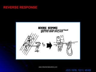
REVERSE RESPONSE
JCO 1978; 12(1): 48-69
www.indiandentalacademy.comwww.indiandentalacademy.com
Expansive Responses With Headgears
From the mechanical standpoint a progressive widening and tipping of the
alveolar base is accomplished by a widening of the inner bow of the face bow.
This expansive process provides for several distinct considerations:
1. Reciprocal expansion of the lower arch.
This can be observed as an anterior movement of the lower incisor and in the
horizontal plane increases in arch width occur.
2. Preventing impacted second molars.
When the upper first molar is translated distally without expansion, the incline
planes of that tooth start to reciprocally constrict the lower molars, carrying
them to the lingual. This tends to either impact the lower second molar or force
them buccally.
JCO 1978; 12(1): 48-69
www.indiandentalacademy.comwww.indiandentalacademy.com
A B
Soft Tissue Esthetic Changes
With normal growth (left), the soft
tissue nose is seen to grow
concentrically approximately 1 mm per
year at the tip. The cant of the nares
remains unchanged.
Following headgear therapy (right),
the nose is seen to cross over at the
bridge, lengthen vertically, and the
upward cant to the nares is tipped down
to a more horizontal position.
JCO 1978; 12(1): 48-69
www.indiandentalacademy.comwww.indiandentalacademy.com
Generalized Response With Combination Type
Headgears
In dolichofacial patterns, it often is desirable to create a rotational
orthopedic effect in the maxillae and at the same time maintain
mandibular stability.
 Long-term directional headgear therapy (part time wear), where
the force is applied below the center of resistance of the maxillae,
again allows the classical orthopedic response, but without the
upper molar extrusion.
If the force applied moves the maxillae distally without overriding
musculature, and is in conjunction with mandibular growth, the
lower face height can be closed or maintained while achieving a
reduction of the maxillary protrusion.
JCO 1978; 12(1): 48-69
www.indiandentalacademy.comwww.indiandentalacademy.com
Forces applied to the maxillae through
the face bow are either
a. Restrictive (retard downward
and forward growth) (A)
b. Rotational (B)
a. Restrictive forces occur when the
vectoral sum of forces lies above
the centre of resistance of the
maxillae
b. Rotational forces occur when the
vectoral sum of forces lie below the
centre of the resistance of the
maxillae.
Force Direction
JCO 1978; 12(1): 48-69
www.indiandentalacademy.comwww.indiandentalacademy.com
Mechanical Application of The Cervical Headgear
1. Force Level - A force level above 400 grams is ideal. In most patients, forces up
to 1000 grams can easily be tolerated and should be applied when possible.
2. Intermittent Wear
(a) sclerotic condition of molar roots- orthopedic effect
(b) Rebound
(c) Patient acceptability is enhanced
3. Outer Bow Length and Position
A rigid outer bow extending beyond the molars and tipped up 15° to the ala of
the ear will prevent propping open the bite by excessive tipping at the molars
and will maximize orthopedic effect by pitting the roots against cortical bone.
4. Expansion-Rotation
It is essential to continually expand the inner bow of the cervical headgear, not
only to correct the tendency to crossbite but also to allow a functional
development of the lower arch.
5. Freedom of Movement of the Maxillae
JCO 1978; 12(1): 48-69
www.indiandentalacademy.comwww.indiandentalacademy.com
Factors Causing Excessive Mandibular Rotation
1. Weak Muscular Pattern
2. Not Retarding Effective Eruption of The Lower Molars
Retarding the normal upward forward development of the lower molar will
have a tendency to counteract the overall rotational effect on the mandible.
3. Severe Tipping of Upper Molars
Maintaining a slight upward cant to the outer bow will minimize this tipping
effect. Severe tipping also is seen in those cases where effective growth has
been completed.
4. Full Arch Therapy Without Freeing Anterior Occlusion— Incisal Trauma
5. Fulltime Cervical Headgear Therapy
JCO 1978; 12(1): 48-69
www.indiandentalacademy.comwww.indiandentalacademy.com
UTILITY AND SECTIONAL ARCHES
The most recognizable single entity in BPT is the utility arch.
It forms the base unit around which the mechanics in all types of cases can be
employed.
Historical Perspective and Development of the Utility Arch
JCO 1978; 12(3): 192-207
www.indiandentalacademy.comwww.indiandentalacademy.com
Construction specifications of the mandibular utility arch
The mandibular utility arch is best fabricated from 0.016” x
0.016” blue elgiloy wire in order to create a force system that
delivers a continuous force that is light enough to be in the range
of 50-75 gms.
Design Principle
The principle of the long lever arm, from the molars to the
incisors is applied to deliver a light continuous force.
The utility arch is stepped down to avoid interference from the
forces of occlusion.
The buccal bridge section is flared bucally to prevent tissue
irritation, opposite the vertical steps as the arch approaches the
tissue and as the incisor teeth are intruded.
JCO 1978; 12(3): 192-207
www.indiandentalacademy.comwww.indiandentalacademy.com
Fabrication of the Mandibular Utility Arch
1) Vertical Step Height
In the lower arch it is 3-5mm
The only function of the vertical step is
to bring the malleable 0.016 x 0.016
elgiloy wire out of the occlusion to avoid
deformation with functional movements.
It is usually formed with a hoe plier.
The anterior vertical step should be
extended far enough beyond the lateral
incisor brackets (2-3 mm_ to allow
unraveling and alignment of the
incisors.
2) Placement of Labial Root Torque.
When the wire is bent at the anterior
vertical step 10° - 15° of lanial root
torque is incorporated.
The anterior arch form is then
contoured using a small turret/arch
forming plier.
JCO 1978; 12(3): 192-207
www.indiandentalacademy.comwww.indiandentalacademy.com
3) Finishing the Opposite Side.
The same procedure is continued in
reverse order after lacing into anterior
brackets.
5) Activation of the Distal Legs.
The molar section that extends into the
molar tube has a 45° buccal root torque,
30°-45° distal lingual rotation with a 30°-45°
tip back bend. Molar uprighting and incisor
intrusion
6) Final Arch Form and Activation
Characteristics.
The precisely contoured anterior arch
form will allow the incisors to intrude
without advancing.
5°-10° labial root torque will counteract
the forward tipping action and allow the
incisor roots to avoid cortical bone.
The posterior legs are parallel to each
other and 45° buccal root torque has been
placed to maintain the buccal cortical
support in the lower molar region.
JCO 1978; 12(3): 192-207
www.indiandentalacademy.comwww.indiandentalacademy.com
COMPLETED MANDIBULAR UTILITY ARCH
JCO 1978; 12(3): 192-207
www.indiandentalacademy.comwww.indiandentalacademy.com
Placement of the mandibular utility arch
When the lower utility arch is engaged In the lower Incisors, approximately 50 to
75 grams (A) of Intrusive force should be applied.
Slight labial root torque (5° to 10°) allows the lower Incisor to avoid cortical
bone in its intrusive movement (B).
JCO 1978; 12(3): 192-207
www.indiandentalacademy.comwww.indiandentalacademy.com
INTRA ORAL ADJUSTMENTS
JCO 1978; 12(3): 192-207
www.indiandentalacademy.comwww.indiandentalacademy.com
Roles and Functions of the lower utility arch
A. Position of the lower arch to allow
for cortical anchorage
In their normal eruptive positions, the
lower molars do not need to be moved
bucally or torqued bucally to put them in
their ideal anchorage positions.
Distal uprighting of the molars is done
to enhance anchorage.
Torquing of the molar roots bucally
under the oblique ridge of the cortical
bone.
www.indiandentalacademy.comwww.indiandentalacademy.com
B. Manipulation and alignment of the lower incisor segment
Intrusion/extrusion of the incisors to the level of the buccal functioning
occlusion
Advancement/retraction of the incisors in either expansion or non expansion
cases.
Leveling and rotational control of the individual incisor teeth.
Axial inclinational control by labial or lingual crown torque.
C. Stabilization of the lower arch allowing segmental treatment of the buccal
segment
Acts to maintain arch stability while canines are intruded and positioned
separately.
Allows use of segmented arch mechanics with cuspid retraction against
anchorage of all other teeth.
Stabilizes the lower arch for Class II elastics to upper segmented or utility
arches.
Allows rotation and alignment of the teeth in the buccal segment.
D. Physiological roles of the lower utility arch
Buccal arm acts as a cheek bumper causing expansion of the buccal occlusion.
Allow better buccal teeth eruption by removing functional interferences.
Corrects overbite before overjet thus avoiding incisor interference
Maintains the physiologic arch form and/ or molar width.
JCO 1978; 12(3): 192-207
www.indiandentalacademy.comwww.indiandentalacademy.com
E. Over treatment
Allows end to end incisor relationship as over treatment in deep bite cases.
Over treatment of buccal occlusion and cuspid relationships via segmented arch
treatment.
Over treatment of rotations in buccal occlusion
F. Role in mixed Dentition
Incisor and molar control during transitional stage of buccal dentition.
Allows distal eruption of the lower second bicuspid when deciduous molars are
uprighted.
Rotational correction of the bicuspids and cuspids during eruption.
G. Arch length control
1. Uprighting the lower molars: using the tip back bend of the utility arch
uprighting of the molar results in a 2mm gain of the arch length on each side
along with leveling of the curve of Spee.
2. Advancement of the lower incisors when lingually placed: Steiner’s rule would
dictate that for each 1mm that the lower incisors are brought forward 2mm of arch
length is gained.
3. Expansion in the buccal segment: Ricketts rule dictates that for each 1mm of
expansion across the bicuspids or deciduous molars, ½ mm of arch length is
gained and for each 1 mm of expansion across the molars 1/3 mm of arch length
is gained.
4. Saving E space: Space gained when the lower deciduous molars are lost.
JCO 1978; 12(3): 192-207
www.indiandentalacademy.comwww.indiandentalacademy.com
MODIFICATIONS OF THE MANDIBULAR UTILITY ARCH
JCO 1978; 12(3): 192-207
www.indiandentalacademy.comwww.indiandentalacademy.com
BIOPROGRESSIVE MIXED DENTITION TREATMENT
BPT in the mixed dentition aims at the natural tendency to alleviate the
problem when it is noticed and the somewhat overstated concept of
“interception versus correction”.
Objectives of early treatment
I. Resolve Functional Problems: The practical definition of a functional problem
is anything that disturbs the growth, health and function of the tempero-
mandibular joint complex.
II. Resolve arch length discrepancy: so that those cases within the bounds of
non extraction therapy can be approached in a manner that allows for their
successful conclusion without removal of permanent teeth.
III. Correct Vertical Problems:
IV. Correct Overjet Problems:
JCO 1978; 12(4): 279-298
www.indiandentalacademy.comwww.indiandentalacademy.com
GROWTH CONCEPTS
 The wide variety of the research involving the growth of the condyle
and the mandible the following conclusions may be derived:
1. Cases with stronger mandibular growth turgor have a propensity
for upward/forward growth of the condyle.
2. Cases with a weak growth turgor demonstrate a more
upward/backward growth of the condyle
3. Morphology alone suggests that the upward/forward cant or bend
of the condyle and neck in brachyfacial types and the
upward/backward cant and bend of the condyle and neck in
dolicofacial types delineates ultimate vertical growth and forward
posture of the chin in the face.
 Anything which jeopardizes the normal upward and forward growth of
the condyle resulting in a temperomandibular joint dysfunction is
worthy of intervening treatment, this forms the basis of treatment in
the mixed dentition.
JCO 1978; 12(4): 279-298
www.indiandentalacademy.comwww.indiandentalacademy.com
JCO 1978; 12(4): 279-298AJO 1955; June
www.indiandentalacademy.comwww.indiandentalacademy.com
I. RESOLVE FUNCTIONAL PROBLEMS
Nine general categories of functional problems can be detected
by clinical or roentgenographic examination of the patient at an
early age:
1. Cross-mouth interferences
2. Anterior cross bite
3. Open bite- Lack of incisal guidance
4. Excessive range of function
5. Distal Displacement
6. Loss of posterior support – Superior displacement
7. Finger Sucking/ Lip sucking/ Tongue thrusting
8. Breathing and Airway problems
9. True Class III Growth patterns
JCO 1978; 12(4): 279-298
www.indiandentalacademy.comwww.indiandentalacademy.com
1. Cross- mouth interferences
A. Clinical Evaluation: Cases where one or more teeth cause shunting of
the mandible in a lateral direction upon final closure.
Typically there will be a lateral shunt a ‘comfort occlusion”, or a broad arc
of closure toward one side or the other.
In the wide open posture usually the midline will align at wide open, and
upon closure there will be a midline shift as guided by neuro- muscular
reflexes.
B. Laminagraphic Evaluation: The
condyle is typically brought down on
the eminence on one side and is either
ideally seated or distally positioned on
the opposite side.
The opposite side from the shift acts in
a translatory manner while the shifting
side condyle is brought into apposition
with the greatest height of the
eminence.
JCO 1978; 12(4): 279-298
www.indiandentalacademy.comwww.indiandentalacademy.com
C. Resultant growth changes:
The translatory condyle may remain normal in growth but the opposite
side condyle will commonly demonstrate restricted growth on its antero-
superior surface and increased growth in the posterosuperior surface will
ensue.
Long term growth effects will demonstrate
•a cant in the occlusal plane,
•abnormal ramal heights, abnormal alveolar process heights,
•abnormal chin positioning.
D. Timing and method of treatment:
Cross mouth interference should be removed as soon as it is noted.
In deciduous dentition, this may mean an equilibration of a posterior tooth,
or canine, to alleviate the shunting.
If the problem is due to bilateral constriction of the maxillae, expansion
therapy is indicated usually when the upper first molars have erupted
sufficiently to allow placement of the expansion appliance.
JCO 1978; 12(4): 279-298
www.indiandentalacademy.comwww.indiandentalacademy.com
2. Anterior crossbite
A. Clinical evaluation: When one or more anterior teeth are severely
malposed, the mandible may be guided forward by the anterior interference.
Clinically, when the mandible is nudged gently in a distal direction and closed,
the area of anterior interference can easily be detected. It is not uncommon to
experience anterior displacement in cases with extreme crowding and/or
situations of ectopic eruption of incisors.
B. Laminagraphic evaluation:
When anterior mandibular
shunting occurs, often both
condyles are brought down
toward the apex of the eminence
(i.e., out of the fossae) and, quite
commonly, articular space
superior and posterior to the
condyles is evidenced.
JCO 1978; 12(4): 279-298
www.indiandentalacademy.comwww.indiandentalacademy.com
C. Resultant growth changes: As both condyles have been brought down
on the eminence, upward-backward growth of the condyles is bilaterally
enhanced. This can increase effective mandibular length and is believed to
be a contributing factor in Class III malocclusion.
D. Timing and method of treatment:
It should be determined whether the individual case is a true Class III
malocclusion or simply an anterior interference.
When the case is simply an anterior interference, alignment of one or more
teeth to prevent the interference is ideal.
This is most easily accomplished prior to full eruption of the incisors or
before incisal trauma damages the teeth at the site of interference.
JCO 1978; 12(4): 279-298
www.indiandentalacademy.comwww.indiandentalacademy.com
3. Open bite— Lack of incisal guidance
A. Clinical evaluation: During active eruptive phases, all cases at one point or
another exhibit either anterior or posterior open bite. Once the eruptive process
of the upper and lower incisors has been abbreviated (usually by contact with
the soft tissue lip or tongue) and active eruption no longer exists, lack of
proprioceptive guidance from the anterior teeth to position the condyles in the
fossae allows for excessive mobility of the mandible. Clinically, these patients
commonly show difficulty in finding centric occlusion. There is generally a
forward shunt of the mandible (to reach out for incisal proprioception) and quite
commonly the mandible can be manipulated distally by extending the thumb
from the lower incisors to the upper incisor teeth.
B. Laminagraphic evaluation: The
condyles are usually forward in the
fossae, down on the eminence, and
often there is flattening and irregularity
of the antero-superior surfaces of the
condyles.
JCO 1978; 12(4): 279-298
www.indiandentalacademy.comwww.indiandentalacademy.com
C. Resultant growth changes: Loss of guidance of the condyle in the
fossa causes abrasion or wear due to the excessive anteroposterior
slide. This can result in growth at the apex of the condyle and increase
upward/backward growth.
D. Timing and method of treatment: This is certainly the most difficult
of all functional problems to correct early, as the etiologies of open bite
are multiple. At this point, there are several basic areas to explore in
early correction of open bite:
1) Evaluate airway for possible tonsillectomy and/or adenoidectomy;
2) Orthopedically expand and rotate the maxillae to improve tongue
space, increase vertical height to the nasal complex, and change
inclination of the maxillae, especially in severe Class II malocclusions;
3)Evaluate allergy symptoms;
4) Early alleviation of severe anterior crowding to allow normal incisor
eruption;
5) Evaluate tongue size, posture, and tongue thrusting pattern.
JCO 1978; 12(4): 279-298
www.indiandentalacademy.comwww.indiandentalacademy.com
4. Excessive range of function
A. Clinical evaluation: Extreme maxillary prognathism causes the mandible to "reach"
forward in order to create a "comfort" centric occlusion. These cases are referred to
as "super Class II" malocclusions, as the mandible must reach forward to gain even a
Class II molar relationship. Clinically, severe Class II malocclusion in which the
mandible can be nudged gently back into centric relation and, upon closure, shows a
more severe maxillomandibular dental relationship, is evidence of abnormal range of
function.
B. Laminagraphic evaluation:
Upon centric occlusion, the
condyles will be forward in the
fossa, downward and forward on
the eminence, and will quite often
reveal flattening of the
anterosuperior surface of the
condyle. Excessive joint space
superior and distal to the condyles
will be evidenced and, frequently,
an upward/backward bend to the
neck and the condyles will be seen.
JCO 1978; 12(4): 279-298
www.indiandentalacademy.comwww.indiandentalacademy.com
C. Resultant growth changes: Pressure atrophy and sclerotic changes
at the antero-superior surface of the condyles enhances the
upward/backward growth and produces a more dolicofacial type of
growth experience.
D. Timing and method of treatment: Although it is not critical that the
entire Class II malocclusion be corrected, it is important that the maxillae
and/or teeth be moved distally enough to allow the mandible to close
without bringing the condyles downward and forward on the eminence. It
is not unusual, following initial headgear therapy, to be able to
cephalometrically measure a distal movement of the maxillae without
appreciable correction of the Class II molar relation. This can be the
result of a distal movement of the mandible, as the condyles drop back
into the fossae. This may be the most important functional change which
occurs with headgear therapy.
JCO 1978; 12(4): 279-298
www.indiandentalacademy.comwww.indiandentalacademy.com
5. Distal displacement
A. Clinical evaluation: The true distal displacement, in which the condyle is
located in the posterior aspect of the temporomandibular joint, is quite
commonly caused by a vertical inclination of the upper and lower incisor teeth,
especially evidenced in Class II Division II malocclusion. Although it is possible
for distal displacement to exist due to the inclines of the functioning buccal
occlusion, incisal interferences are usually the culprits. These are typically the
first functional problems to demonstrate pain in the temporomandibular joint
complex and it is possible to have crepitation, tinnitus, and early loss of mobility
in a relatively young child.
B. Laminagraphic evaluation: The
condyles are seated distally in the
fossae with excessive space anterior
and superior to the condyles. The
posterior portion of the condyles is
often seen to abut the tympanic
plates and petrotympanic fissure of
the temporal bone. Usually no
irregularities in the condyles are
evidenced. JCO 1978; 12(4): 279-298
www.indiandentalacademy.comwww.indiandentalacademy.com
C. Resultant growth changes: Since there is no interference with the antero-superior
portion of the condyles, these cases most often demonstrate normal growth turgor in the
condyles. It is felt by some that it is the lack of normal articulatory pressure at the antero-
superior portions of the condyles that enhances the brachyfacial aspect of these
particular cases.
D. Timing of treatment: As the distal displacement is often caused by the vertical
eruptive pattern of the upper and lower incisors, clinical factors which cause this eruptive
pose should be avoided.
Early removal of deciduous cuspids in the deep bite, brachyfacial type cases will free the
anterior teeth to move in a lingual direction. This will further deepen the bite and the
incisal trauma will slowly seat the condyles distally in the fossae.
When a vertical inclination of the incisors already exists, early advancement of the upper
incisors to create overjet often will allow the protracting musculature of the mandible to
react, dominate, and free the condyles of the distal displacement.
Over closure of the mandible, with excessive freeway space, will also allow the condyle
to seat distally in the fossa. Long-term, gentle, Class II elastics which help protract the
mandible, as well as allow extrusion of the posterior buccal segments, are most helpful
in correction of distal displacement. Where the extreme brachyfacial type exists,
avoidance of extraction is important to assure proper vertical support in the buccal
segment.
JCO 1978; 12(4): 279-298
www.indiandentalacademy.comwww.indiandentalacademy.com
6. Loss of posterior support superior displacement
A. Clinical evaluation: a superior and distal movement of the condyles, as in
distal displacements, can have an early onset of pain. Although this functional
problem is seldom seen in the mixed dentition, ankylosis of numerous
deciduous teeth and/or numerous congenitally missing teeth can create
superior displacement. Superior displacement is most commonly seen,
however, in the adult patient where anterior teeth have been retained, posterior
teeth have been extracted, and proper vertical support in the buccal segments
has not been maintained. Superior displacements are also seen in open bite
cases where only a posterior occlusion exists. The condyles are seated
superiorly in the fossae as the mandible pivots off of the limited posterior
contacts.
B. Laminagraphic evaluation:
The superior portion of the
condyles seat near the apex of
the fossae and excessive space
is seen mesial to the condyle.
JCO 1978; 12(4): 279-298
www.indiandentalacademy.comwww.indiandentalacademy.com
C. Resultant growth changes: As in the posterior displacements, there
do not appear to be any early signs of growth alteration due to superior
displacement.
D. Timing and method of treatment: Since the superior displacement
can be caused by loss of posterior support, early removal of carious
deciduous teeth without proper vertical support can be influential in
creating this abnormal position to the condyles. When a stronger
muscular pattern exists, and numerous deciduous teeth must, by
necessity, be removed, replacement of these teeth in a retainer is
important.
The over closure syndrome can take some time to develop and it is quite
difficult to restore once the posterior vertical dimension has been
diminished and the retained anterior teeth have adapted to the abnormal
positions of the condyles.
JCO 1978; 12(4): 279-298
www.indiandentalacademy.comwww.indiandentalacademy.com
7. Finger sucking /Lip sucking/Tongue thrust
A. Clinical evaluation: An open bite syndrome that is commonly initiated by
the finger, aggravated by the lip, and maintained by the tongue can be
considered a functional problem in that these habits may cause the
development of, or accentuate, an open bite. It is not unusual for youngsters
to suck on digits up to five or six years of age. However, when the
permanent incisors start to erupt, deformation of the anterior alveolar
process with dental protrusion and open bite can occur. Once the open bite
occurs, the tongue and lip oppose during the act of swallowing, aggravating
and continuing the open bite pattern.
B. Timing and method of treatment: The approach toward the functional
muscular problem should begin as a conservative suggestion to the child
that the activity should be ceased. If the child is unable to control the habit
pattern, expansion/thumb appliances should be placed when the upper and
lower incisors and first molars are erupting. Due to the fact that these habit
problems often cause constriction and posterior crossbite, expansion
appliances should be incorporated at the same time the digit habit is being
alleviated.
JCO 1978; 12(4): 279-298
www.indiandentalacademy.comwww.indiandentalacademy.com
8. Breathing and airway problems
A. Resultant growth changes:
Because the tongue is held low in the oral cavity to increase air uptake, these cases
are prone to maxillary collapse and crossbite.
While holding the tongue low and the mouth open, the condyles are cantilevered
down on the eminence, allowing the suprahyoid musculature to dominate, holding the
chin down and back.
This action creates wear on the upward/forward portion of the condyle and,
again, allows upward/backward growth to dominate.
Dominant upward/backward growth allows for a more receded chin posture in the
face, worsening the open bite, and accentuating the functional muscular
aberration.
B. Timing and method of treatment: Although the oral and nasal passages increase in
size as the child grows, and tonsils and adenoids atrophy with age, long-term
breathing problems that create open bite and potentially affect condylar growth,
should be evaluated at an early age. It is not unusual to suggest tonsillectomy and/or
adenoidectomy, allergy evaluation, and early orthodontic therapy to increase the size
of the nasal airway.
JCO 1978; 12(4): 279-298
www.indiandentalacademy.comwww.indiandentalacademy.com
9. True Class III Growth Patterns
A. Clinical evaluation:
True Class III growth patterns represent the epitome in functional problems.
They quite often exhibit a genetic propensity for extreme upward/backward
condylar growth, increasing the overall effective length of the mandible.
This, in conjunction with maxillary deficiency, can be mistaken for the simple
anterior crossbite or vice versa.
When true Class III is suspected, a family history as well as early
cephalometric evaluation is warranted. Several cephalometric
measurements can be utilized to evaluate the possibility that a Class III
growth pattern exists.
B. Laminagraphic evaluation:
When the mandibular teeth have bypassed the maxillary incisors, the
condyles are often downward and forward on the eminence, with excessive
space superior and distal to the condyles in the fossae.
A long, thin condylar neck and long, thin ramus is often noted.
Where the lower incisors are locked beneath the upper incisors or the
patient physically restrains the mandible, distal displacement may be noted
in the true Class III.
JCO 1978; 12(4): 279-298
www.indiandentalacademy.comwww.indiandentalacademy.com
C. Resultant growth changes: The true Class III has an inherent
tendency for functional displacement and genetic overgrowth.
D. Timing and method of treatment: When the true Class III growth
pattern is detected early, it is usual to treat only the maxillary deficiency.
Quite often early dental treatment of true Class III results in linguoversion
of the lower incisors and proversion of the upper incisors, which can make
successful surgery at a later time difficult without retreatment.
Relatively few true Class III's lend themselves to purely orthodontic
treatment alone. Maxillary expansion and advancement, in an attempt to
reduce maxillary deficiency, is the usual treatment of choice.
JCO 1978; 12(4): 279-298
www.indiandentalacademy.comwww.indiandentalacademy.com
II. Resolve Arch Length Discrepancy
Arch length gain in the lower arch occurs three ways.
1. Lateral expansion of the lower buccal segments
A. Expansion primarily by change in axial inclination
B. Expansion by midpalatal disjunction
2. Advancement or forward movement of the lower incisors
3. Uprighting and/or distal movement of the lower molars
JCO 1978; 12(4): 279-298
www.indiandentalacademy.comwww.indiandentalacademy.com
Many cases, especially those of a Class II nature, demonstrate the possibility for
arch length gain by lateral expansion of the lower buccal segments. This is a
functional type of expansion, which proceeds in a slow, meticulous manner.
As the upper arch is expanded and moved distally (and held in its expanded form
for a long period of time), the lower arch responds, through muscular adaptation
and function, reciprocally to expand.
1. Lateral expansion of the lower buccal segments
Lingual tipping of upper molar (above)
must be distinguished from true maxillary
deficiency (below).
JCO 1978; 12(4): 279-298
www.indiandentalacademy.comwww.indiandentalacademy.com
A. Expansion primarily by change in axial inclination
The appliance used to change arch form in most cases is the quad-helix or
W expansion appliance (Ricketts).
Following expansion with the W
appliance the following should
occur,
The upper molars should be
rotated distally
The upper buccal segments
expanded,
A more normal upper arch form
created
Increased space for erupting upper
central and lateral incisor teeth.
On frontal head film some
midpalatal disjunction will also be
noted.
B. Expansion by midpalatal disjunction
Where the axial inclination of the upper buccal segments is more ideal and
yet crossbite exists, palatally borne appliances are typically used to enhance
midpalatal disjunction. A Haas-type or modified Nance appliance is used to
gain these changes.
JCO 1978; 12(4): 279-298
www.indiandentalacademy.comwww.indiandentalacademy.com
2. Advancement or forward movement of the lower
incisors
When the visual treatment objectives and physiologic factors
warrant (i.e., symphysis size, shape, and form; muscle position;
esthetic considerations), retruded lower incisors can be gently
intruded and advanced to reach a more favorable esthetic
relationship to the APo line.
This type of forward movement of the lower incisors is attempted in
the brachyfacial type case, where bite opening should partially occur
by virtue of incisor intrusion, as well as change in axial inclination of
these teeth.
Each 1mm of forward movement of the lower incisors will yield
2mm of arch length gain (Steiner).
JCO 1978; 12(4): 279-298
www.indiandentalacademy.comwww.indiandentalacademy.com
3. Uprighting and/or distal movement of the lower molars
With routine use of the utility arch in deep bite situations, the simple
uprighting of the lower molars will allow the roots of these teeth to
come forward while yielding space in the arch.
 When mesial tipping of the lower molars is evident, 2mm per side
of arch length is gained by this simple uprighting effect. Further distal
movement or intrusion of the lower molars can create problems with
the erupting second molars.
It is usually ideal to stabilize the lower molar once it has reached a
normal position upright at 5° to the occlusal plane.
JCO 1978; 12(4): 279-298
www.indiandentalacademy.comwww.indiandentalacademy.com
RETENTION PROCEDURES
The retainer that is most
commonly used after first phase
therapy is the Hawley retainer with
an inclined plane.
The Hawley bow acts to hold
upper incisor alignment and
position, while the inclined plane
holds the lower incisor alignment
both from the labial (by the upper
incisors) and the lingual (by the
incline plane).
The patients are instructed to
wear the upper Hawley retainer full
time during the first year after
treatment and usually are
instructed to wear the retainer at
night time during the second
and/or third year of retention
therapy.
JCO 1978; 12(4): 279-298
www.indiandentalacademy.comwww.indiandentalacademy.com
THE BRACKET SYSTEM
With the advent of pre formed bands band
material was designed and bracket
angulations were considered so that ‘second
order’ moves were built in by angulating the
brackets.
In the original design it was decided that a
bracket should be angulated to 5° or not at all.
This accounts for the original prescription of
5° on all canines and 5° on the lower molar
tubes and brackets. In addition it was decide
on 8° for the maxillary laterals.
All the rest were straight on to the margin of
the band leaving to the orthodontist the 1° to
4° changes in angulation of the bracket by
fitting the band as required for the individual
patient needs.
1. Rickett’s Standard Bioprogressive
It soon became evident that control of torque simultaneously with placement of
loops was difficult so Rickett’s incorporated torque values of Jarabak and Holdaway
into the brackets. Upper incisor of 22°, laterals 14° and cuspids at 7° of lingual root
torque.
JCO 1978; 12(8): 569-586
www.indiandentalacademy.comwww.indiandentalacademy.com
With the standard bioprogressive system
difficulty was experienced in placing
enough torque on the lower molar area,
particularly in view of the need for
anchorage.
The same angulations of 7°, 14°, 21°
were incorporated into the lower anterior
segment, in addition the lower first molar
had a rotation of 12°.
Torque was also incorporated into the
lower premolar bracket.
2. Rickett’s Full Torque Bioprogressive
JCO 1978; 12(8): 569-586
www.indiandentalacademy.comwww.indiandentalacademy.com
3. Rickett’s Triple Control Bioprogressive
Raised bracket bases of canines
and bicuspids
+7° torque on the canines
Upper and lower molar offsets
Second molar convertible tubes
In the extraction series the torque
on the second premolar was reduced
to 7° to the lingual, the canine torque
was 0 and the bracket was not raised
to assist in the transition from the
anterior to the buccal segment.
JCO 1978; 12(8): 569-586
www.indiandentalacademy.comwww.indiandentalacademy.com
ARCH FORMS
Factors taken into account in the research of arch forms included:
 Arch correlation
 Consideration of size
 Arch length
 Where the arch was to be measured
 Contact details
 Final determination of form at the bracket location
Twelve arch forms were originally identified, which were narrowed down
to nine by computer work.
Studies of other normal and stable treated patients resulted in five arch
forms.
JCO 1978; 12(8): 569-586
www.indiandentalacademy.comwww.indiandentalacademy.com
Penta Morphic Arches
1. Narrow ovoid
2. Tapered
3. Normal ideal
4. Ovoid
5. Narrow tapered
JCO 1978; 12(8): 569-586
www.indiandentalacademy.comwww.indiandentalacademy.com
MECHANICS SEQUENCE FOR EXTRACTION CASES
The mechanics for extraction cases in BPT can best be organized into
four general procedures that can be individually evaluated and
analyzed as to the needs of the specific case.
I) Stabilization of upper and lower molar anchorage
II) Retraction and uprighting of cuspids with sectional arch
mechanics
III) Retraction and consolidation of upper and lower incisors
IV) Continuous arches for details of ideal and finishing
occlusion
JCO 1978; 12(5): 334-357
www.indiandentalacademy.comwww.indiandentalacademy.com
I) Stabilization of the upper and lower molar anchorage.
Upper molar anchorage
Minimum anchorage
Maximum
anchorage
JCO 1978; 12(5): 334-357
www.indiandentalacademy.comwww.indiandentalacademy.com
Lower molar anchorage
JCO 1978; 12(5): 334-357
www.indiandentalacademy.comwww.indiandentalacademy.com
II) Retraction and uprighting of cuspids with sectional arch
mechanics.
90° gable and 90° offset antirotation bends
100 to 150 gms of force
2-3 mm of activation
JCO 1978; 12(5): 334-357
www.indiandentalacademy.comwww.indiandentalacademy.com
Three dimensional cuspid control
www.indiandentalacademy.comwww.indiandentalacademy.com
III. Retraction and consolidation of upper and lower
incisors.
Contraction utility arch
Torquing contracting
utility arch
Utility arch with stepped
up delta loop
Utility arch with regular
delta loop
JCO 1978; 12(5): 334-357
www.indiandentalacademy.comwww.indiandentalacademy.com
Finishing wires
JCO 1978; 12(5): 334-357
0.016 x 0.022
5/16” heavy class II elastics
www.indiandentalacademy.comwww.indiandentalacademy.com
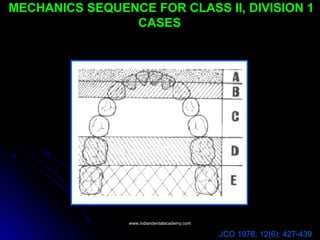
MECHANICS SEQUENCE FOR CLASS II, DIVISION 1
CASES
JCO 1978; 12(6): 427-439
www.indiandentalacademy.comwww.indiandentalacademy.com
Leveling of the Lower arch
JCO 1978; 12(6): 427-439
www.indiandentalacademy.comwww.indiandentalacademy.com
Upper Buccal segment alignment
JCO 1978; 12(6): 427-439
www.indiandentalacademy.comwww.indiandentalacademy.com
Class II elastics and complete lower arch alignment
Segmental correction of the Class II with
elastics.
Pitting upper and lower arches with
continuous archwire against each other
has several detrimental effects.
1. Skidding effect that simply throws the
lower arch forward while extruding and
retracting the upper arch.
2. With a tendency for deep bite the class II
elastics can bring the upper incisors
back and start ‘jamming’ the lower
incisors as they are retracted.
3. It is difficult to overcorrect the upper
buccal segment without bringing the
upper anterior teeth into lingual cross
bite.
JCO 1978; 12(6): 427-439
www.indiandentalacademy.comwww.indiandentalacademy.com
Traction sections
The tendency for the downward pull of
the Class II elastics to extrude and throw
the root of the canine mesially is
countered by placing a small closed
helix distal to the upper cuspid teeth with
a gable or tipback of 30°.
The anterior portion of the segment
should also be rotated mesially 45° and
often a horizontal closed helix is placed
at the molar region to maintain or
accentuate distal molar rotation.
The traction section also stabilizes the
upper buccal segments against the
impending intrusion and torque in the
upper incisors.
JCO 1978; 12(6): 427-439
www.indiandentalacademy.comwww.indiandentalacademy.com
Upper incisor intrusion and retraction
JCO 1978; 12(6): 427-439
www.indiandentalacademy.comwww.indiandentalacademy.com
Idealization of arches and finishing details
JCO 1978; 12(6): 427-439
www.indiandentalacademy.comwww.indiandentalacademy.com
MECHANICS SEQUENCE FOR CLASS II, DIVISION 2
CASES
In general there are three treatment possibilities in a Class II, Division 2
malocclusion:
1. Distalizing the upper arch
2. Advancing the lower arch
3. A reciprocal movement, advancing the lower arch and the
distalizing the upper arch at the same time.
There are six functions necessary in treating Class II, division 2
malocclusions, which are general considerations for evaluating the
mechanics sequence:
I) Advancement, torque control and intrusion of the upper incisors.
II) Intrusion of the lower incisors and cuspids.
III) Alignment of the buccal segments and Class II correction.
IV) Consolidation of the upper incisors.
V) Idealizing the arches
VI) Finishing.
JCO 1978; 12(7): 505-521
www.indiandentalacademy.comwww.indiandentalacademy.com
I) Advancement, torque control and intrusion of
the upper incisors.
JCO 1978; 12(7): 505-521
www.indiandentalacademy.comwww.indiandentalacademy.com
II) Intrusion of the lower incisors and
cuspids.
JCO 1978; 12(7): 505-521
www.indiandentalacademy.comwww.indiandentalacademy.com
III) Alignment of the buccal segments and Class II
correction.
JCO 1978; 12(7): 505-521
www.indiandentalacademy.comwww.indiandentalacademy.com
IV) Consolidation of the upper incisors.
JCO 1978; 12(7): 505-521
www.indiandentalacademy.comwww.indiandentalacademy.com
V) Idealizing the arches and Finishing.
JCO 1978; 12(7): 505-521
www.indiandentalacademy.comwww.indiandentalacademy.com
FINISHING PROCEDURES IN BIOPROGRESSIVE
THERAPY
Ricketts interpreted Angle's
line of occlusion to include a
line drawn through the contact
points of the posterior teeth
and slightly below them
through the contact
embrasures of the anteriors.
The line is suggested as the
line to which our brackets can
be placed on the individual
teeth in order to allow the
cusp/marginal ridge function
that our occlusal stops
produce.
JCO 1978; 12(8): 569-586
www.indiandentalacademy.comwww.indiandentalacademy.com
1) Ideal occlusion, perhaps not quite ever found in Nature,
represents an occlusion in which there is perfect size and fit of
the individual teeth and the teeth are in ideal arch form,
balance, and harmony; an occlusion in which every incline and
stop is perfect and every tooth is in an ideal location within its
arch and functions perfectly with its opponent teeth in the
opposite arch.
2) Normal occlusion would be an untreated natural occlusion that
is within an expected normal range of variation in all of the
measurements thought to be critical in evaluating occlusion.
The normal range of variation represents two-thirds of the
population and eliminates those extremes on either end of the
normal bell curve distribution.
Concepts of Occlusion
JCO 1978; 12(8): 569-586
www.indiandentalacademy.comwww.indiandentalacademy.com
3) Reconstructed occlusion represents those
occlusions that are being restored, where the ability to
critically record the various jaw movements is
essential. The occlusion is designed to accommodate
to the pathways of function recorded for the individual
case and the teeth can be "constructed" to function
properly in all movements in the specific case.
4) Orthodontic finishing occlusion is represented by
the occlusion that is desired at the time of band or
active appliance removal.
JCO 1978; 12(8): 569-586
www.indiandentalacademy.comwww.indiandentalacademy.com
Function Influences Finishing and Retention
The proper location and function of the condyle in the
temporomandibular joint is essential to the health and stability of
the occlusion.
A normal airway which effects the basic respiratory process and
influences the tongue posture and function is important to the
stability of the denture.
Lip function and its variations have an influence upon the incisor
alignment and stability.
The buccal and facial musculature along with the muscles of
mastication, which are reflected in the facial type as described by
our cephalometrics, are also critical influences, and are
considered during the original diagnostic criteria.
JCO 1978; 12(8): 569-586
www.indiandentalacademy.comwww.indiandentalacademy.com
Occlusal Check List in Finishing
An occlusal check list including eight areas in each arch is used
in establishing the ideal finishing arch configuration and
individualized tooth rotation in our overtreated orthodontic finishing
occlusion.
The patient at this stage is seen at two-week appointments, for
the adjustments are more delicate and controlled.
During the final two-week adjustment the cuspid and bicuspid
bands may be removed to allow closing of the band space. New
bonding procedures that eliminate the interproximal band material
may not require the stage of final finishing.
JCO 1978; 12(8): 569-586
www.indiandentalacademy.comwww.indiandentalacademy.com
1. Arch width across second molars.
2. Distal of first molar rotated lingually
until the distobuccal cusp approximates
mesial sluiceway on second molar.
3. Large buccal offset at mesial of first
molar.
4. Check inter-bicuspid width for
necessary expansion.
5. Proper buccal arch form and contour.
6. Premolar offset to bring it in contact
with distal lingual incline of upper canine
(2-3mm).
7. Mesial of cuspid tucked slightly behind
lateral incisor distal of the cuspid buccal.
8. Over-rotation of incisors; smooth arc
Occlusal Check list for finishing in mandibular arch
JCO 1978; 12(8): 569-586
www.indiandentalacademy.comwww.indiandentalacademy.com
Occlusal Check list for finishing in maxillary arch
1. Width across first and second molars.
2. Distal rotation of first molar so that line
drawn through distobuccal and
mesiolingual cusps points to the mesial
third of the opposite side cuspid .
3. Mesial offset (large) on molar.
4. Mesial rotation of lingual cusp of first
bicuspid to seat in distal fossa of lower first
bicuspid.
5. Premolar offset (2-3mm) to avoid first
area of prematurity.
6. Cuspid brought into contact with lower
cuspid and premolar to establish cuspid
rise.
7. Lateral left labial (until retainer) to allow
overtreatment of buccal segments; then
tucked in.
8. Smooth arc across incisors.
JCO 1978; 12(8): 569-586
www.indiandentalacademy.comwww.indiandentalacademy.com
RETENTION IN BIOPROGRESSIVE THERAPY
Retention in Bioprogressive Therapy is the process that
sustains and guides the settling from the overtreated or
orthodontic occlusion into the final functioning occlusion.
It first guides these changes during the initial adjustments, and
then supports the bony sutural and muscular accommodations to
the changing environment.
Finally, retention should consider the long range influences
which involve changes created by growth, tooth eruption, and
function, characterized by the different facial types.
JCO 1978; 12(8): 569-586
www.indiandentalacademy.comwww.indiandentalacademy.com
The Initial Stage of Retention
The initial stage of retention, perhaps the most obvious and critical,
occurs during the first six weeks following the conclusion of the active
phase of treatment when the appliances are removed and the teeth are
"turned loose" to erupt along their normal eruptive paths into the
functioning occlusion.
Retainers inserted at this initial phase are not designed to hold, but to
assist in guiding this settling process.
The adjustments in the upper retainer include
relieving the lingual to:
(1) close the anterior band space between the
central and lateral incisors (buccal band space
is closed with finishing arches),
(2) allow the tucking in of the distal of the
upper cuspids following their expansion and
overtreatment, and
(3) sustain the settling distal rotation of the
upper molar as it functions with the lower
rotated molar occlusion.Upper Arch Retainer
JCO 1978; 12(8): 569-586
www.indiandentalacademy.comwww.indiandentalacademy.com
In the lower arch, a fixed first bicuspid
retainer is placed in order to
(1)maintain the cross arch bicuspid width
and support the first bicuspid against the
upper cuspid and bicuspid function,
(2) allow the lower cuspids the freedom of
adjustment against the upper occlusion,
and
(3) place a lingual bar against the incisal
third of the lower incisors to maintain
their alignment and rotational
connection.
The fixed lower retainer being back on the
bicuspids is easily acceptable to the
patient and can be maintained longer.
JCO 1978; 12(8): 569-586
www.indiandentalacademy.comwww.indiandentalacademy.com
The Stabilizing Stage of Retention
The stabilizing stage of retention involves the ongoing phase
over the first year following active treatment when the sutural
adjustment, transseptal fibers, functioning occlusion and muscle
physiology need to be considered in supporting the new
occlusion.
During this period the lower fixed retainer is kept in place and
the upper retainer is worn most of the time.
Following the 1st year, if the functioning occlusion remains
stable, the retainer is worn only part time, during sleeping .
JCO 1978; 12(8): 569-586
www.indiandentalacademy.comwww.indiandentalacademy.com
This seminar attempted to present the basic tenets of the Bioprogressive
Therapy.
It began with a systems approach diagnosis and treatment planning and
an overview of the management procedures used to implement and carry
out the logic process employed in our treatment.
Various treatment sequences were suggested that could be applied to a
total course of therapy, rather than a cookbook technique blindly followed in
every case.
Orthopedic alteration, optimum orthodontic forces and combination of
mechanics were suggested that would unlock the malocclusion in a
progressive sequence in order to establish more normal function for
optimum health and stability of the denture.
Bioprogressive Therapy approaches an in-depth analysis of the basic
malocclusion, the underlying morphology with its functional variations, then
attempts to treat them to as normal a function and esthetic relationship as is
possible for the long range health and stability of the denture.
Each case is approached individually because of its individual
morphology, physiology and malocclusion and the prescribed treatment
sequence is selected to accomplish quality results with efficiency.
CONCLUSION
www.indiandentalacademy.comwww.indiandentalacademy.com
THANK YOUwww.indiandentalacademy.comwww.indiandentalacademy.com

More Related Content

What's hot

Construction of bite for various functional orthodontic appliances
Construction of bite for various functional orthodontic appliancesConstruction of bite for various functional orthodontic appliances
Construction of bite for various functional orthodontic appliancesIndian dental academy
 
Steiner's Annalysis - Acceptable Deviation
Steiner's Annalysis - Acceptable DeviationSteiner's Annalysis - Acceptable Deviation
Steiner's Annalysis - Acceptable DeviationPam Fabie
 
Begg’s philosophy and technique
Begg’s philosophy and techniqueBegg’s philosophy and technique
Begg’s philosophy and techniqueDr Susna Paul
 
Segmented arch technique /certified fixed orthodontic courses by Indian denta...
Segmented arch technique /certified fixed orthodontic courses by Indian denta...Segmented arch technique /certified fixed orthodontic courses by Indian denta...
Segmented arch technique /certified fixed orthodontic courses by Indian denta...Indian dental academy
 
Tissue response functional appliance
Tissue response  functional applianceTissue response  functional appliance
Tissue response functional applianceIndian dental academy
 
Indirect bonding in lingual orthodontics parag
Indirect bonding in lingual orthodontics paragIndirect bonding in lingual orthodontics parag
Indirect bonding in lingual orthodontics paragParag Deshmukh
 
Straight wire appliance /certified fixed orthodontic courses by Indian dental...
Straight wire appliance /certified fixed orthodontic courses by Indian dental...Straight wire appliance /certified fixed orthodontic courses by Indian dental...
Straight wire appliance /certified fixed orthodontic courses by Indian dental...Indian dental academy
 
Torque in pre adjusted e.w.a /certified fixed orthodontic courses by Indian...
Torque in pre adjusted e.w.a   /certified fixed orthodontic courses by Indian...Torque in pre adjusted e.w.a   /certified fixed orthodontic courses by Indian...
Torque in pre adjusted e.w.a /certified fixed orthodontic courses by Indian...Indian dental academy
 
Edgewise appliance- evolution and technique
Edgewise appliance- evolution and techniqueEdgewise appliance- evolution and technique
Edgewise appliance- evolution and techniqueIndian dental academy
 
Traditional begg technique stage 1 and stage 2
Traditional begg technique stage 1 and stage 2Traditional begg technique stage 1 and stage 2
Traditional begg technique stage 1 and stage 2Indian dental academy
 
Cephalometric superimposition methods
Cephalometric superimposition methodsCephalometric superimposition methods
Cephalometric superimposition methodsIndian dental academy
 

What's hot (20)

Construction of bite for various functional orthodontic appliances
Construction of bite for various functional orthodontic appliancesConstruction of bite for various functional orthodontic appliances
Construction of bite for various functional orthodontic appliances
 
Steiner's Annalysis - Acceptable Deviation
Steiner's Annalysis - Acceptable DeviationSteiner's Annalysis - Acceptable Deviation
Steiner's Annalysis - Acceptable Deviation
 
Begg’s philosophy and technique
Begg’s philosophy and techniqueBegg’s philosophy and technique
Begg’s philosophy and technique
 
Intrusion arches
Intrusion archesIntrusion arches
Intrusion arches
 
Segmented arch technique /certified fixed orthodontic courses by Indian denta...
Segmented arch technique /certified fixed orthodontic courses by Indian denta...Segmented arch technique /certified fixed orthodontic courses by Indian denta...
Segmented arch technique /certified fixed orthodontic courses by Indian denta...
 
S begg’s
S begg’s S begg’s
S begg’s
 
Tweed philosophy
Tweed philosophyTweed philosophy
Tweed philosophy
 
Tissue response functional appliance
Tissue response  functional applianceTissue response  functional appliance
Tissue response functional appliance
 
Brackets in ortho final
Brackets in ortho finalBrackets in ortho final
Brackets in ortho final
 
Roth philosophy
Roth philosophyRoth philosophy
Roth philosophy
 
Indirect bonding in lingual orthodontics parag
Indirect bonding in lingual orthodontics paragIndirect bonding in lingual orthodontics parag
Indirect bonding in lingual orthodontics parag
 
Arnetts analysis
Arnetts analysisArnetts analysis
Arnetts analysis
 
Straight wire appliance /certified fixed orthodontic courses by Indian dental...
Straight wire appliance /certified fixed orthodontic courses by Indian dental...Straight wire appliance /certified fixed orthodontic courses by Indian dental...
Straight wire appliance /certified fixed orthodontic courses by Indian dental...
 
Andrew's (2)
Andrew's (2)Andrew's (2)
Andrew's (2)
 
Torque in pre adjusted e.w.a /certified fixed orthodontic courses by Indian...
Torque in pre adjusted e.w.a   /certified fixed orthodontic courses by Indian...Torque in pre adjusted e.w.a   /certified fixed orthodontic courses by Indian...
Torque in pre adjusted e.w.a /certified fixed orthodontic courses by Indian...
 
beggs technique bends
 beggs technique bends beggs technique bends
beggs technique bends
 
Edgewise appliance- evolution and technique
Edgewise appliance- evolution and techniqueEdgewise appliance- evolution and technique
Edgewise appliance- evolution and technique
 
Dental VTO
Dental VTODental VTO
Dental VTO
 
Traditional begg technique stage 1 and stage 2
Traditional begg technique stage 1 and stage 2Traditional begg technique stage 1 and stage 2
Traditional begg technique stage 1 and stage 2
 
Cephalometric superimposition methods
Cephalometric superimposition methodsCephalometric superimposition methods
Cephalometric superimposition methods
 

Similar to Bpt (2)

Bio progressive therapy
Bio progressive therapyBio progressive therapy
Bio progressive therapyMiliya Parveen
 
Bioprogressive therapy /certified fixed orthodontic courses by Indian dental ...
Bioprogressive therapy /certified fixed orthodontic courses by Indian dental ...Bioprogressive therapy /certified fixed orthodontic courses by Indian dental ...
Bioprogressive therapy /certified fixed orthodontic courses by Indian dental ...Indian dental academy
 
Bioprogressive theraphy
Bioprogressive theraphy Bioprogressive theraphy
Bioprogressive theraphy Mothi Krishna
 
SECONDARY PREPROSTHETIC SURGE.pptx
SECONDARY PREPROSTHETIC SURGE.pptxSECONDARY PREPROSTHETIC SURGE.pptx
SECONDARY PREPROSTHETIC SURGE.pptxVikramRaj87
 
Clinical tips to increase consistency using a selfligathing
Clinical tips to increase consistency using a selfligathingClinical tips to increase consistency using a selfligathing
Clinical tips to increase consistency using a selfligathingUniversidad de Cuenca
 
Orthopedic biomechanics /certified fixed orthodontic courses by Indian dental...
Orthopedic biomechanics /certified fixed orthodontic courses by Indian dental...Orthopedic biomechanics /certified fixed orthodontic courses by Indian dental...
Orthopedic biomechanics /certified fixed orthodontic courses by Indian dental...Indian dental academy
 
loading of dental implants / academy of fixed orthodontics
loading of dental implants  / academy of fixed orthodonticsloading of dental implants  / academy of fixed orthodontics
loading of dental implants / academy of fixed orthodonticsIndian dental academy
 

Similar to Bpt (2) (20)

Bio progressive therapy
Bio progressive therapyBio progressive therapy
Bio progressive therapy
 
Bpt
Bpt Bpt
Bpt
 
Bio 1
Bio 1Bio 1
Bio 1
 
Bioprogressive therap yy
Bioprogressive therap yyBioprogressive therap yy
Bioprogressive therap yy
 
Bioprogressive therapy /certified fixed orthodontic courses by Indian dental ...
Bioprogressive therapy /certified fixed orthodontic courses by Indian dental ...Bioprogressive therapy /certified fixed orthodontic courses by Indian dental ...
Bioprogressive therapy /certified fixed orthodontic courses by Indian dental ...
 
Part 1
Part 1Part 1
Part 1
 
Bioprogressive therapy
Bioprogressive therapyBioprogressive therapy
Bioprogressive therapy
 
Bioprogressive therapy
Bioprogressive therapyBioprogressive therapy
Bioprogressive therapy
 
Bioprogressive theraphy
Bioprogressive theraphy Bioprogressive theraphy
Bioprogressive theraphy
 
3rd publication JCDR-8th name.pdf
3rd publication JCDR-8th name.pdf3rd publication JCDR-8th name.pdf
3rd publication JCDR-8th name.pdf
 
SECONDARY PREPROSTHETIC SURGE.pptx
SECONDARY PREPROSTHETIC SURGE.pptxSECONDARY PREPROSTHETIC SURGE.pptx
SECONDARY PREPROSTHETIC SURGE.pptx
 
3RD PUBLICATION - JCDR - Dr. RAHUL VC TIWARI, SIBAR INSTITUTE OF DENTAL SCIE...
3RD  PUBLICATION - JCDR - Dr. RAHUL VC TIWARI, SIBAR INSTITUTE OF DENTAL SCIE...3RD  PUBLICATION - JCDR - Dr. RAHUL VC TIWARI, SIBAR INSTITUTE OF DENTAL SCIE...
3RD PUBLICATION - JCDR - Dr. RAHUL VC TIWARI, SIBAR INSTITUTE OF DENTAL SCIE...
 
Clinical tips to increase consistency using a selfligathing
Clinical tips to increase consistency using a selfligathingClinical tips to increase consistency using a selfligathing
Clinical tips to increase consistency using a selfligathing
 
fmr
 fmr fmr
fmr
 
fmr
 fmr fmr
fmr
 
BPT part 1.ppt
BPT part 1.pptBPT part 1.ppt
BPT part 1.ppt
 
Orthopedic biomechanics /certified fixed orthodontic courses by Indian dental...
Orthopedic biomechanics /certified fixed orthodontic courses by Indian dental...Orthopedic biomechanics /certified fixed orthodontic courses by Indian dental...
Orthopedic biomechanics /certified fixed orthodontic courses by Indian dental...
 
loading of dental implants / academy of fixed orthodontics
loading of dental implants  / academy of fixed orthodonticsloading of dental implants  / academy of fixed orthodontics
loading of dental implants / academy of fixed orthodontics
 
Biomechanics in orthopedics
Biomechanics in orthopedicsBiomechanics in orthopedics
Biomechanics in orthopedics
 
Biomechanics in orthopedics
Biomechanics in orthopedicsBiomechanics in orthopedics
Biomechanics in orthopedics
 

More from Indian dental academy

Indian Dentist - relocate to united kingdom
Indian Dentist - relocate to united kingdomIndian Dentist - relocate to united kingdom
Indian Dentist - relocate to united kingdomIndian dental academy
 
1ST, 2ND AND 3RD ORDER BENDS IN STANDARD EDGEWISE APPLIANCE SYSTEM /Fixed ort...
1ST, 2ND AND 3RD ORDER BENDS IN STANDARD EDGEWISE APPLIANCE SYSTEM /Fixed ort...1ST, 2ND AND 3RD ORDER BENDS IN STANDARD EDGEWISE APPLIANCE SYSTEM /Fixed ort...
1ST, 2ND AND 3RD ORDER BENDS IN STANDARD EDGEWISE APPLIANCE SYSTEM /Fixed ort...Indian dental academy
 
Invisalign -invisible aligners course in india
Invisalign -invisible aligners course in india Invisalign -invisible aligners course in india
Invisalign -invisible aligners course in india Indian dental academy
 
Invisible aligners for your orthodontics pratice
Invisible aligners for your orthodontics praticeInvisible aligners for your orthodontics pratice
Invisible aligners for your orthodontics praticeIndian dental academy
 
Development of muscles of mastication / dental implant courses
Development of muscles of mastication / dental implant coursesDevelopment of muscles of mastication / dental implant courses
Development of muscles of mastication / dental implant coursesIndian dental academy
 
Corticosteriods uses in dentistry/ oral surgery courses  
Corticosteriods uses in dentistry/ oral surgery courses  Corticosteriods uses in dentistry/ oral surgery courses  
Corticosteriods uses in dentistry/ oral surgery courses  Indian dental academy
 
Cytotoxicity of silicone materials used in maxillofacial prosthesis / dental ...
Cytotoxicity of silicone materials used in maxillofacial prosthesis / dental ...Cytotoxicity of silicone materials used in maxillofacial prosthesis / dental ...
Cytotoxicity of silicone materials used in maxillofacial prosthesis / dental ...Indian dental academy
 
Diagnosis and treatment planning in completely endntulous arches/dental courses
Diagnosis and treatment planning in completely endntulous arches/dental coursesDiagnosis and treatment planning in completely endntulous arches/dental courses
Diagnosis and treatment planning in completely endntulous arches/dental coursesIndian dental academy
 
Properties of Denture base materials /rotary endodontic courses
Properties of Denture base materials /rotary endodontic coursesProperties of Denture base materials /rotary endodontic courses
Properties of Denture base materials /rotary endodontic coursesIndian dental academy
 
Use of modified tooth forms in complete denture occlusion / dental implant...
Use of modified  tooth forms  in  complete denture occlusion / dental implant...Use of modified  tooth forms  in  complete denture occlusion / dental implant...
Use of modified tooth forms in complete denture occlusion / dental implant...Indian dental academy
 
Dental luting cements / oral surgery courses  
Dental   luting cements / oral surgery courses  Dental   luting cements / oral surgery courses  
Dental luting cements / oral surgery courses  Indian dental academy
 
Dental casting alloys/ oral surgery courses  
Dental casting alloys/ oral surgery courses  Dental casting alloys/ oral surgery courses  
Dental casting alloys/ oral surgery courses  Indian dental academy
 
Dental casting investment materials/endodontic courses
Dental casting investment materials/endodontic coursesDental casting investment materials/endodontic courses
Dental casting investment materials/endodontic coursesIndian dental academy
 
Dental casting waxes/ oral surgery courses  
Dental casting waxes/ oral surgery courses  Dental casting waxes/ oral surgery courses  
Dental casting waxes/ oral surgery courses  Indian dental academy
 
Dental ceramics/prosthodontic courses
Dental ceramics/prosthodontic coursesDental ceramics/prosthodontic courses
Dental ceramics/prosthodontic coursesIndian dental academy
 
Dental implant/ oral surgery courses  
Dental implant/ oral surgery courses  Dental implant/ oral surgery courses  
Dental implant/ oral surgery courses  Indian dental academy
 
Dental perspective/cosmetic dentistry courses
Dental perspective/cosmetic dentistry coursesDental perspective/cosmetic dentistry courses
Dental perspective/cosmetic dentistry coursesIndian dental academy
 
Dental tissues and their replacements/ oral surgery courses  
Dental tissues and their replacements/ oral surgery courses  Dental tissues and their replacements/ oral surgery courses  
Dental tissues and their replacements/ oral surgery courses  Indian dental academy
 

More from Indian dental academy (20)

Indian Dentist - relocate to united kingdom
Indian Dentist - relocate to united kingdomIndian Dentist - relocate to united kingdom
Indian Dentist - relocate to united kingdom
 
1ST, 2ND AND 3RD ORDER BENDS IN STANDARD EDGEWISE APPLIANCE SYSTEM /Fixed ort...
1ST, 2ND AND 3RD ORDER BENDS IN STANDARD EDGEWISE APPLIANCE SYSTEM /Fixed ort...1ST, 2ND AND 3RD ORDER BENDS IN STANDARD EDGEWISE APPLIANCE SYSTEM /Fixed ort...
1ST, 2ND AND 3RD ORDER BENDS IN STANDARD EDGEWISE APPLIANCE SYSTEM /Fixed ort...
 
Invisalign -invisible aligners course in india
Invisalign -invisible aligners course in india Invisalign -invisible aligners course in india
Invisalign -invisible aligners course in india
 
Invisible aligners for your orthodontics pratice
Invisible aligners for your orthodontics praticeInvisible aligners for your orthodontics pratice
Invisible aligners for your orthodontics pratice
 
online fixed orthodontics course
online fixed orthodontics courseonline fixed orthodontics course
online fixed orthodontics course
 
online orthodontics course
online orthodontics courseonline orthodontics course
online orthodontics course
 
Development of muscles of mastication / dental implant courses
Development of muscles of mastication / dental implant coursesDevelopment of muscles of mastication / dental implant courses
Development of muscles of mastication / dental implant courses
 
Corticosteriods uses in dentistry/ oral surgery courses  
Corticosteriods uses in dentistry/ oral surgery courses  Corticosteriods uses in dentistry/ oral surgery courses  
Corticosteriods uses in dentistry/ oral surgery courses  
 
Cytotoxicity of silicone materials used in maxillofacial prosthesis / dental ...
Cytotoxicity of silicone materials used in maxillofacial prosthesis / dental ...Cytotoxicity of silicone materials used in maxillofacial prosthesis / dental ...
Cytotoxicity of silicone materials used in maxillofacial prosthesis / dental ...
 
Diagnosis and treatment planning in completely endntulous arches/dental courses
Diagnosis and treatment planning in completely endntulous arches/dental coursesDiagnosis and treatment planning in completely endntulous arches/dental courses
Diagnosis and treatment planning in completely endntulous arches/dental courses
 
Properties of Denture base materials /rotary endodontic courses
Properties of Denture base materials /rotary endodontic coursesProperties of Denture base materials /rotary endodontic courses
Properties of Denture base materials /rotary endodontic courses
 
Use of modified tooth forms in complete denture occlusion / dental implant...
Use of modified  tooth forms  in  complete denture occlusion / dental implant...Use of modified  tooth forms  in  complete denture occlusion / dental implant...
Use of modified tooth forms in complete denture occlusion / dental implant...
 
Dental luting cements / oral surgery courses  
Dental   luting cements / oral surgery courses  Dental   luting cements / oral surgery courses  
Dental luting cements / oral surgery courses  
 
Dental casting alloys/ oral surgery courses  
Dental casting alloys/ oral surgery courses  Dental casting alloys/ oral surgery courses  
Dental casting alloys/ oral surgery courses  
 
Dental casting investment materials/endodontic courses
Dental casting investment materials/endodontic coursesDental casting investment materials/endodontic courses
Dental casting investment materials/endodontic courses
 
Dental casting waxes/ oral surgery courses  
Dental casting waxes/ oral surgery courses  Dental casting waxes/ oral surgery courses  
Dental casting waxes/ oral surgery courses  
 
Dental ceramics/prosthodontic courses
Dental ceramics/prosthodontic coursesDental ceramics/prosthodontic courses
Dental ceramics/prosthodontic courses
 
Dental implant/ oral surgery courses  
Dental implant/ oral surgery courses  Dental implant/ oral surgery courses  
Dental implant/ oral surgery courses  
 
Dental perspective/cosmetic dentistry courses
Dental perspective/cosmetic dentistry coursesDental perspective/cosmetic dentistry courses
Dental perspective/cosmetic dentistry courses
 
Dental tissues and their replacements/ oral surgery courses  
Dental tissues and their replacements/ oral surgery courses  Dental tissues and their replacements/ oral surgery courses  
Dental tissues and their replacements/ oral surgery courses  
 

Recently uploaded

EPANDING THE CONTENT OF AN OUTLINE using notes.pptx
EPANDING THE CONTENT OF AN OUTLINE using notes.pptxEPANDING THE CONTENT OF AN OUTLINE using notes.pptx
EPANDING THE CONTENT OF AN OUTLINE using notes.pptxRaymartEstabillo3
 
How to Configure Email Server in Odoo 17
How to Configure Email Server in Odoo 17How to Configure Email Server in Odoo 17
How to Configure Email Server in Odoo 17Celine George
 
CELL CYCLE Division Science 8 quarter IV.pptx
CELL CYCLE Division Science 8 quarter IV.pptxCELL CYCLE Division Science 8 quarter IV.pptx
CELL CYCLE Division Science 8 quarter IV.pptxJiesonDelaCerna
 
Procuring digital preservation CAN be quick and painless with our new dynamic...
Procuring digital preservation CAN be quick and painless with our new dynamic...Procuring digital preservation CAN be quick and painless with our new dynamic...
Procuring digital preservation CAN be quick and painless with our new dynamic...Jisc
 
Solving Puzzles Benefits Everyone (English).pptx
Solving Puzzles Benefits Everyone (English).pptxSolving Puzzles Benefits Everyone (English).pptx
Solving Puzzles Benefits Everyone (English).pptxOH TEIK BIN
 
Hierarchy of management that covers different levels of management
Hierarchy of management that covers different levels of managementHierarchy of management that covers different levels of management
Hierarchy of management that covers different levels of managementmkooblal
 
Computed Fields and api Depends in the Odoo 17
Computed Fields and api Depends in the Odoo 17Computed Fields and api Depends in the Odoo 17
Computed Fields and api Depends in the Odoo 17Celine George
 
MARGINALIZATION (Different learners in Marginalized Group
MARGINALIZATION (Different learners in Marginalized GroupMARGINALIZATION (Different learners in Marginalized Group
MARGINALIZATION (Different learners in Marginalized GroupJonathanParaisoCruz
 
Painted Grey Ware.pptx, PGW Culture of India
Painted Grey Ware.pptx, PGW Culture of IndiaPainted Grey Ware.pptx, PGW Culture of India
Painted Grey Ware.pptx, PGW Culture of IndiaVirag Sontakke
 
Introduction to AI in Higher Education_draft.pptx
Introduction to AI in Higher Education_draft.pptxIntroduction to AI in Higher Education_draft.pptx
Introduction to AI in Higher Education_draft.pptxpboyjonauth
 
Alper Gobel In Media Res Media Component
Alper Gobel In Media Res Media ComponentAlper Gobel In Media Res Media Component
Alper Gobel In Media Res Media ComponentInMediaRes1
 
Presiding Officer Training module 2024 lok sabha elections
Presiding Officer Training module 2024 lok sabha electionsPresiding Officer Training module 2024 lok sabha elections
Presiding Officer Training module 2024 lok sabha electionsanshu789521
 
Proudly South Africa powerpoint Thorisha.pptx
Proudly South Africa powerpoint Thorisha.pptxProudly South Africa powerpoint Thorisha.pptx
Proudly South Africa powerpoint Thorisha.pptxthorishapillay1
 
Blooming Together_ Growing a Community Garden Worksheet.docx
Blooming Together_ Growing a Community Garden Worksheet.docxBlooming Together_ Growing a Community Garden Worksheet.docx
Blooming Together_ Growing a Community Garden Worksheet.docxUnboundStockton
 
Introduction to ArtificiaI Intelligence in Higher Education
Introduction to ArtificiaI Intelligence in Higher EducationIntroduction to ArtificiaI Intelligence in Higher Education
Introduction to ArtificiaI Intelligence in Higher Educationpboyjonauth
 
Roles & Responsibilities in Pharmacovigilance
Roles & Responsibilities in PharmacovigilanceRoles & Responsibilities in Pharmacovigilance
Roles & Responsibilities in PharmacovigilanceSamikshaHamane
 
Employee wellbeing at the workplace.pptx
Employee wellbeing at the workplace.pptxEmployee wellbeing at the workplace.pptx
Employee wellbeing at the workplace.pptxNirmalaLoungPoorunde1
 
Types of Journalistic Writing Grade 8.pptx
Types of Journalistic Writing Grade 8.pptxTypes of Journalistic Writing Grade 8.pptx
Types of Journalistic Writing Grade 8.pptxEyham Joco
 

Recently uploaded (20)

EPANDING THE CONTENT OF AN OUTLINE using notes.pptx
EPANDING THE CONTENT OF AN OUTLINE using notes.pptxEPANDING THE CONTENT OF AN OUTLINE using notes.pptx
EPANDING THE CONTENT OF AN OUTLINE using notes.pptx
 
How to Configure Email Server in Odoo 17
How to Configure Email Server in Odoo 17How to Configure Email Server in Odoo 17
How to Configure Email Server in Odoo 17
 
CELL CYCLE Division Science 8 quarter IV.pptx
CELL CYCLE Division Science 8 quarter IV.pptxCELL CYCLE Division Science 8 quarter IV.pptx
CELL CYCLE Division Science 8 quarter IV.pptx
 
Procuring digital preservation CAN be quick and painless with our new dynamic...
Procuring digital preservation CAN be quick and painless with our new dynamic...Procuring digital preservation CAN be quick and painless with our new dynamic...
Procuring digital preservation CAN be quick and painless with our new dynamic...
 
Solving Puzzles Benefits Everyone (English).pptx
Solving Puzzles Benefits Everyone (English).pptxSolving Puzzles Benefits Everyone (English).pptx
Solving Puzzles Benefits Everyone (English).pptx
 
Hierarchy of management that covers different levels of management
Hierarchy of management that covers different levels of managementHierarchy of management that covers different levels of management
Hierarchy of management that covers different levels of management
 
Computed Fields and api Depends in the Odoo 17
Computed Fields and api Depends in the Odoo 17Computed Fields and api Depends in the Odoo 17
Computed Fields and api Depends in the Odoo 17
 
MARGINALIZATION (Different learners in Marginalized Group
MARGINALIZATION (Different learners in Marginalized GroupMARGINALIZATION (Different learners in Marginalized Group
MARGINALIZATION (Different learners in Marginalized Group
 
Painted Grey Ware.pptx, PGW Culture of India
Painted Grey Ware.pptx, PGW Culture of IndiaPainted Grey Ware.pptx, PGW Culture of India
Painted Grey Ware.pptx, PGW Culture of India
 
Introduction to AI in Higher Education_draft.pptx
Introduction to AI in Higher Education_draft.pptxIntroduction to AI in Higher Education_draft.pptx
Introduction to AI in Higher Education_draft.pptx
 
9953330565 Low Rate Call Girls In Rohini Delhi NCR
9953330565 Low Rate Call Girls In Rohini  Delhi NCR9953330565 Low Rate Call Girls In Rohini  Delhi NCR
9953330565 Low Rate Call Girls In Rohini Delhi NCR
 
Alper Gobel In Media Res Media Component
Alper Gobel In Media Res Media ComponentAlper Gobel In Media Res Media Component
Alper Gobel In Media Res Media Component
 
ESSENTIAL of (CS/IT/IS) class 06 (database)
ESSENTIAL of (CS/IT/IS) class 06 (database)ESSENTIAL of (CS/IT/IS) class 06 (database)
ESSENTIAL of (CS/IT/IS) class 06 (database)
 
Presiding Officer Training module 2024 lok sabha elections
Presiding Officer Training module 2024 lok sabha electionsPresiding Officer Training module 2024 lok sabha elections
Presiding Officer Training module 2024 lok sabha elections
 
Proudly South Africa powerpoint Thorisha.pptx
Proudly South Africa powerpoint Thorisha.pptxProudly South Africa powerpoint Thorisha.pptx
Proudly South Africa powerpoint Thorisha.pptx
 
Blooming Together_ Growing a Community Garden Worksheet.docx
Blooming Together_ Growing a Community Garden Worksheet.docxBlooming Together_ Growing a Community Garden Worksheet.docx
Blooming Together_ Growing a Community Garden Worksheet.docx
 
Introduction to ArtificiaI Intelligence in Higher Education
Introduction to ArtificiaI Intelligence in Higher EducationIntroduction to ArtificiaI Intelligence in Higher Education
Introduction to ArtificiaI Intelligence in Higher Education
 
Roles & Responsibilities in Pharmacovigilance
Roles & Responsibilities in PharmacovigilanceRoles & Responsibilities in Pharmacovigilance
Roles & Responsibilities in Pharmacovigilance
 
Employee wellbeing at the workplace.pptx
Employee wellbeing at the workplace.pptxEmployee wellbeing at the workplace.pptx
Employee wellbeing at the workplace.pptx
 
Types of Journalistic Writing Grade 8.pptx
Types of Journalistic Writing Grade 8.pptxTypes of Journalistic Writing Grade 8.pptx
Types of Journalistic Writing Grade 8.pptx
 

Bpt (2)

  • 2. Introduction The Management Umbrella Principles of Bioprogressive Therapy Visual Treatment Objective The Use of Superimposition Areas Orthopedics in Bioprogressive Therapy The Utility and Sectional Arches Bioprogressive Mixed Dentition Treatment Mechanics Sequence for Extraction Cases Mechanics Sequence for Class II Division 1 Cases Mechanics Sequence for Class II Division 2 Cases Finishing Procedures and Retentionwww.indiandentalacademy.comwww.indiandentalacademy.com
  • 3. Bioprogressive Therapy was originated by Drs. Robert Ricketts and Ruel Bench who combined contemporary edgewise mechanics with solid diagnostic principles and an innovative approach to sectional mechanics. Bioprogressive Therapy accepts as its mission the treatment of the total face rather than the narrower objective of the teeth or the occlusion. Although the teeth and the occlusion are of critical importance in achieving the broader goal of treating and improving the face, orthodontic therapies must be designed to be applied appropriately to specific facial types, muscular patterns, and functional needs of individuals. The relationship of the jaws to each other, with the resulting convexity or concavity of the profile, suggests the orthopedic alteration that will be required to achieve the desired result. The progressive unfolding of these arches, in conjunction with the purposeful alterations resulting from orthodontic therapy, combine to produce the desired outcomes as they relate to aesthetic effect, occlusal and respiratory function. www.indiandentalacademy.comwww.indiandentalacademy.com
  • 4. Basic to an understanding of these potential changes is the dynamics of growth and function under normal relationships with an appreciation for a range of variation from the normal as applied to the individual with his specific needs and potential. Dr. Ricketts' orthodontic philosophy and therapy involves a broad concept of total treatment, rather than a sequence of technical and mechanical steps. Referred to as Bio- Progressive Therapy, it takes advantage of biological progressions including growth, development, and function, and directs them in a fashion that normalizes function and enhances aesthetic effect. www.indiandentalacademy.comwww.indiandentalacademy.com
  • 5. THE MANAGEMENT UMBRELLA CONCEPT The management umbrella comprises of the following  Planning  Organizing  Leading Controlling Allen management system JCO 1977; 11(9): 616-627 www.indiandentalacademy.comwww.indiandentalacademy.com
  • 6. JCO 1977; 11(9): 616-627 www.indiandentalacademy.comwww.indiandentalacademy.com
  • 7. JCO 1977; 11(9): 616-627 www.indiandentalacademy.comwww.indiandentalacademy.com
  • 8. JCO 1977; 11(9): 616-627 www.indiandentalacademy.comwww.indiandentalacademy.com
  • 9. JCO 1977; 11(9): 616-627 www.indiandentalacademy.comwww.indiandentalacademy.com
  • 10. JCO 1977; 11(9): 616-627 www.indiandentalacademy.comwww.indiandentalacademy.com
  • 11. PRINCIPLES OF BIOPROGRESSIVE THERAPY 1.The use of a systems approach to diagnosis and treatment by the application of the visual treatment objective in planning treatment, evaluating anchorage and monitoring results. 2. Torque control throughout treatment. 3. Muscular and cortical bone anchorage. 4. Movement of all teeth in any direction with the proper application of pressure. 5. Orthopedic alteration. JCO 1977; 11(10): 661-682 www.indiandentalacademy.comwww.indiandentalacademy.com
  • 12. 6. Treat the overbite before the overjet correction. 7. Sectional arch therapy. 8. Concept of overtreatment. 9. Unlocking the malocclusion in a progressive sequence of treatment in order to establish or restore more normal function. 10. Efficiency in treatment with quality results, utilizing a concept of prefabrication of appliances. JCO 1977; 11(10): 661-682 www.indiandentalacademy.comwww.indiandentalacademy.com
  • 13. #1. The use of a systems approach to diagnosis and treatment by the application of the visual treatment objective in planning treatment, evaluating anchorage, and monitoring results. TREATMENT MANAGEMENT CYCLE This management cycle when applied to treatment, demonstrates the many values in treatment design and specific treatment planning; execution, feedback and analysis. All are interrelated in establishing procedures for continual improvement and progress JCO 1977; 11(10): 661-682 www.indiandentalacademy.comwww.indiandentalacademy.com
  • 14. TREATMENT DESIGN Descriptive Analysis+Examination = Diagnosis VTO Five Position growth and treatment analysis Design progressive sequence of treatment ANALYSIS OF RESULTS 1.Retention tracing evaluated with •Projected non Rx tracing •Goals of the VTO tracing 2. Identify growth from Rx changes •To identify abnormal growth variations •To acknowledge unexpected Rx •To verify expected growth and Rx EXECUTION OF TREATMENT Functional correction Orthopedic correction Arch length requirements Extraction anchorage needs Sequence of progressive mechanics Programmed treatment TREATMENT PROGRESS FEEDBACK Mid treatment tracing to monitor progress Mid treatment tracing superimposed between beginning and VTO tracings Mid treatment evaluation and correction if necessary JCO 1977; 11(10): 661-682 www.indiandentalacademy.comwww.indiandentalacademy.com
  • 15. #2. Torque control throughout treatment. Bio Progressive Therapy mentions four treatment situations where torque control of the root movement is necessary: 1. Keep roots in vascular trabecular bone— for efficient movement. For beginning movements, such as incisor intrusion or cuspid retraction— where movement through a less dense trabecular bone structure is desired because it is more efficient— torque control allows us to steer the roots away from the denser, thicker cortical bone, and through the less dense channels of the vascular trabecular bone. 2. Place roots against dense cortical bone— for anchorage. Torque control of teeth being anchored or stabilized against movement is done by placing their roots in juxtaposition against the more dense cortical bone. JCO 1977; 11(10): 661-682 www.indiandentalacademy.comwww.indiandentalacademy.com
  • 16. 3. Torque to remodel cortical bone Repositioning of the teeth often require that the roots must be moved into the dense, less vascular cortical bone structure. Examples of such situations are: a. Upper and lower incisor retraction through the dense lingual cortical plates; b. Upper incisor root torquing movements; c. Impacted upper cuspids, either in the palate or high in the labial vestibule; d. Forward movement of lower molars to close spaces created by missing or extracted teeth. 4. Torque used to position teeth in final occlusion details. The fourth situation where torque control of the root is desired is during the final stages of treatment where the final details of occlusion are being established, where fit and mesh of the teeth require proper root alignment for proper function and better stability. JCO 1977; 11(10): 661-682 www.indiandentalacademy.comwww.indiandentalacademy.com
  • 17. #3. Muscular and cortical bone anchorage Muscular Anchorage Stabilizing the teeth against the horizontal movements and also against vertical or extruding forces produced by a cervical headgear to the upper molars is countered by the posterior muscles of mastication, primarily the masseters and temporalis. Treatment procedures in individuals with weaker muscular support should be monitored and modified to compensate for weaker anchorage support. JCO 1977; 11(10): 661-682 www.indiandentalacademy.comwww.indiandentalacademy.com
  • 18. Cortical Bone Anchorage Tooth movement can be further delayed where excess forces against the cortical bone can press out the blood supply and limit the physiology and the tooth movement. Bio-Progressive Therapy applies this principle of cortical bone anchorage in stabilizing the teeth in those areas where it desires to limit their movement. Lower molar anchorage is enhanced by expanding the molar roots into the dense cortical bone on their buccal surface. Excessive buccal root torque and expansion is placed in the arch wires to locate the roots into the cortical bone. The upper molar that is adjacent to the zygomatic ridge, the maxillary sinus, and the cortical bone shelves of the alveolar process needs to be anchored and stabilized for use in orthopedic alterations JCO 1977; 11(10): 661-682 www.indiandentalacademy.comwww.indiandentalacademy.com
  • 19. #4.Movement of any tooth in any direction with the proper application of pressure BPT maintains that forces that are lighter allow for the blood supply to sustain cell physiology enabling more efficient tooth movement as compared to heavier forces. Brian Lee, following the work of Storey and Smith in Australia, has suggested that the most efficient force for tooth movement is based upon the size of the root surface of the tooth to be moved, which he called the enface root surface or the portion of the root that is in the direction of movement. BPT suggests that the force can be reduced by one half, to 100gms/cm2 of enface root surface. Density of the supportive bone is also an influencing factor in the rate of tooth movement. JCO 1977; 11(10): 661-682 www.indiandentalacademy.comwww.indiandentalacademy.com
  • 20. #5. Orthopedic alteration Orthopedic alteration changes the relationship of the basic supporting jaw structure, as contrasted to tooth movement in the more localized area of the alveolar process. Orthopedic change or alteration of the supporting structure usually is associated with treatment of the younger child. Orthopedic alteration brings about changes in the maxilla and compensatory changes in the mandible and TMJ. Expected mandibular rotation and facial type usually dictate the kind of headgear prescribed. JCO 1977; 11(10): 661-682 www.indiandentalacademy.comwww.indiandentalacademy.com
  • 21. #6 Treat the overbite before the overjet. Incisor overbite correction can be accomplished by two methods. 1. Extrusion of posterior teeth, which increases the lower face height by mandibular rotation. 2. Intrusion of the upper or lower incisor teeth, with little or no mandibular rotation.  BPT mechanics finds that incisor intrusion is the treatment of choice for the best results not only during treatment, but also for stability of results and optimizing function Often another complication of overbite interference during treatment is the distal displacement of the condyle in the fossa resulting in temperomandibular joint dysfunction and incisor instability due to traumatic interference of the incisor deep bite occlusion. JCO 1977; 11(10): 661-682 www.indiandentalacademy.comwww.indiandentalacademy.com
  • 22. #7 Sectional arch treatment. Sectional arch treatment is a basic treatment procedure of BPT in which the arches are broken into sections or segments in order that the application of force in direction and amount will be of more benefit in the efficient movements of the teeth. There are four benefits of sectional arch treatment: 1. It allows lighter continuous forces to be directed to the individual teeth (for their efficient movement). 2. More effective root control in the basic tooth movements. 3. It supplements maxillary orthopedic alteration. 4. It reduces the binding and friction of the brackets as they slide along the arch wire. JCO 1977; 11(10): 661-682 www.indiandentalacademy.comwww.indiandentalacademy.com
  • 23. #8 Concept of overtreatment. BPT suggests four areas where the concept of overtreatment may help compensate for the anticipated post-treatment adjustments: 1. To overcome muscular forces against the tooth surfaces. a)Over expansion also encourages the tongue to elevate and function in support of the dental arches. b) Overclosure of an anterior open bite. c) Overtreatment of the incisor overjet and interincisal angle in lip sucking habits, 2. Root movements needed for stability. Incisor deep overbite treatment benefits in its stability by over intrusion and overtorquing.  Paralleling of the roots of the teeth adjacent to extraction sites is important to the stability of space closure. Severe rotation, where periodontal ligaments exhibit elastic action that can have prolonged post-treatment influence, needs over-rotation of the roots to help compensate for the relapse effect. JCO 1977; 11(10): 661-682 www.indiandentalacademy.comwww.indiandentalacademy.com
  • 24. 3. To overcome orthopedic rebound. Rebound of orthopedic corrections may be beneficial or may compound the problem. In Class II treatment the rebound effect which closes the bite and rotates the chin forward will help in Class II correction. In Class III treatment correction this rotation would compound the problem. 4. To allow settling in retention. Overtreatment of the individual teeth within the arches allows them to "settle" into a functioning occlusion. In BPT, retainers then are considered active appliances and are adjusted to allow this settling action to take place, rather than to just hold or maintain teeth. Overtreatment of the typical Class II correction begins with the molars by overtreating them into a "super Class I" through distal rotation of the upper first molar behind an uprighted distally rotated lower molar. JCO 1977; 11(10): 661-682 www.indiandentalacademy.comwww.indiandentalacademy.com
  • 25. #9 Unlocking the malocclusion in a progressive sequence of treatment in order to establish or restore more normal function. Bio-Progressive Therapy maintains that many malocclusions have resulted because of abnormal function, and that the present malocclusion, while stable under its present abnormal function, may never have had the opportunity for normal development. Planning for the unlocking of the malocclusion begins at the initial exam and evaluation. 1. To describe the malocclusion and visualize the position of the teeth in terms of what functional influences have been responsible for their present alignment. 2. To describe the facial type and skeletal structure from the cephalometric x- rays, and the implied description of function. 3. To describe the present abnormal functional influences upon the dental arches; if not abnormal, then lack of normal development by default. JCO 1977; 11(10): 661-682 www.indiandentalacademy.comwww.indiandentalacademy.com
  • 26. The following process of evaluation is used in setting up a treatment plan and prescribing the various appliances and treatment: I. Functional influences and their correction. II. Orthopedic alterations that may be necessary. III. Arch form— arch length, extraction needs. IV. Tooth movements and anchorage planning. V. Case management, with key factors to monitor during treatment. Situations where treatment changes alter the environment, which then allow an improved function to support it. 1. Upper Arch Expansion. 2. Incisor Protrusion Correction. 3. Temperomandibular Joint Dysfunction. Further restriction of a collapsed upper arch can develop into a functional crossbite where occlusal interference now blocks upper arch development and produces condylar shifts and changes in the temporomandibular joint function and development JCO 1977; 11(10): 661-682 www.indiandentalacademy.comwww.indiandentalacademy.com
  • 27. #10 Efficiency in treatment with quality results utilizing a concept of prefabrication of appliances. In an attempt to relieve some of the burden imposed by the myriad of procedures that are required in the construction and fabrication of orthodontic appliances, Bio-Progressive Therapy utilizes the concept of prefabrication and has appliances ready- made for clinical application, so that the clinician directs his expertise to diagnosis and treatment planning. JCO 1977; 11(10): 661-682 www.indiandentalacademy.comwww.indiandentalacademy.com
  • 28. THE VISUAL TREATMENT OBJECTIVE It is a visual plan to forecast the normal growth of the patient and the anticipated influences of treatment, to establish the individuals objectives we want to achieve for that patient. CONSTRUCTION OF THE VTO The VTO construction is divided into the following steps: I. The cranial base prediction II. The mandibular growth prediction III. The maxillary growth prediction IV. The occlusal plane position V. The location of the dentition VI. The soft tissue of the face JCO 1977; 11(11): 744-763 www.indiandentalacademy.comwww.indiandentalacademy.com
  • 29. ORIGINAL TRACING JCO 1977; 11(11): 744-763 www.indiandentalacademy.comwww.indiandentalacademy.com
  • 30. I. VTO — Cranial Base Prediction Grow Nasion 1mm/year (average normal growth) for 2 years (estimated treatment time). Grow Basion 1mm/year (average normal growth) for 2 years (estimated treatment time). JCO 1977; 11(11): 744-763 www.indiandentalacademy.comwww.indiandentalacademy.com
  • 31. II.VTO — Mandibular Growth Prediction Rotation Condylar Axis Growth & Corpus Axis Growth Symphysis Construction JCO 1977; 11(11): 744-763 www.indiandentalacademy.comwww.indiandentalacademy.com
  • 32. II.VTO — Mandibular Growth Prediction — Rotation The average such effect of mechanics on mandibular rotation is as follows: 1. Convexity Reduction— Facial Axis opens 1°/ 5mm. 2. Molar Correction — Facial Axis opens 1°/ 3mm. 3. Overbite Correction — Facial Axis opens 1°/ 4mm. 4. Crossbite Correction— Facial Axis opens 1°-1½°. Recovers half the distance 5. Facial Pattern— Facial Axis opens 1°/1 S.D. dolichofacial; 1° closing effect against mechanics if brachyfacial. JCO 1977; 11(11): 744-763 www.indiandentalacademy.comwww.indiandentalacademy.com
  • 33. Rotate "up" at Nasion to close the bite and "down" at Nasion to open the bite using point DC as the fulcrum. JCO 1977; 11(11): 744-763 www.indiandentalacademy.comwww.indiandentalacademy.com
  • 34. ii. VTO — Mandibular Growth Prediction—Condylar Axis Growth & Corpus Axis Growth 1mm per year down from point DC PM moves forward 2mm/year JCO 1977; 11(11): 744-763 www.indiandentalacademy.comwww.indiandentalacademy.com
  • 35. ii. VTO — Mandibular Growth Prediction — Symphysis Construction JCO 1977; 11(11): 744-763 www.indiandentalacademy.comwww.indiandentalacademy.com
  • 36. III. VTO — Maxillary Growth Prediction JCO 1977; 11(11): 744-763 www.indiandentalacademy.comwww.indiandentalacademy.com
  • 37. iii. VTO — Maxillary Growth Prediction — Point A Change Related to BA-NA Mechanics Maximum range 1. HG -8 mm 2. Class II elastics -3 mm 3. Activator -2 mm 4. Class III elastics +2-3 mm 5. Face Mask +2-4 mm For every 1mm distal movement of Point A there is a downward movement of 0.5mm JCO 1977; 11(11): 744-763 www.indiandentalacademy.comwww.indiandentalacademy.com
  • 38. IV. VTO — Occlusal Plane Position JCO 1977; 11(11): 744-763 www.indiandentalacademy.comwww.indiandentalacademy.com
  • 39. V. VTO — Dentition — Lower Incisor JCO 1977; 11(11): 744-763 For each 1mm of forward compromise the angle increases by 2° www.indiandentalacademy.comwww.indiandentalacademy.com
  • 40. v. VTO — Dentition — Lower Molar JCO 1977; 11(11): 744-763 www.indiandentalacademy.comwww.indiandentalacademy.com
  • 41. V. VTO — Dentition — Upper Molar JCO 1977; 11(11): 744-763 www.indiandentalacademy.comwww.indiandentalacademy.com
  • 42. V. VTO — Dentition — Upper Incisor JCO 1977; 11(11): 744-763 www.indiandentalacademy.comwww.indiandentalacademy.com
  • 43. VI. VTO — Soft Tissue — Nose Move 1mm back for every year along palatal plane JCO 1977; 11(11): 744-763 www.indiandentalacademy.comwww.indiandentalacademy.com
  • 44. VI. VTO — Soft Tissue — Point A and Upper Lip JCO 1977; 11(11): 744-763 www.indiandentalacademy.comwww.indiandentalacademy.com
  • 45. VI. VTO — Soft Tissue — Lower Lip, Point B, Mid points of overjet and overbite JCO 1977; 11(11): 744-763 www.indiandentalacademy.comwww.indiandentalacademy.com
  • 46. VI. VTO — Soft Tissue — Chin Soft tissue chin is evenly distributed taking into consideration reduction of strain and bite opening JCO 1977; 11(11): 744-763 www.indiandentalacademy.comwww.indiandentalacademy.com
  • 47. VTO — Completed Visual Treatment Objective JCO 1977; 11(11): 744-763 www.indiandentalacademy.comwww.indiandentalacademy.com
  • 48. USE OF SUPERIMPOSITION AREAS TO ESTABLISH TREATMENT DESIGN It is necessary to understand the following to draw up an effective treatment plan. 1. Describe the basic facial, skeletal and dental structures 2. Understand the anticipated normal growth in amount and direction in various areas of the face and jaws. 3. Understand the response of individual skeletal and facial structures to various treatment mechanics. Eleven factors of the basic facial and skeletal structures are recorded from the cephalometric tracing to describe the chin, maxilla teeth and soft tissue profile. Five areas of superimposition within which seven areas of evaluation are used to evaluate. JCO 1977; 11(12): 820-834 www.indiandentalacademy.comwww.indiandentalacademy.com
  • 49. THE ELEVEN FACTOR SUMMARY ANALYSIS www.indiandentalacademy.comwww.indiandentalacademy.com
  • 50. DESCRIBING THE FACE 1. Facial Axis Angle: Gives the direction of growth of the chin and expresses the ratio of facial height to the facial depth 2. Facial Angle: This locates the chin horizontally in the face. It is a facial depth indicator; and it determines if a skeletal Class II or Class III is due to the mandible. 3. Mandibular Plane Angle: A high mandibular plane angle implies that a skeletal open bite is due to the mandible. A low mandibular plane angle implies that a skeletal deep bite is due to the mandible. www.indiandentalacademy.comwww.indiandentalacademy.com
  • 51. Lower Face Height Mandibular Arc Lower face height describes the divergence of the oral cavity Mandibular arc describes the mandible and tells us the direction of growth of the mandible www.indiandentalacademy.comwww.indiandentalacademy.com
  • 52. Superimposition Area 1 (Evaluation Area 1) (Basion-Nasion at CC Point) Evaluate the amount of growth of the chin in millimeters; Any change in chin in an opening or closing direction that may result from our mechanics; Any change in upper molar. JCO 1977; 11(12): 820-834 www.indiandentalacademy.comwww.indiandentalacademy.com
  • 53. Superimposition Area 2 (Evaluation Area 2) (Basion-Nasion at Nasion) To show any change in the maxilla (Point A). With Evaluation Area 2, we determine whether we wish to use an orthodontic or an orthopedic force on the maxilla with a headgear. JCO 1977; 11(12): 820-834 www.indiandentalacademy.comwww.indiandentalacademy.com
  • 54. Superimposition Area 3 (Evaluation Areas 3 and 4) (Corpus Axis at PM) In Evaluation Area 3, we evaluate whether we are going to intrude, extrude, advance or retract the lower incisors, which helps us determine what type of utility arch we will use. In Evaluation Area 4, we evaluate the lower molars to determine what type of anchorage we need and whether we wish to advance, upright or hold the lower molars. JCO 1977; 11(12): 820-834 www.indiandentalacademy.comwww.indiandentalacademy.com
  • 55. Superimposition Area 4 (Evaluation Areas 5 and 6) (Palate at ANS) In Evaluation Area 5, we evaluate what we are going to do with the upper molars — hold, intrude, extrude, distallize or bring them forward. In Evaluation Area 6, we evaluate what we are going to do with the upper incisors— intrude, extrude, retract, advance, torque or tip them. JCO 1977; 11(12): 820-834 www.indiandentalacademy.comwww.indiandentalacademy.com
  • 56. 5th Superimposition Area (Evaluation Area 7) (Esthetic plane at the crossing of the occlusal plane) Evaluate the soft tissue profile. In normal growth, the face becomes less protrusive with reference to the esthetic plane. JCO 1977; 11(12): 820-834 www.indiandentalacademy.comwww.indiandentalacademy.com
  • 57. ORTHOPEDICS IN BIOPROGRESSIVE THERAPY By definition orthopedics implies any manipulation that alters the skeletal system and associated motor organs. From a practical standpoint in a growing individual orthopedic alteration would be any manipulation which would change the normal growth of the dentofacial complex in either direction or amount. It is important to describe the basic facial and dental characteristics of the classical orthopedic problem. Bimler described Class II skeletal malocclusion as Micro Rhino Dysplasia JCO 1978; 12(1): 48-69 www.indiandentalacademy.comwww.indiandentalacademy.com
  • 58. MICRO RHINO DYSPLASIA 1. Upward tilt of the palate 2. Short Vertical height of the nose 3. Upward cant of the nares 4. High convexity (+6mm or more) 5. Excessive anterior overjet 6. Finger, tongue or lip habits 7. Hypertonic lower lip 8. Retruded Lower Arch 9. Fractured Upper Incisors 10.Hypotonic Upper Lip 11.Blocked Upper Laterals and Canines 12.Mandible apparently unrelated JCO 1978; 12(1): 48-69 www.indiandentalacademy.comwww.indiandentalacademy.com
  • 59. (A) protrusion and tapering of the anterior arch form that quite often blocks eruption of the upper lateral incisors. The upper cuspid width (B) is narrow and confined by the caninus muscle. Arch widths across the upper bicuspids (C) and upper molars (D) are also narrow in response to their mesial position over the lower arch. The upper molars (E) are in mesial rotation. The lower bicuspids (F) are quite often lingually inclined The anterior mandibular arch form is flattened (G) with the lower incisors positioned lingual to the lower canines. JCO 1978; 12(1): 48-69 www.indiandentalacademy.comwww.indiandentalacademy.com
  • 60. CLASSICAL RESPONSES WITH DIFFERENTIAL HEADGEAR THERAPY Generalized Orthopedic Response With Cervical Headgear Alone The general orthopedic response in the mandible is highly variable, depending upon facial growth type, the maxillae invariably respond in a highly predictable way to a line of force directed at the level of, or below, the rotational center of the maxillae. At a point which roughly approximates the top of the pterygomaxillary fissure, the maxillary complex rotates in a clockwise direction This rotational effect accounts for the reduction in maxillary protrusion, a downward canting of the palatal plane and concomitant nasal changes JCO 1978; 12(1): 48-69 www.indiandentalacademy.comwww.indiandentalacademy.com
  • 61. Generalized Orthodontic Response With Cervical Headgear Alone Extrusion of the upper molars occur, the effect of which is primarily dictated by the facial growth pattern. The upper incisor will tip lingually (from its apex) - after overjet has been reduced enough to allow the everted lower lip to close over the upper incisor The lower molars upright and often move distally when carried by the incline planes of the extruded upper molar. The lower incisor, without the inhibiting effect of the lower lip, will quite often tip labially as the upper and lower lips start to reach equilibrium, and the tongue starts to dominate the labial positioning of these teeth. JCO 1978; 12(1): 48-69 www.indiandentalacademy.comwww.indiandentalacademy.com
  • 62. The net effect is that in weak muscular growth patterns the extrusive effects of cervical headgear are elicited as a negative response in the mandible (orthopedic in nature) In strong muscular patterns the extrusive forces of the cervical headgear are seen as responses in the dentition (orthodontic in nature) www.indiandentalacademy.comwww.indiandentalacademy.com
  • 63. REVERSE RESPONSE JCO 1978; 12(1): 48-69 www.indiandentalacademy.comwww.indiandentalacademy.com
  • 64. Expansive Responses With Headgears From the mechanical standpoint a progressive widening and tipping of the alveolar base is accomplished by a widening of the inner bow of the face bow. This expansive process provides for several distinct considerations: 1. Reciprocal expansion of the lower arch. This can be observed as an anterior movement of the lower incisor and in the horizontal plane increases in arch width occur. 2. Preventing impacted second molars. When the upper first molar is translated distally without expansion, the incline planes of that tooth start to reciprocally constrict the lower molars, carrying them to the lingual. This tends to either impact the lower second molar or force them buccally. JCO 1978; 12(1): 48-69 www.indiandentalacademy.comwww.indiandentalacademy.com
  • 65. A B Soft Tissue Esthetic Changes With normal growth (left), the soft tissue nose is seen to grow concentrically approximately 1 mm per year at the tip. The cant of the nares remains unchanged. Following headgear therapy (right), the nose is seen to cross over at the bridge, lengthen vertically, and the upward cant to the nares is tipped down to a more horizontal position. JCO 1978; 12(1): 48-69 www.indiandentalacademy.comwww.indiandentalacademy.com
  • 66. Generalized Response With Combination Type Headgears In dolichofacial patterns, it often is desirable to create a rotational orthopedic effect in the maxillae and at the same time maintain mandibular stability.  Long-term directional headgear therapy (part time wear), where the force is applied below the center of resistance of the maxillae, again allows the classical orthopedic response, but without the upper molar extrusion. If the force applied moves the maxillae distally without overriding musculature, and is in conjunction with mandibular growth, the lower face height can be closed or maintained while achieving a reduction of the maxillary protrusion. JCO 1978; 12(1): 48-69 www.indiandentalacademy.comwww.indiandentalacademy.com
  • 67. Forces applied to the maxillae through the face bow are either a. Restrictive (retard downward and forward growth) (A) b. Rotational (B) a. Restrictive forces occur when the vectoral sum of forces lies above the centre of resistance of the maxillae b. Rotational forces occur when the vectoral sum of forces lie below the centre of the resistance of the maxillae. Force Direction JCO 1978; 12(1): 48-69 www.indiandentalacademy.comwww.indiandentalacademy.com
  • 68. Mechanical Application of The Cervical Headgear 1. Force Level - A force level above 400 grams is ideal. In most patients, forces up to 1000 grams can easily be tolerated and should be applied when possible. 2. Intermittent Wear (a) sclerotic condition of molar roots- orthopedic effect (b) Rebound (c) Patient acceptability is enhanced 3. Outer Bow Length and Position A rigid outer bow extending beyond the molars and tipped up 15° to the ala of the ear will prevent propping open the bite by excessive tipping at the molars and will maximize orthopedic effect by pitting the roots against cortical bone. 4. Expansion-Rotation It is essential to continually expand the inner bow of the cervical headgear, not only to correct the tendency to crossbite but also to allow a functional development of the lower arch. 5. Freedom of Movement of the Maxillae JCO 1978; 12(1): 48-69 www.indiandentalacademy.comwww.indiandentalacademy.com
  • 69. Factors Causing Excessive Mandibular Rotation 1. Weak Muscular Pattern 2. Not Retarding Effective Eruption of The Lower Molars Retarding the normal upward forward development of the lower molar will have a tendency to counteract the overall rotational effect on the mandible. 3. Severe Tipping of Upper Molars Maintaining a slight upward cant to the outer bow will minimize this tipping effect. Severe tipping also is seen in those cases where effective growth has been completed. 4. Full Arch Therapy Without Freeing Anterior Occlusion— Incisal Trauma 5. Fulltime Cervical Headgear Therapy JCO 1978; 12(1): 48-69 www.indiandentalacademy.comwww.indiandentalacademy.com
  • 70. UTILITY AND SECTIONAL ARCHES The most recognizable single entity in BPT is the utility arch. It forms the base unit around which the mechanics in all types of cases can be employed. Historical Perspective and Development of the Utility Arch JCO 1978; 12(3): 192-207 www.indiandentalacademy.comwww.indiandentalacademy.com
  • 71. Construction specifications of the mandibular utility arch The mandibular utility arch is best fabricated from 0.016” x 0.016” blue elgiloy wire in order to create a force system that delivers a continuous force that is light enough to be in the range of 50-75 gms. Design Principle The principle of the long lever arm, from the molars to the incisors is applied to deliver a light continuous force. The utility arch is stepped down to avoid interference from the forces of occlusion. The buccal bridge section is flared bucally to prevent tissue irritation, opposite the vertical steps as the arch approaches the tissue and as the incisor teeth are intruded. JCO 1978; 12(3): 192-207 www.indiandentalacademy.comwww.indiandentalacademy.com
  • 72. Fabrication of the Mandibular Utility Arch 1) Vertical Step Height In the lower arch it is 3-5mm The only function of the vertical step is to bring the malleable 0.016 x 0.016 elgiloy wire out of the occlusion to avoid deformation with functional movements. It is usually formed with a hoe plier. The anterior vertical step should be extended far enough beyond the lateral incisor brackets (2-3 mm_ to allow unraveling and alignment of the incisors. 2) Placement of Labial Root Torque. When the wire is bent at the anterior vertical step 10° - 15° of lanial root torque is incorporated. The anterior arch form is then contoured using a small turret/arch forming plier. JCO 1978; 12(3): 192-207 www.indiandentalacademy.comwww.indiandentalacademy.com
  • 73. 3) Finishing the Opposite Side. The same procedure is continued in reverse order after lacing into anterior brackets. 5) Activation of the Distal Legs. The molar section that extends into the molar tube has a 45° buccal root torque, 30°-45° distal lingual rotation with a 30°-45° tip back bend. Molar uprighting and incisor intrusion 6) Final Arch Form and Activation Characteristics. The precisely contoured anterior arch form will allow the incisors to intrude without advancing. 5°-10° labial root torque will counteract the forward tipping action and allow the incisor roots to avoid cortical bone. The posterior legs are parallel to each other and 45° buccal root torque has been placed to maintain the buccal cortical support in the lower molar region. JCO 1978; 12(3): 192-207 www.indiandentalacademy.comwww.indiandentalacademy.com
  • 74. COMPLETED MANDIBULAR UTILITY ARCH JCO 1978; 12(3): 192-207 www.indiandentalacademy.comwww.indiandentalacademy.com
  • 75. Placement of the mandibular utility arch When the lower utility arch is engaged In the lower Incisors, approximately 50 to 75 grams (A) of Intrusive force should be applied. Slight labial root torque (5° to 10°) allows the lower Incisor to avoid cortical bone in its intrusive movement (B). JCO 1978; 12(3): 192-207 www.indiandentalacademy.comwww.indiandentalacademy.com
  • 76. INTRA ORAL ADJUSTMENTS JCO 1978; 12(3): 192-207 www.indiandentalacademy.comwww.indiandentalacademy.com
  • 77. Roles and Functions of the lower utility arch A. Position of the lower arch to allow for cortical anchorage In their normal eruptive positions, the lower molars do not need to be moved bucally or torqued bucally to put them in their ideal anchorage positions. Distal uprighting of the molars is done to enhance anchorage. Torquing of the molar roots bucally under the oblique ridge of the cortical bone. www.indiandentalacademy.comwww.indiandentalacademy.com
  • 78. B. Manipulation and alignment of the lower incisor segment Intrusion/extrusion of the incisors to the level of the buccal functioning occlusion Advancement/retraction of the incisors in either expansion or non expansion cases. Leveling and rotational control of the individual incisor teeth. Axial inclinational control by labial or lingual crown torque. C. Stabilization of the lower arch allowing segmental treatment of the buccal segment Acts to maintain arch stability while canines are intruded and positioned separately. Allows use of segmented arch mechanics with cuspid retraction against anchorage of all other teeth. Stabilizes the lower arch for Class II elastics to upper segmented or utility arches. Allows rotation and alignment of the teeth in the buccal segment. D. Physiological roles of the lower utility arch Buccal arm acts as a cheek bumper causing expansion of the buccal occlusion. Allow better buccal teeth eruption by removing functional interferences. Corrects overbite before overjet thus avoiding incisor interference Maintains the physiologic arch form and/ or molar width. JCO 1978; 12(3): 192-207 www.indiandentalacademy.comwww.indiandentalacademy.com
  • 79. E. Over treatment Allows end to end incisor relationship as over treatment in deep bite cases. Over treatment of buccal occlusion and cuspid relationships via segmented arch treatment. Over treatment of rotations in buccal occlusion F. Role in mixed Dentition Incisor and molar control during transitional stage of buccal dentition. Allows distal eruption of the lower second bicuspid when deciduous molars are uprighted. Rotational correction of the bicuspids and cuspids during eruption. G. Arch length control 1. Uprighting the lower molars: using the tip back bend of the utility arch uprighting of the molar results in a 2mm gain of the arch length on each side along with leveling of the curve of Spee. 2. Advancement of the lower incisors when lingually placed: Steiner’s rule would dictate that for each 1mm that the lower incisors are brought forward 2mm of arch length is gained. 3. Expansion in the buccal segment: Ricketts rule dictates that for each 1mm of expansion across the bicuspids or deciduous molars, ½ mm of arch length is gained and for each 1 mm of expansion across the molars 1/3 mm of arch length is gained. 4. Saving E space: Space gained when the lower deciduous molars are lost. JCO 1978; 12(3): 192-207 www.indiandentalacademy.comwww.indiandentalacademy.com
  • 80. MODIFICATIONS OF THE MANDIBULAR UTILITY ARCH JCO 1978; 12(3): 192-207 www.indiandentalacademy.comwww.indiandentalacademy.com
  • 81. BIOPROGRESSIVE MIXED DENTITION TREATMENT BPT in the mixed dentition aims at the natural tendency to alleviate the problem when it is noticed and the somewhat overstated concept of “interception versus correction”. Objectives of early treatment I. Resolve Functional Problems: The practical definition of a functional problem is anything that disturbs the growth, health and function of the tempero- mandibular joint complex. II. Resolve arch length discrepancy: so that those cases within the bounds of non extraction therapy can be approached in a manner that allows for their successful conclusion without removal of permanent teeth. III. Correct Vertical Problems: IV. Correct Overjet Problems: JCO 1978; 12(4): 279-298 www.indiandentalacademy.comwww.indiandentalacademy.com
  • 82. GROWTH CONCEPTS  The wide variety of the research involving the growth of the condyle and the mandible the following conclusions may be derived: 1. Cases with stronger mandibular growth turgor have a propensity for upward/forward growth of the condyle. 2. Cases with a weak growth turgor demonstrate a more upward/backward growth of the condyle 3. Morphology alone suggests that the upward/forward cant or bend of the condyle and neck in brachyfacial types and the upward/backward cant and bend of the condyle and neck in dolicofacial types delineates ultimate vertical growth and forward posture of the chin in the face.  Anything which jeopardizes the normal upward and forward growth of the condyle resulting in a temperomandibular joint dysfunction is worthy of intervening treatment, this forms the basis of treatment in the mixed dentition. JCO 1978; 12(4): 279-298 www.indiandentalacademy.comwww.indiandentalacademy.com
  • 83. JCO 1978; 12(4): 279-298AJO 1955; June www.indiandentalacademy.comwww.indiandentalacademy.com
  • 84. I. RESOLVE FUNCTIONAL PROBLEMS Nine general categories of functional problems can be detected by clinical or roentgenographic examination of the patient at an early age: 1. Cross-mouth interferences 2. Anterior cross bite 3. Open bite- Lack of incisal guidance 4. Excessive range of function 5. Distal Displacement 6. Loss of posterior support – Superior displacement 7. Finger Sucking/ Lip sucking/ Tongue thrusting 8. Breathing and Airway problems 9. True Class III Growth patterns JCO 1978; 12(4): 279-298 www.indiandentalacademy.comwww.indiandentalacademy.com
  • 85. 1. Cross- mouth interferences A. Clinical Evaluation: Cases where one or more teeth cause shunting of the mandible in a lateral direction upon final closure. Typically there will be a lateral shunt a ‘comfort occlusion”, or a broad arc of closure toward one side or the other. In the wide open posture usually the midline will align at wide open, and upon closure there will be a midline shift as guided by neuro- muscular reflexes. B. Laminagraphic Evaluation: The condyle is typically brought down on the eminence on one side and is either ideally seated or distally positioned on the opposite side. The opposite side from the shift acts in a translatory manner while the shifting side condyle is brought into apposition with the greatest height of the eminence. JCO 1978; 12(4): 279-298 www.indiandentalacademy.comwww.indiandentalacademy.com
  • 86. C. Resultant growth changes: The translatory condyle may remain normal in growth but the opposite side condyle will commonly demonstrate restricted growth on its antero- superior surface and increased growth in the posterosuperior surface will ensue. Long term growth effects will demonstrate •a cant in the occlusal plane, •abnormal ramal heights, abnormal alveolar process heights, •abnormal chin positioning. D. Timing and method of treatment: Cross mouth interference should be removed as soon as it is noted. In deciduous dentition, this may mean an equilibration of a posterior tooth, or canine, to alleviate the shunting. If the problem is due to bilateral constriction of the maxillae, expansion therapy is indicated usually when the upper first molars have erupted sufficiently to allow placement of the expansion appliance. JCO 1978; 12(4): 279-298 www.indiandentalacademy.comwww.indiandentalacademy.com
  • 87. 2. Anterior crossbite A. Clinical evaluation: When one or more anterior teeth are severely malposed, the mandible may be guided forward by the anterior interference. Clinically, when the mandible is nudged gently in a distal direction and closed, the area of anterior interference can easily be detected. It is not uncommon to experience anterior displacement in cases with extreme crowding and/or situations of ectopic eruption of incisors. B. Laminagraphic evaluation: When anterior mandibular shunting occurs, often both condyles are brought down toward the apex of the eminence (i.e., out of the fossae) and, quite commonly, articular space superior and posterior to the condyles is evidenced. JCO 1978; 12(4): 279-298 www.indiandentalacademy.comwww.indiandentalacademy.com
  • 88. C. Resultant growth changes: As both condyles have been brought down on the eminence, upward-backward growth of the condyles is bilaterally enhanced. This can increase effective mandibular length and is believed to be a contributing factor in Class III malocclusion. D. Timing and method of treatment: It should be determined whether the individual case is a true Class III malocclusion or simply an anterior interference. When the case is simply an anterior interference, alignment of one or more teeth to prevent the interference is ideal. This is most easily accomplished prior to full eruption of the incisors or before incisal trauma damages the teeth at the site of interference. JCO 1978; 12(4): 279-298 www.indiandentalacademy.comwww.indiandentalacademy.com
  • 89. 3. Open bite— Lack of incisal guidance A. Clinical evaluation: During active eruptive phases, all cases at one point or another exhibit either anterior or posterior open bite. Once the eruptive process of the upper and lower incisors has been abbreviated (usually by contact with the soft tissue lip or tongue) and active eruption no longer exists, lack of proprioceptive guidance from the anterior teeth to position the condyles in the fossae allows for excessive mobility of the mandible. Clinically, these patients commonly show difficulty in finding centric occlusion. There is generally a forward shunt of the mandible (to reach out for incisal proprioception) and quite commonly the mandible can be manipulated distally by extending the thumb from the lower incisors to the upper incisor teeth. B. Laminagraphic evaluation: The condyles are usually forward in the fossae, down on the eminence, and often there is flattening and irregularity of the antero-superior surfaces of the condyles. JCO 1978; 12(4): 279-298 www.indiandentalacademy.comwww.indiandentalacademy.com
  • 90. C. Resultant growth changes: Loss of guidance of the condyle in the fossa causes abrasion or wear due to the excessive anteroposterior slide. This can result in growth at the apex of the condyle and increase upward/backward growth. D. Timing and method of treatment: This is certainly the most difficult of all functional problems to correct early, as the etiologies of open bite are multiple. At this point, there are several basic areas to explore in early correction of open bite: 1) Evaluate airway for possible tonsillectomy and/or adenoidectomy; 2) Orthopedically expand and rotate the maxillae to improve tongue space, increase vertical height to the nasal complex, and change inclination of the maxillae, especially in severe Class II malocclusions; 3)Evaluate allergy symptoms; 4) Early alleviation of severe anterior crowding to allow normal incisor eruption; 5) Evaluate tongue size, posture, and tongue thrusting pattern. JCO 1978; 12(4): 279-298 www.indiandentalacademy.comwww.indiandentalacademy.com
  • 91. 4. Excessive range of function A. Clinical evaluation: Extreme maxillary prognathism causes the mandible to "reach" forward in order to create a "comfort" centric occlusion. These cases are referred to as "super Class II" malocclusions, as the mandible must reach forward to gain even a Class II molar relationship. Clinically, severe Class II malocclusion in which the mandible can be nudged gently back into centric relation and, upon closure, shows a more severe maxillomandibular dental relationship, is evidence of abnormal range of function. B. Laminagraphic evaluation: Upon centric occlusion, the condyles will be forward in the fossa, downward and forward on the eminence, and will quite often reveal flattening of the anterosuperior surface of the condyle. Excessive joint space superior and distal to the condyles will be evidenced and, frequently, an upward/backward bend to the neck and the condyles will be seen. JCO 1978; 12(4): 279-298 www.indiandentalacademy.comwww.indiandentalacademy.com
  • 92. C. Resultant growth changes: Pressure atrophy and sclerotic changes at the antero-superior surface of the condyles enhances the upward/backward growth and produces a more dolicofacial type of growth experience. D. Timing and method of treatment: Although it is not critical that the entire Class II malocclusion be corrected, it is important that the maxillae and/or teeth be moved distally enough to allow the mandible to close without bringing the condyles downward and forward on the eminence. It is not unusual, following initial headgear therapy, to be able to cephalometrically measure a distal movement of the maxillae without appreciable correction of the Class II molar relation. This can be the result of a distal movement of the mandible, as the condyles drop back into the fossae. This may be the most important functional change which occurs with headgear therapy. JCO 1978; 12(4): 279-298 www.indiandentalacademy.comwww.indiandentalacademy.com
  • 93. 5. Distal displacement A. Clinical evaluation: The true distal displacement, in which the condyle is located in the posterior aspect of the temporomandibular joint, is quite commonly caused by a vertical inclination of the upper and lower incisor teeth, especially evidenced in Class II Division II malocclusion. Although it is possible for distal displacement to exist due to the inclines of the functioning buccal occlusion, incisal interferences are usually the culprits. These are typically the first functional problems to demonstrate pain in the temporomandibular joint complex and it is possible to have crepitation, tinnitus, and early loss of mobility in a relatively young child. B. Laminagraphic evaluation: The condyles are seated distally in the fossae with excessive space anterior and superior to the condyles. The posterior portion of the condyles is often seen to abut the tympanic plates and petrotympanic fissure of the temporal bone. Usually no irregularities in the condyles are evidenced. JCO 1978; 12(4): 279-298 www.indiandentalacademy.comwww.indiandentalacademy.com
  • 94. C. Resultant growth changes: Since there is no interference with the antero-superior portion of the condyles, these cases most often demonstrate normal growth turgor in the condyles. It is felt by some that it is the lack of normal articulatory pressure at the antero- superior portions of the condyles that enhances the brachyfacial aspect of these particular cases. D. Timing of treatment: As the distal displacement is often caused by the vertical eruptive pattern of the upper and lower incisors, clinical factors which cause this eruptive pose should be avoided. Early removal of deciduous cuspids in the deep bite, brachyfacial type cases will free the anterior teeth to move in a lingual direction. This will further deepen the bite and the incisal trauma will slowly seat the condyles distally in the fossae. When a vertical inclination of the incisors already exists, early advancement of the upper incisors to create overjet often will allow the protracting musculature of the mandible to react, dominate, and free the condyles of the distal displacement. Over closure of the mandible, with excessive freeway space, will also allow the condyle to seat distally in the fossa. Long-term, gentle, Class II elastics which help protract the mandible, as well as allow extrusion of the posterior buccal segments, are most helpful in correction of distal displacement. Where the extreme brachyfacial type exists, avoidance of extraction is important to assure proper vertical support in the buccal segment. JCO 1978; 12(4): 279-298 www.indiandentalacademy.comwww.indiandentalacademy.com
  • 95. 6. Loss of posterior support superior displacement A. Clinical evaluation: a superior and distal movement of the condyles, as in distal displacements, can have an early onset of pain. Although this functional problem is seldom seen in the mixed dentition, ankylosis of numerous deciduous teeth and/or numerous congenitally missing teeth can create superior displacement. Superior displacement is most commonly seen, however, in the adult patient where anterior teeth have been retained, posterior teeth have been extracted, and proper vertical support in the buccal segments has not been maintained. Superior displacements are also seen in open bite cases where only a posterior occlusion exists. The condyles are seated superiorly in the fossae as the mandible pivots off of the limited posterior contacts. B. Laminagraphic evaluation: The superior portion of the condyles seat near the apex of the fossae and excessive space is seen mesial to the condyle. JCO 1978; 12(4): 279-298 www.indiandentalacademy.comwww.indiandentalacademy.com
  • 96. C. Resultant growth changes: As in the posterior displacements, there do not appear to be any early signs of growth alteration due to superior displacement. D. Timing and method of treatment: Since the superior displacement can be caused by loss of posterior support, early removal of carious deciduous teeth without proper vertical support can be influential in creating this abnormal position to the condyles. When a stronger muscular pattern exists, and numerous deciduous teeth must, by necessity, be removed, replacement of these teeth in a retainer is important. The over closure syndrome can take some time to develop and it is quite difficult to restore once the posterior vertical dimension has been diminished and the retained anterior teeth have adapted to the abnormal positions of the condyles. JCO 1978; 12(4): 279-298 www.indiandentalacademy.comwww.indiandentalacademy.com
  • 97. 7. Finger sucking /Lip sucking/Tongue thrust A. Clinical evaluation: An open bite syndrome that is commonly initiated by the finger, aggravated by the lip, and maintained by the tongue can be considered a functional problem in that these habits may cause the development of, or accentuate, an open bite. It is not unusual for youngsters to suck on digits up to five or six years of age. However, when the permanent incisors start to erupt, deformation of the anterior alveolar process with dental protrusion and open bite can occur. Once the open bite occurs, the tongue and lip oppose during the act of swallowing, aggravating and continuing the open bite pattern. B. Timing and method of treatment: The approach toward the functional muscular problem should begin as a conservative suggestion to the child that the activity should be ceased. If the child is unable to control the habit pattern, expansion/thumb appliances should be placed when the upper and lower incisors and first molars are erupting. Due to the fact that these habit problems often cause constriction and posterior crossbite, expansion appliances should be incorporated at the same time the digit habit is being alleviated. JCO 1978; 12(4): 279-298 www.indiandentalacademy.comwww.indiandentalacademy.com
  • 98. 8. Breathing and airway problems A. Resultant growth changes: Because the tongue is held low in the oral cavity to increase air uptake, these cases are prone to maxillary collapse and crossbite. While holding the tongue low and the mouth open, the condyles are cantilevered down on the eminence, allowing the suprahyoid musculature to dominate, holding the chin down and back. This action creates wear on the upward/forward portion of the condyle and, again, allows upward/backward growth to dominate. Dominant upward/backward growth allows for a more receded chin posture in the face, worsening the open bite, and accentuating the functional muscular aberration. B. Timing and method of treatment: Although the oral and nasal passages increase in size as the child grows, and tonsils and adenoids atrophy with age, long-term breathing problems that create open bite and potentially affect condylar growth, should be evaluated at an early age. It is not unusual to suggest tonsillectomy and/or adenoidectomy, allergy evaluation, and early orthodontic therapy to increase the size of the nasal airway. JCO 1978; 12(4): 279-298 www.indiandentalacademy.comwww.indiandentalacademy.com
  • 99. 9. True Class III Growth Patterns A. Clinical evaluation: True Class III growth patterns represent the epitome in functional problems. They quite often exhibit a genetic propensity for extreme upward/backward condylar growth, increasing the overall effective length of the mandible. This, in conjunction with maxillary deficiency, can be mistaken for the simple anterior crossbite or vice versa. When true Class III is suspected, a family history as well as early cephalometric evaluation is warranted. Several cephalometric measurements can be utilized to evaluate the possibility that a Class III growth pattern exists. B. Laminagraphic evaluation: When the mandibular teeth have bypassed the maxillary incisors, the condyles are often downward and forward on the eminence, with excessive space superior and distal to the condyles in the fossae. A long, thin condylar neck and long, thin ramus is often noted. Where the lower incisors are locked beneath the upper incisors or the patient physically restrains the mandible, distal displacement may be noted in the true Class III. JCO 1978; 12(4): 279-298 www.indiandentalacademy.comwww.indiandentalacademy.com
  • 100. C. Resultant growth changes: The true Class III has an inherent tendency for functional displacement and genetic overgrowth. D. Timing and method of treatment: When the true Class III growth pattern is detected early, it is usual to treat only the maxillary deficiency. Quite often early dental treatment of true Class III results in linguoversion of the lower incisors and proversion of the upper incisors, which can make successful surgery at a later time difficult without retreatment. Relatively few true Class III's lend themselves to purely orthodontic treatment alone. Maxillary expansion and advancement, in an attempt to reduce maxillary deficiency, is the usual treatment of choice. JCO 1978; 12(4): 279-298 www.indiandentalacademy.comwww.indiandentalacademy.com
  • 101. II. Resolve Arch Length Discrepancy Arch length gain in the lower arch occurs three ways. 1. Lateral expansion of the lower buccal segments A. Expansion primarily by change in axial inclination B. Expansion by midpalatal disjunction 2. Advancement or forward movement of the lower incisors 3. Uprighting and/or distal movement of the lower molars JCO 1978; 12(4): 279-298 www.indiandentalacademy.comwww.indiandentalacademy.com
  • 102. Many cases, especially those of a Class II nature, demonstrate the possibility for arch length gain by lateral expansion of the lower buccal segments. This is a functional type of expansion, which proceeds in a slow, meticulous manner. As the upper arch is expanded and moved distally (and held in its expanded form for a long period of time), the lower arch responds, through muscular adaptation and function, reciprocally to expand. 1. Lateral expansion of the lower buccal segments Lingual tipping of upper molar (above) must be distinguished from true maxillary deficiency (below). JCO 1978; 12(4): 279-298 www.indiandentalacademy.comwww.indiandentalacademy.com
  • 103. A. Expansion primarily by change in axial inclination The appliance used to change arch form in most cases is the quad-helix or W expansion appliance (Ricketts). Following expansion with the W appliance the following should occur, The upper molars should be rotated distally The upper buccal segments expanded, A more normal upper arch form created Increased space for erupting upper central and lateral incisor teeth. On frontal head film some midpalatal disjunction will also be noted. B. Expansion by midpalatal disjunction Where the axial inclination of the upper buccal segments is more ideal and yet crossbite exists, palatally borne appliances are typically used to enhance midpalatal disjunction. A Haas-type or modified Nance appliance is used to gain these changes. JCO 1978; 12(4): 279-298 www.indiandentalacademy.comwww.indiandentalacademy.com
  • 104. 2. Advancement or forward movement of the lower incisors When the visual treatment objectives and physiologic factors warrant (i.e., symphysis size, shape, and form; muscle position; esthetic considerations), retruded lower incisors can be gently intruded and advanced to reach a more favorable esthetic relationship to the APo line. This type of forward movement of the lower incisors is attempted in the brachyfacial type case, where bite opening should partially occur by virtue of incisor intrusion, as well as change in axial inclination of these teeth. Each 1mm of forward movement of the lower incisors will yield 2mm of arch length gain (Steiner). JCO 1978; 12(4): 279-298 www.indiandentalacademy.comwww.indiandentalacademy.com
  • 105. 3. Uprighting and/or distal movement of the lower molars With routine use of the utility arch in deep bite situations, the simple uprighting of the lower molars will allow the roots of these teeth to come forward while yielding space in the arch.  When mesial tipping of the lower molars is evident, 2mm per side of arch length is gained by this simple uprighting effect. Further distal movement or intrusion of the lower molars can create problems with the erupting second molars. It is usually ideal to stabilize the lower molar once it has reached a normal position upright at 5° to the occlusal plane. JCO 1978; 12(4): 279-298 www.indiandentalacademy.comwww.indiandentalacademy.com
  • 106. RETENTION PROCEDURES The retainer that is most commonly used after first phase therapy is the Hawley retainer with an inclined plane. The Hawley bow acts to hold upper incisor alignment and position, while the inclined plane holds the lower incisor alignment both from the labial (by the upper incisors) and the lingual (by the incline plane). The patients are instructed to wear the upper Hawley retainer full time during the first year after treatment and usually are instructed to wear the retainer at night time during the second and/or third year of retention therapy. JCO 1978; 12(4): 279-298 www.indiandentalacademy.comwww.indiandentalacademy.com
  • 107. THE BRACKET SYSTEM With the advent of pre formed bands band material was designed and bracket angulations were considered so that ‘second order’ moves were built in by angulating the brackets. In the original design it was decided that a bracket should be angulated to 5° or not at all. This accounts for the original prescription of 5° on all canines and 5° on the lower molar tubes and brackets. In addition it was decide on 8° for the maxillary laterals. All the rest were straight on to the margin of the band leaving to the orthodontist the 1° to 4° changes in angulation of the bracket by fitting the band as required for the individual patient needs. 1. Rickett’s Standard Bioprogressive It soon became evident that control of torque simultaneously with placement of loops was difficult so Rickett’s incorporated torque values of Jarabak and Holdaway into the brackets. Upper incisor of 22°, laterals 14° and cuspids at 7° of lingual root torque. JCO 1978; 12(8): 569-586 www.indiandentalacademy.comwww.indiandentalacademy.com
  • 108. With the standard bioprogressive system difficulty was experienced in placing enough torque on the lower molar area, particularly in view of the need for anchorage. The same angulations of 7°, 14°, 21° were incorporated into the lower anterior segment, in addition the lower first molar had a rotation of 12°. Torque was also incorporated into the lower premolar bracket. 2. Rickett’s Full Torque Bioprogressive JCO 1978; 12(8): 569-586 www.indiandentalacademy.comwww.indiandentalacademy.com
  • 109. 3. Rickett’s Triple Control Bioprogressive Raised bracket bases of canines and bicuspids +7° torque on the canines Upper and lower molar offsets Second molar convertible tubes In the extraction series the torque on the second premolar was reduced to 7° to the lingual, the canine torque was 0 and the bracket was not raised to assist in the transition from the anterior to the buccal segment. JCO 1978; 12(8): 569-586 www.indiandentalacademy.comwww.indiandentalacademy.com
  • 110. ARCH FORMS Factors taken into account in the research of arch forms included:  Arch correlation  Consideration of size  Arch length  Where the arch was to be measured  Contact details  Final determination of form at the bracket location Twelve arch forms were originally identified, which were narrowed down to nine by computer work. Studies of other normal and stable treated patients resulted in five arch forms. JCO 1978; 12(8): 569-586 www.indiandentalacademy.comwww.indiandentalacademy.com
  • 111. Penta Morphic Arches 1. Narrow ovoid 2. Tapered 3. Normal ideal 4. Ovoid 5. Narrow tapered JCO 1978; 12(8): 569-586 www.indiandentalacademy.comwww.indiandentalacademy.com
  • 112. MECHANICS SEQUENCE FOR EXTRACTION CASES The mechanics for extraction cases in BPT can best be organized into four general procedures that can be individually evaluated and analyzed as to the needs of the specific case. I) Stabilization of upper and lower molar anchorage II) Retraction and uprighting of cuspids with sectional arch mechanics III) Retraction and consolidation of upper and lower incisors IV) Continuous arches for details of ideal and finishing occlusion JCO 1978; 12(5): 334-357 www.indiandentalacademy.comwww.indiandentalacademy.com
  • 113. I) Stabilization of the upper and lower molar anchorage. Upper molar anchorage Minimum anchorage Maximum anchorage JCO 1978; 12(5): 334-357 www.indiandentalacademy.comwww.indiandentalacademy.com
  • 114. Lower molar anchorage JCO 1978; 12(5): 334-357 www.indiandentalacademy.comwww.indiandentalacademy.com
  • 115. II) Retraction and uprighting of cuspids with sectional arch mechanics. 90° gable and 90° offset antirotation bends 100 to 150 gms of force 2-3 mm of activation JCO 1978; 12(5): 334-357 www.indiandentalacademy.comwww.indiandentalacademy.com
  • 116. Three dimensional cuspid control www.indiandentalacademy.comwww.indiandentalacademy.com
  • 117. III. Retraction and consolidation of upper and lower incisors. Contraction utility arch Torquing contracting utility arch Utility arch with stepped up delta loop Utility arch with regular delta loop JCO 1978; 12(5): 334-357 www.indiandentalacademy.comwww.indiandentalacademy.com
  • 118. Finishing wires JCO 1978; 12(5): 334-357 0.016 x 0.022 5/16” heavy class II elastics www.indiandentalacademy.comwww.indiandentalacademy.com
  • 119. MECHANICS SEQUENCE FOR CLASS II, DIVISION 1 CASES JCO 1978; 12(6): 427-439 www.indiandentalacademy.comwww.indiandentalacademy.com
  • 120. Leveling of the Lower arch JCO 1978; 12(6): 427-439 www.indiandentalacademy.comwww.indiandentalacademy.com
  • 121. Upper Buccal segment alignment JCO 1978; 12(6): 427-439 www.indiandentalacademy.comwww.indiandentalacademy.com
  • 122. Class II elastics and complete lower arch alignment Segmental correction of the Class II with elastics. Pitting upper and lower arches with continuous archwire against each other has several detrimental effects. 1. Skidding effect that simply throws the lower arch forward while extruding and retracting the upper arch. 2. With a tendency for deep bite the class II elastics can bring the upper incisors back and start ‘jamming’ the lower incisors as they are retracted. 3. It is difficult to overcorrect the upper buccal segment without bringing the upper anterior teeth into lingual cross bite. JCO 1978; 12(6): 427-439 www.indiandentalacademy.comwww.indiandentalacademy.com
  • 123. Traction sections The tendency for the downward pull of the Class II elastics to extrude and throw the root of the canine mesially is countered by placing a small closed helix distal to the upper cuspid teeth with a gable or tipback of 30°. The anterior portion of the segment should also be rotated mesially 45° and often a horizontal closed helix is placed at the molar region to maintain or accentuate distal molar rotation. The traction section also stabilizes the upper buccal segments against the impending intrusion and torque in the upper incisors. JCO 1978; 12(6): 427-439 www.indiandentalacademy.comwww.indiandentalacademy.com
  • 124. Upper incisor intrusion and retraction JCO 1978; 12(6): 427-439 www.indiandentalacademy.comwww.indiandentalacademy.com
  • 125. Idealization of arches and finishing details JCO 1978; 12(6): 427-439 www.indiandentalacademy.comwww.indiandentalacademy.com
  • 126. MECHANICS SEQUENCE FOR CLASS II, DIVISION 2 CASES In general there are three treatment possibilities in a Class II, Division 2 malocclusion: 1. Distalizing the upper arch 2. Advancing the lower arch 3. A reciprocal movement, advancing the lower arch and the distalizing the upper arch at the same time. There are six functions necessary in treating Class II, division 2 malocclusions, which are general considerations for evaluating the mechanics sequence: I) Advancement, torque control and intrusion of the upper incisors. II) Intrusion of the lower incisors and cuspids. III) Alignment of the buccal segments and Class II correction. IV) Consolidation of the upper incisors. V) Idealizing the arches VI) Finishing. JCO 1978; 12(7): 505-521 www.indiandentalacademy.comwww.indiandentalacademy.com
  • 127. I) Advancement, torque control and intrusion of the upper incisors. JCO 1978; 12(7): 505-521 www.indiandentalacademy.comwww.indiandentalacademy.com
  • 128. II) Intrusion of the lower incisors and cuspids. JCO 1978; 12(7): 505-521 www.indiandentalacademy.comwww.indiandentalacademy.com
  • 129. III) Alignment of the buccal segments and Class II correction. JCO 1978; 12(7): 505-521 www.indiandentalacademy.comwww.indiandentalacademy.com
  • 130. IV) Consolidation of the upper incisors. JCO 1978; 12(7): 505-521 www.indiandentalacademy.comwww.indiandentalacademy.com
  • 131. V) Idealizing the arches and Finishing. JCO 1978; 12(7): 505-521 www.indiandentalacademy.comwww.indiandentalacademy.com
  • 132. FINISHING PROCEDURES IN BIOPROGRESSIVE THERAPY Ricketts interpreted Angle's line of occlusion to include a line drawn through the contact points of the posterior teeth and slightly below them through the contact embrasures of the anteriors. The line is suggested as the line to which our brackets can be placed on the individual teeth in order to allow the cusp/marginal ridge function that our occlusal stops produce. JCO 1978; 12(8): 569-586 www.indiandentalacademy.comwww.indiandentalacademy.com
  • 133. 1) Ideal occlusion, perhaps not quite ever found in Nature, represents an occlusion in which there is perfect size and fit of the individual teeth and the teeth are in ideal arch form, balance, and harmony; an occlusion in which every incline and stop is perfect and every tooth is in an ideal location within its arch and functions perfectly with its opponent teeth in the opposite arch. 2) Normal occlusion would be an untreated natural occlusion that is within an expected normal range of variation in all of the measurements thought to be critical in evaluating occlusion. The normal range of variation represents two-thirds of the population and eliminates those extremes on either end of the normal bell curve distribution. Concepts of Occlusion JCO 1978; 12(8): 569-586 www.indiandentalacademy.comwww.indiandentalacademy.com
  • 134. 3) Reconstructed occlusion represents those occlusions that are being restored, where the ability to critically record the various jaw movements is essential. The occlusion is designed to accommodate to the pathways of function recorded for the individual case and the teeth can be "constructed" to function properly in all movements in the specific case. 4) Orthodontic finishing occlusion is represented by the occlusion that is desired at the time of band or active appliance removal. JCO 1978; 12(8): 569-586 www.indiandentalacademy.comwww.indiandentalacademy.com
  • 135. Function Influences Finishing and Retention The proper location and function of the condyle in the temporomandibular joint is essential to the health and stability of the occlusion. A normal airway which effects the basic respiratory process and influences the tongue posture and function is important to the stability of the denture. Lip function and its variations have an influence upon the incisor alignment and stability. The buccal and facial musculature along with the muscles of mastication, which are reflected in the facial type as described by our cephalometrics, are also critical influences, and are considered during the original diagnostic criteria. JCO 1978; 12(8): 569-586 www.indiandentalacademy.comwww.indiandentalacademy.com
  • 136. Occlusal Check List in Finishing An occlusal check list including eight areas in each arch is used in establishing the ideal finishing arch configuration and individualized tooth rotation in our overtreated orthodontic finishing occlusion. The patient at this stage is seen at two-week appointments, for the adjustments are more delicate and controlled. During the final two-week adjustment the cuspid and bicuspid bands may be removed to allow closing of the band space. New bonding procedures that eliminate the interproximal band material may not require the stage of final finishing. JCO 1978; 12(8): 569-586 www.indiandentalacademy.comwww.indiandentalacademy.com
  • 137. 1. Arch width across second molars. 2. Distal of first molar rotated lingually until the distobuccal cusp approximates mesial sluiceway on second molar. 3. Large buccal offset at mesial of first molar. 4. Check inter-bicuspid width for necessary expansion. 5. Proper buccal arch form and contour. 6. Premolar offset to bring it in contact with distal lingual incline of upper canine (2-3mm). 7. Mesial of cuspid tucked slightly behind lateral incisor distal of the cuspid buccal. 8. Over-rotation of incisors; smooth arc Occlusal Check list for finishing in mandibular arch JCO 1978; 12(8): 569-586 www.indiandentalacademy.comwww.indiandentalacademy.com
  • 138. Occlusal Check list for finishing in maxillary arch 1. Width across first and second molars. 2. Distal rotation of first molar so that line drawn through distobuccal and mesiolingual cusps points to the mesial third of the opposite side cuspid . 3. Mesial offset (large) on molar. 4. Mesial rotation of lingual cusp of first bicuspid to seat in distal fossa of lower first bicuspid. 5. Premolar offset (2-3mm) to avoid first area of prematurity. 6. Cuspid brought into contact with lower cuspid and premolar to establish cuspid rise. 7. Lateral left labial (until retainer) to allow overtreatment of buccal segments; then tucked in. 8. Smooth arc across incisors. JCO 1978; 12(8): 569-586 www.indiandentalacademy.comwww.indiandentalacademy.com
  • 139. RETENTION IN BIOPROGRESSIVE THERAPY Retention in Bioprogressive Therapy is the process that sustains and guides the settling from the overtreated or orthodontic occlusion into the final functioning occlusion. It first guides these changes during the initial adjustments, and then supports the bony sutural and muscular accommodations to the changing environment. Finally, retention should consider the long range influences which involve changes created by growth, tooth eruption, and function, characterized by the different facial types. JCO 1978; 12(8): 569-586 www.indiandentalacademy.comwww.indiandentalacademy.com
  • 140. The Initial Stage of Retention The initial stage of retention, perhaps the most obvious and critical, occurs during the first six weeks following the conclusion of the active phase of treatment when the appliances are removed and the teeth are "turned loose" to erupt along their normal eruptive paths into the functioning occlusion. Retainers inserted at this initial phase are not designed to hold, but to assist in guiding this settling process. The adjustments in the upper retainer include relieving the lingual to: (1) close the anterior band space between the central and lateral incisors (buccal band space is closed with finishing arches), (2) allow the tucking in of the distal of the upper cuspids following their expansion and overtreatment, and (3) sustain the settling distal rotation of the upper molar as it functions with the lower rotated molar occlusion.Upper Arch Retainer JCO 1978; 12(8): 569-586 www.indiandentalacademy.comwww.indiandentalacademy.com
  • 141. In the lower arch, a fixed first bicuspid retainer is placed in order to (1)maintain the cross arch bicuspid width and support the first bicuspid against the upper cuspid and bicuspid function, (2) allow the lower cuspids the freedom of adjustment against the upper occlusion, and (3) place a lingual bar against the incisal third of the lower incisors to maintain their alignment and rotational connection. The fixed lower retainer being back on the bicuspids is easily acceptable to the patient and can be maintained longer. JCO 1978; 12(8): 569-586 www.indiandentalacademy.comwww.indiandentalacademy.com
  • 142. The Stabilizing Stage of Retention The stabilizing stage of retention involves the ongoing phase over the first year following active treatment when the sutural adjustment, transseptal fibers, functioning occlusion and muscle physiology need to be considered in supporting the new occlusion. During this period the lower fixed retainer is kept in place and the upper retainer is worn most of the time. Following the 1st year, if the functioning occlusion remains stable, the retainer is worn only part time, during sleeping . JCO 1978; 12(8): 569-586 www.indiandentalacademy.comwww.indiandentalacademy.com
  • 143. This seminar attempted to present the basic tenets of the Bioprogressive Therapy. It began with a systems approach diagnosis and treatment planning and an overview of the management procedures used to implement and carry out the logic process employed in our treatment. Various treatment sequences were suggested that could be applied to a total course of therapy, rather than a cookbook technique blindly followed in every case. Orthopedic alteration, optimum orthodontic forces and combination of mechanics were suggested that would unlock the malocclusion in a progressive sequence in order to establish more normal function for optimum health and stability of the denture. Bioprogressive Therapy approaches an in-depth analysis of the basic malocclusion, the underlying morphology with its functional variations, then attempts to treat them to as normal a function and esthetic relationship as is possible for the long range health and stability of the denture. Each case is approached individually because of its individual morphology, physiology and malocclusion and the prescribed treatment sequence is selected to accomplish quality results with efficiency. CONCLUSION www.indiandentalacademy.comwww.indiandentalacademy.com