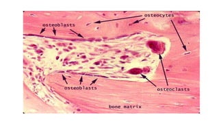
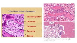
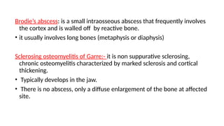
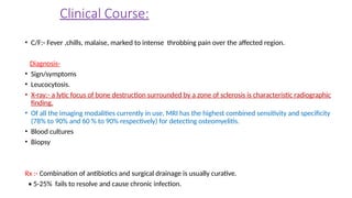
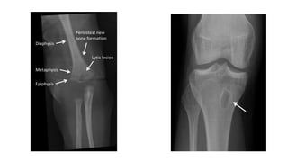
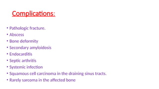
BONE
Dr. NIKHIL KUMAR
ASSISTANT PROFESSOR
Parts of a young bone
• Epiphysis:- End of bone, ossifies
from secondary center of
ossification.
• Diaphysis:- Shaft of a long bone,
ossify by primary center of
ossification.
• Metaphysis:- epiphyseal end of
diaphysis, zone of active growth.
• Epiphyseal plate of cartilage :-
separates epiphysis from diaphysis.
It causes increased length of bone.
Functions of bones-
• Mechanical support
• Force transmission
• Internal organ protection
• Mineral homeostasis
Constituents of bone
• An extracellular matrix and specialized cells responsible for production
and maintenance of the matrix.
• Bone matrix :- composed of an organic component known as osteoid
(35%) and a mineral component (65%).
• The unique feature of bone matrix, its hardness, is imparted by the
inorganic moiety hydroxyapatite [Ca10(PO4)6(OH)2 ], which also serves as a
repository for 99% of the body’s calcium and 85% of its phosphorus.
• Osteoid :- type I collagen + GAGs + other proteins
• Osteopontin/ Osteocalcin :- a protein, produced by osteoblasts &
contributes to the regulation of bone formation, mineralization & calcium
homeostasis.
• Its levels are used as a specific marker of osteoblast activity.
• Bone matrix can be woven or lamellar.
• Woven bone is produced rapidly (during fetal development or fracture
repair) and is always abnormal in adults, but its presence is not specific for
any particular disease.
Cellular components of mature bone
• It consists of bone synthesizing Osteoblasts, Osteocytes, and bone-
resorbing Osteoclasts.
• Osteoblasts:- located on the surface of the matrix, synthesize, transport
and assemble the matrix and regulate its mineralization.
→ Derived from primitive bone marrow stromal cells known as
osteoprogenitor cells under the influence of growth factors such as bone
morphogenic proteins (BMPs).
• Osteocytes:- help to control calcium and phosphate levels.
• Osteoclasts:- specialized multinucleated macrophages derived from
circulating monocytes that are responsible for bone resorption.
Osteoclasts lie in the shallow depressions (Howship’s lacunae) on the
surface of bone.
Proteins of bone matrix
Osteoblast-derived Proteins:-
• Type I collagen
• Calcium-binding protein- Osteonectin, bone sialoprotein
• Cell adhesion Proteins:- Osteopontin, fibronectin, thrombospondin.
• Cytokines:- IL-1, IL-6, RANKL
• Enzymes:- Collagenase, alkaline phosphatase
• Growth factors:- IGF-1, TGF-β, PDGF
• Proteins involved in mineralization:- Osteocalcin.
Proteins Concentrated from serum:- Albumin, β2 microglobulin
Important topics from this system
• 1) Osteomyelitis :- Pyogenic and tuberculous
• 2) Classification of bone tumors
• 3) Osteosarcoma and chondrosarcoma
• 4) Ewing’s sarcoma
• 5) Giant cell tumor
• 6) Osteoporosis and rickets.
OSTEOMYELITIS
• It denotes inflammation of bone and marrow virtually always
secondary to infection.
• All types of organisms, including viruses, parasites, fungi and bacteria,
can produce osteomyelitis, but infections caused by certain pyogenic
bacteria and mycobacteria are the most common.
Classification of Osteomyelitis
• Two major classification schemes for osteomyelitis-
• The first is by Lew and Waldvogel, while the other is by Cierny and
Mader.
• Lew and Waldvogel classified osteomyelitis based on the duration of
illness as acute or chronic and by the mechanism of infection (either
hematogenous or contiguous infection).
• The Cierny and Mader scheme provides guidance in patient
management. In this scheme, the classification of osteomyelitis is
by anatomic stage and the host health status.
• The categories and corresponding anatomic types are:
• Stage 1: Disease confined to the medullary of the bone
• Stage 2: Superficial disease
• Stage 3: Localized spread
• Stage 4: Diffuse disease
Pyogenic Osteomyelitis
• Almost always caused by bacterial infections.
• Organisms may reach the bone by -
A) Haematogenous spread (in children, through mucosal injury or skin infection)
B) Extension from a contiguous site (from soft tissue or joint infection), and
C) Direct implantation (e.g, in traumatic injury)
•In children, most osteomyelitis is hematogenous in origin and develops in the
long bones. (Epiphyseal infection can cause articular damage and permanent
disability).
•In adults, osteomyelitis more often occurs as a complication of open fractures,
surgical procedures and infections of the feet in diabetics .
Causative organisms of Pyogenic osteomyelitis
• Staphylococcus aureus is responsible for 80-90% of the cases of culture
positive pyogenic osteomyelitis.
• Others:- E.coli, Pseudomonas, and klebsiella in patients with genito-urinary
infection or IV drug users.
• In Drug addicts and immunocompromised:- Pseudomonas.
• In the neonatal period, Haemophilus influenzae and Group B streptococci are
frequent pathogens.
• In Sickle cell anemia:- Salmonella infection is more common.
Pathology of osteomyelitis
Most common mode of infection is hematogenous.
•In neonates, metaphyseal vessels penetrate the growth plate, resulting in
frequent infection of the metaphysis, epiphysis or both.
• In children, metaphysis of long bone (usually lower end femur > upper end
tibia) is earliest and most commonly involved site.
• The cause of involvement of metaphysis is because of stasis of blood due
to hair pin arrangement of blood vessels and its proneness to trauma.
• The pathological sequence is inflammation, suppuration, necrosis,
reactive bone formation & ultimately resolution and healing.
• In adults commonest site of infection is thoracolumbar spine.
SEQUENCE OF INFECTION:-
• Once localized in bone, the bacteria proliferate and induce an acute
inflammatory reaction and cause cell death.
• Necrosis of the bone cells and marrow occurs within first 48hrs.
• Spread of bacteria and inflammation within the shaft of the bone and may
percolate through the Haversian systems to reach the periosteum.
• In children ,the periosteum is loosely attached to the cortex; therefore sizable
subperiosteal abscess formation occurs (may rupture and form sinuses).
• Further ischemia and bone necrosis occurs.
• Dead pieces of bone are known as the sequestrum.
• Rupture of the periosteum → soft tissue abscess formation → draining
sinuses.
• In infants, epiphyseal infection may spread to the adjacent joint and
causes septic or suppurative arthritis.; may lead to permanent disability.
• After the first week chronic inflammatory cells become more
numerous with the release of cytokines that stimulates osteoclastic
bone resorption, ingrowth of fibrous tissue and deposition of new
bone formation occur at the periphery.
• Newly deposited bone can form a shell of living tissue, known as an
Involucrum around the segment of devitalized infected bone.
Brodie’s abscess: is a small intraosseous abscess that frequently involves
the cortex and is walled off by reactive bone.
• it usually involves long bones (metaphysis or diaphysis)
Sclerosing osteomyelitis of Garre:- it is non suppurative sclerosing,
chronic osteomyelitis characterized by marked sclerosis and cortical
thickening.
• Typically develops in the jaw.
• There is no abscess, only a diffuse enlargement of the bone at affected
site.
Clinical Course:
• C/F:- Fever ,chills, malaise, marked to intense throbbing pain over the affected region.
Diagnosis-
• Sign/symptoms
• Leucocytosis.
• X-ray:- a lytic focus of bone destruction surrounded by a zone of sclerosis is characteristic radiographic
finding.
• Of all the imaging modalities currently in use, MRI has the highest combined sensitivity and specificity
(78% to 90% and 60 % to 90% respectively) for detecting osteomyelitis.
• Blood cultures
• Biopsy
Rx :- Combination of antibiotics and surgical drainage is usually curative.
• 5-25% fails to resolve and cause chronic infection.
Complications:
• Pathologic fracture.
• Abscess
• Bone deformity
• Secondary amyloidosis
• Endocarditis
• Septic arthritis
• Systemic infection
• Squamous cell carcinoma in the draining sinus tracts.
• Rarely sarcoma in the affected bone
Tuberculous osteomyelitis
• More destructive and more resistant to control.
• Increased risk in patients with pulmonary and extrapulmonary TB.
Routes of entry:-
• Usually blood borne and originate from a focus of active visceral disease
during the initial stages of primary infection.
• Direct extension (e.g. from a pulmonary focus into a rib or from
tracheobronchial nodes into adjacent vertebrae) or spread via draining
lymphatics.
• In patients with AIDS, it is frequently multifocal or disseminated.
• Pott disease (Tuberculous Spondylitis) is the involvement of spine.
• Histology:- granulomatous inflammation & caseous necrosis.
• Thoracic and lumber vertebrae followed by the knees and hips are the
most common sites of skeletal involvement.
• The infection breaks through the intervertebral discs to involve multiple
vertebrae & and extends into the soft tissues forming abscesses.
Clinical features and complications:
• Represents a cold fluctuant psoas abscess.
• Bone destruction.
• Compression fractures may lead to scoliosis, kyphosis and neurologic deficit.
• Tuberculous arthritis.
• Sinus tract formation
• Amyloidosis
• The histologic findings, namely caseous necrosis and granulomas are typical of
tuberculosis.
• Mycobacterial osteomyelitis tends to be more destructive and resistant to
control than pyogenic osteomyelitis.
Q- When osteomyelitis disseminates by hematogenous
way, the most affected part of bone is-
( AIIMS Nov 10, Jipmer 95, DNB 97)
A) Metaphyses
B) Epiphyses
C) Diaphyses
D) Any of the above
Q- The most common organism causing osteomyelitis in
drug abusers is-
( PGI 97)
A) E. Coli
B) Pseudomonas
C) Klebsiella
D) Staph. aureus
• A 5-year-old boy presents with the acute onset of fever, chills, and severe,
throbbing pain over the metaphysis of his left femur. His peripheral
leukocyte count is increased, and an x-ray of his left femur reveals a lytic
focus of bone surrounded by a zone of sclerosis. Which of the following is
the most likely diagnosis?
a. Avascular necrosis due to infarction of the femoral head
b. Acute pyogenic osteomyelitis due to infection with Staphylococcus
aureus
c. Pott disease due to infection with Mycobacterium tuberculosis
d. Chronic osteomyelitis due to infection with Pasteurella multocida
e. Osteitis deformans due to a viral infection of osteoclasts
BONE & soft tissue- Osteomyelitis, pathology.pptx

BONE & soft tissue- Osteomyelitis, pathology.pptx

  • 1.
  • 2.
    Parts of ayoung bone • Epiphysis:- End of bone, ossifies from secondary center of ossification. • Diaphysis:- Shaft of a long bone, ossify by primary center of ossification. • Metaphysis:- epiphyseal end of diaphysis, zone of active growth. • Epiphyseal plate of cartilage :- separates epiphysis from diaphysis. It causes increased length of bone.
  • 3.
    Functions of bones- •Mechanical support • Force transmission • Internal organ protection • Mineral homeostasis
  • 4.
    Constituents of bone •An extracellular matrix and specialized cells responsible for production and maintenance of the matrix. • Bone matrix :- composed of an organic component known as osteoid (35%) and a mineral component (65%). • The unique feature of bone matrix, its hardness, is imparted by the inorganic moiety hydroxyapatite [Ca10(PO4)6(OH)2 ], which also serves as a repository for 99% of the body’s calcium and 85% of its phosphorus. • Osteoid :- type I collagen + GAGs + other proteins
  • 5.
    • Osteopontin/ Osteocalcin:- a protein, produced by osteoblasts & contributes to the regulation of bone formation, mineralization & calcium homeostasis. • Its levels are used as a specific marker of osteoblast activity. • Bone matrix can be woven or lamellar. • Woven bone is produced rapidly (during fetal development or fracture repair) and is always abnormal in adults, but its presence is not specific for any particular disease.
  • 7.
    Cellular components ofmature bone • It consists of bone synthesizing Osteoblasts, Osteocytes, and bone- resorbing Osteoclasts. • Osteoblasts:- located on the surface of the matrix, synthesize, transport and assemble the matrix and regulate its mineralization. → Derived from primitive bone marrow stromal cells known as osteoprogenitor cells under the influence of growth factors such as bone morphogenic proteins (BMPs). • Osteocytes:- help to control calcium and phosphate levels. • Osteoclasts:- specialized multinucleated macrophages derived from circulating monocytes that are responsible for bone resorption. Osteoclasts lie in the shallow depressions (Howship’s lacunae) on the surface of bone.
  • 11.
    Proteins of bonematrix Osteoblast-derived Proteins:- • Type I collagen • Calcium-binding protein- Osteonectin, bone sialoprotein • Cell adhesion Proteins:- Osteopontin, fibronectin, thrombospondin. • Cytokines:- IL-1, IL-6, RANKL • Enzymes:- Collagenase, alkaline phosphatase • Growth factors:- IGF-1, TGF-β, PDGF • Proteins involved in mineralization:- Osteocalcin. Proteins Concentrated from serum:- Albumin, β2 microglobulin
  • 12.
    Important topics fromthis system • 1) Osteomyelitis :- Pyogenic and tuberculous • 2) Classification of bone tumors • 3) Osteosarcoma and chondrosarcoma • 4) Ewing’s sarcoma • 5) Giant cell tumor • 6) Osteoporosis and rickets.
  • 13.
    OSTEOMYELITIS • It denotesinflammation of bone and marrow virtually always secondary to infection. • All types of organisms, including viruses, parasites, fungi and bacteria, can produce osteomyelitis, but infections caused by certain pyogenic bacteria and mycobacteria are the most common.
  • 14.
    Classification of Osteomyelitis •Two major classification schemes for osteomyelitis- • The first is by Lew and Waldvogel, while the other is by Cierny and Mader. • Lew and Waldvogel classified osteomyelitis based on the duration of illness as acute or chronic and by the mechanism of infection (either hematogenous or contiguous infection).
  • 15.
    • The Ciernyand Mader scheme provides guidance in patient management. In this scheme, the classification of osteomyelitis is by anatomic stage and the host health status. • The categories and corresponding anatomic types are: • Stage 1: Disease confined to the medullary of the bone • Stage 2: Superficial disease • Stage 3: Localized spread • Stage 4: Diffuse disease
  • 16.
    Pyogenic Osteomyelitis • Almostalways caused by bacterial infections. • Organisms may reach the bone by - A) Haematogenous spread (in children, through mucosal injury or skin infection) B) Extension from a contiguous site (from soft tissue or joint infection), and C) Direct implantation (e.g, in traumatic injury) •In children, most osteomyelitis is hematogenous in origin and develops in the long bones. (Epiphyseal infection can cause articular damage and permanent disability). •In adults, osteomyelitis more often occurs as a complication of open fractures, surgical procedures and infections of the feet in diabetics .
  • 17.
    Causative organisms ofPyogenic osteomyelitis • Staphylococcus aureus is responsible for 80-90% of the cases of culture positive pyogenic osteomyelitis. • Others:- E.coli, Pseudomonas, and klebsiella in patients with genito-urinary infection or IV drug users. • In Drug addicts and immunocompromised:- Pseudomonas. • In the neonatal period, Haemophilus influenzae and Group B streptococci are frequent pathogens. • In Sickle cell anemia:- Salmonella infection is more common.
  • 18.
    Pathology of osteomyelitis Mostcommon mode of infection is hematogenous. •In neonates, metaphyseal vessels penetrate the growth plate, resulting in frequent infection of the metaphysis, epiphysis or both. • In children, metaphysis of long bone (usually lower end femur > upper end tibia) is earliest and most commonly involved site. • The cause of involvement of metaphysis is because of stasis of blood due to hair pin arrangement of blood vessels and its proneness to trauma.
  • 19.
    • The pathologicalsequence is inflammation, suppuration, necrosis, reactive bone formation & ultimately resolution and healing. • In adults commonest site of infection is thoracolumbar spine.
  • 20.
    SEQUENCE OF INFECTION:- •Once localized in bone, the bacteria proliferate and induce an acute inflammatory reaction and cause cell death. • Necrosis of the bone cells and marrow occurs within first 48hrs. • Spread of bacteria and inflammation within the shaft of the bone and may percolate through the Haversian systems to reach the periosteum. • In children ,the periosteum is loosely attached to the cortex; therefore sizable subperiosteal abscess formation occurs (may rupture and form sinuses). • Further ischemia and bone necrosis occurs.
  • 21.
    • Dead piecesof bone are known as the sequestrum. • Rupture of the periosteum → soft tissue abscess formation → draining sinuses. • In infants, epiphyseal infection may spread to the adjacent joint and causes septic or suppurative arthritis.; may lead to permanent disability.
  • 22.
    • After thefirst week chronic inflammatory cells become more numerous with the release of cytokines that stimulates osteoclastic bone resorption, ingrowth of fibrous tissue and deposition of new bone formation occur at the periphery. • Newly deposited bone can form a shell of living tissue, known as an Involucrum around the segment of devitalized infected bone.
  • 25.
    Brodie’s abscess: isa small intraosseous abscess that frequently involves the cortex and is walled off by reactive bone. • it usually involves long bones (metaphysis or diaphysis) Sclerosing osteomyelitis of Garre:- it is non suppurative sclerosing, chronic osteomyelitis characterized by marked sclerosis and cortical thickening. • Typically develops in the jaw. • There is no abscess, only a diffuse enlargement of the bone at affected site.
  • 26.
    Clinical Course: • C/F:-Fever ,chills, malaise, marked to intense throbbing pain over the affected region. Diagnosis- • Sign/symptoms • Leucocytosis. • X-ray:- a lytic focus of bone destruction surrounded by a zone of sclerosis is characteristic radiographic finding. • Of all the imaging modalities currently in use, MRI has the highest combined sensitivity and specificity (78% to 90% and 60 % to 90% respectively) for detecting osteomyelitis. • Blood cultures • Biopsy Rx :- Combination of antibiotics and surgical drainage is usually curative. • 5-25% fails to resolve and cause chronic infection.
  • 28.
    Complications: • Pathologic fracture. •Abscess • Bone deformity • Secondary amyloidosis • Endocarditis • Septic arthritis • Systemic infection • Squamous cell carcinoma in the draining sinus tracts. • Rarely sarcoma in the affected bone
  • 29.
    Tuberculous osteomyelitis • Moredestructive and more resistant to control. • Increased risk in patients with pulmonary and extrapulmonary TB. Routes of entry:- • Usually blood borne and originate from a focus of active visceral disease during the initial stages of primary infection. • Direct extension (e.g. from a pulmonary focus into a rib or from tracheobronchial nodes into adjacent vertebrae) or spread via draining lymphatics.
  • 30.
    • In patientswith AIDS, it is frequently multifocal or disseminated. • Pott disease (Tuberculous Spondylitis) is the involvement of spine. • Histology:- granulomatous inflammation & caseous necrosis. • Thoracic and lumber vertebrae followed by the knees and hips are the most common sites of skeletal involvement. • The infection breaks through the intervertebral discs to involve multiple vertebrae & and extends into the soft tissues forming abscesses.
  • 31.
    Clinical features andcomplications: • Represents a cold fluctuant psoas abscess. • Bone destruction. • Compression fractures may lead to scoliosis, kyphosis and neurologic deficit. • Tuberculous arthritis. • Sinus tract formation • Amyloidosis • The histologic findings, namely caseous necrosis and granulomas are typical of tuberculosis. • Mycobacterial osteomyelitis tends to be more destructive and resistant to control than pyogenic osteomyelitis.
  • 32.
    Q- When osteomyelitisdisseminates by hematogenous way, the most affected part of bone is- ( AIIMS Nov 10, Jipmer 95, DNB 97) A) Metaphyses B) Epiphyses C) Diaphyses D) Any of the above
  • 33.
    Q- The mostcommon organism causing osteomyelitis in drug abusers is- ( PGI 97) A) E. Coli B) Pseudomonas C) Klebsiella D) Staph. aureus
  • 34.
    • A 5-year-oldboy presents with the acute onset of fever, chills, and severe, throbbing pain over the metaphysis of his left femur. His peripheral leukocyte count is increased, and an x-ray of his left femur reveals a lytic focus of bone surrounded by a zone of sclerosis. Which of the following is the most likely diagnosis? a. Avascular necrosis due to infarction of the femoral head b. Acute pyogenic osteomyelitis due to infection with Staphylococcus aureus c. Pott disease due to infection with Mycobacterium tuberculosis d. Chronic osteomyelitis due to infection with Pasteurella multocida e. Osteitis deformans due to a viral infection of osteoclasts