Serological tests detect antibodies in blood samples and can be used to identify individuals exposed to infectious diseases like COVID-19. Enzyme-linked immunosorbent assays (ELISAs) and lateral flow immunoassays (LFIAs) are commonly used serological test methods. ELISAs are often used in laboratories to detect and quantify antibodies, while LFIAs can provide rapid, point-of-care testing using strips similar to pregnancy tests. Both tests aim to specifically detect antibodies produced against target antigens, helping clinicians understand infection histories.
Serological test for virus identificationPlock Ghosh
This presentation consist of detailed study of serological method of virus identification. Basically ELISA is vastly used for virus detection. Western blot method is used for HIV identification.
Serological test for virus identificationPlock Ghosh
This presentation consist of detailed study of serological method of virus identification. Basically ELISA is vastly used for virus detection. Western blot method is used for HIV identification.
Antibodies, also known as immunoglobulins, are secreted by B cells (plasma cells) to neutralize antigens such as bacteria and viruses. The classical representation of an antibody is a Y-shaped molecule composed of four polypeptides-two heavy chains and two light chains. Each tip of the "Y" contains a paratope (a structure analogous to a lock) that is specific for one particular epitope (similarly analogous to a key) on an antigen, allowing these two structures to bind together with precision. The ability of binding to an antigen has led to their ubiquitous use in a variety of life science and medical science. These antibodies can be classified into two primary types (monoclonal and polyclonal) by the means in which they are created from lymphocytes. Each of them has important role in the immune system, diagnostic exams, and treatments.
Immunity
It can be defined as the resistance to disease, specifically to infectious disease or pathogens. The term “immune” is derived from the Latin word “immunis” that is exempt from charges. In medical term, it refers to the being protected from infectious pathogens.
Immune system
It is adaptive defense system which is able to generate a variety of cell and molecules capable of specifically recognizing and eliminating a variety of limitless foreign invaders into the system.
In 1975 Georges Kohler and Milstein succeeded in making fusions of myeloma cell lines with B cells to create hybridomas that could produce antibodies.
antibody
Also known as immunoglobulin is a large, Y shaped glycoprotein produced mainly by plasma cells that is used by the immune system to neutralize pathogens.
monoclonal antibodies
Antibodies that are made by identical immune cells that are clones of a unique parent cell.
polyclonal antibodies
A polyclonal antibodies represents a collection of antibodies from different B cells that recognize multiple epitopes on the same antigen.
An antibody (Ab), also known as an immunoglobulin (Ig), is a large, Y-shaped protein produced mainly by plasma cells that is used by the immune system to neutralize pathogens such as pathogenic bacteria and viruses.
Monoclonal antibodies are important reagents used in biomedical research, in diagnosis of diseases, and in treatment of such diseases as infections and cancer.
These antibodies are produced by cell lines or clones obtained from animals that have been immunized with the substance that is the subject of study.
The lecture was presented to the students of Saudi board of Community Medicine to help them know about the various serological methods applicable in the diagnosis of infectious diseases in general with attention upon the specificity and sensitivity of various diagnostic modalities. The lecture covers the basic principles of each test and the clinical applications with the advantages and disadvantages of each.
Antigen ,Antibody and Ag-Ab reactions ppt by DR.C.P.PRINCEDR.PRINCE C P
An immunogen refers to a molecule that is capable of eliciting an immune response, whereas an antigen refers to a molecule that is capable of binding to the product of that immune response (Ab).
So, an immunogen is necessarily an antigen, but an antigen may not necessarily be an immunogen
The terms immunogen and antigen are often used interchangeably but the later is more common.
Antibodies are Globulin Protein (Immunoglobulin) that are synthesized in the Serum and Tissue fluids.
It reacts specifically with the antigen that stimulated their production.
There are two types serum proteins: albumin and globulin
There are Three types of globulins .
1. Alpha globulin
2. Beta globulin
3. Gamma globulin (Antibodies)
Gamma globulins are responsible for immunity. So they are called as Immunoglobulin (Ig)
The binding of an antibody with an antigen of the type that stimulated the formation of antibody that results in the following reaction
Agglutination
Precipitation
Complement fixation
Phagocytosis
Neutralization of an exotoxin
Opsonization
Tissue fixation
Chemotaxis
Activation of mast cells and basophils
PPT prepared by:
DR.PRINCE C P
Associate Professor , Department of Microbiology,
Mother Theresa Post Graduate & Research Institute of Health Sciences (Government of Puducherry Institution)
ELISA, principle and method by kk sahuKAUSHAL SAHU
What is ELISA.
Principle.
History.
Types of ELISA method.
1.Direct ELISA.
2.Indirect ELISA.
3.Sandwhich ELISA.
Conclusion.
References.
Antibodies (also known as immunoglobulins abbreviated Ig) are gamma globulin proteins that are found in blood and are used by the immune system to identify and neutralize foreign objects, such as bacteria and viruses.
Applications of immunological functions(Sp13-bty-001) CIIT Abbottabad Zohaib HUSSAIN
Applications of Immunological functions
Introduction
Immunological function is define as biological effects of immune system on antigens during immune responds
Effects may be
1.Pathological effects (By keeping normal homeostasis)
2.Physiological effects (Resulting in diseases)
Main functions of immunity
1.Defense
2.Homeostasis
3.Surveillance
Physiological and pathological representation of immune response
Function Physiological
(advantageous) Pathological
(harmful)
1.Immune defense Resist to pathogen Immunologic deficiency disease
2.Immune homeostasis scavenge damaged Autoalergic disease
3.Immune surveillance Scavenge cells with misreplication Cell cancerization persistent infection
Applications of Immunological functions
1.Treatment (Vaccines)
Immunity to a disease is achieved through the presence of antibodies to that disease in a person system. Antibodies are proteins produced by the body to neutralize or destroy toxins or disease-carrying organisms. Antibodies are disease specific. For example, measles antibody will protect a person who is exposed to measles disease, but will have no effect if he or she is exposed to mumps.
There are two types of immunity: active and passive.
a)Active Immunity
It results when exposure to a disease organism triggers the immune system to produce antibodies to that disease. Exposure to the disease organism can occur through infection with the actual disease (natural immunity), or introduction of a killed or weakened form of the disease organism through vaccination (vaccine-induced immunity). If an immune person comes into contact with that disease in the future, their immune system will recognize it and immediately produce the antibodies needed to fight it. Active immunity is long-lasting, and sometimes life-long.Vaccines are available for all of the following vaccine-preventable diseases Typhoid hepatitis A and B influenza and many more
b)Passive Immunity
It is provided when a person is given antibodies to a disease rather than producing them through his or her own immune system. A newborn baby acquires passive immunity from its mother through the placenta. A major advantage of passive immunity is protection is mediated whereas active immunity takes time usually 2 to 3 weeks but it is for only few weeks for months only active immunity is long lasting e.g. tetanus Pertussis Diphtheria etc. is cure by passive immunity
2.Diagnosis
It use purified antibody solutions (antiserum) to diagnose disease. Diagnostic antibodies can be produced to detect particular microbe
A. In animals (mixed antiserum)
Inject animal with microbe or antigenic fragments
Allow immune response (1-2 weeks)
Harvest blood
Purify antibodies from serum to make antiserum = purified antibody solution to one particular antigen. These preparations will produce multiple Antibody types that recognize
Introduction:
RNA interference (RNAi) or Post-Transcriptional Gene Silencing (PTGS) is an important biological process for modulating eukaryotic gene expression.
It is highly conserved process of posttranscriptional gene silencing by which double stranded RNA (dsRNA) causes sequence-specific degradation of mRNA sequences.
dsRNA-induced gene silencing (RNAi) is reported in a wide range of eukaryotes ranging from worms, insects, mammals and plants.
This process mediates resistance to both endogenous parasitic and exogenous pathogenic nucleic acids, and regulates the expression of protein-coding genes.
What are small ncRNAs?
micro RNA (miRNA)
short interfering RNA (siRNA)
Properties of small non-coding RNA:
Involved in silencing mRNA transcripts.
Called “small” because they are usually only about 21-24 nucleotides long.
Synthesized by first cutting up longer precursor sequences (like the 61nt one that Lee discovered).
Silence an mRNA by base pairing with some sequence on the mRNA.
Discovery of siRNA?
The first small RNA:
In 1993 Rosalind Lee (Victor Ambros lab) was studying a non- coding gene in C. elegans, lin-4, that was involved in silencing of another gene, lin-14, at the appropriate time in the
development of the worm C. elegans.
Two small transcripts of lin-4 (22nt and 61nt) were found to be complementary to a sequence in the 3' UTR of lin-14.
Because lin-4 encoded no protein, she deduced that it must be these transcripts that are causing the silencing by RNA-RNA interactions.
Types of RNAi ( non coding RNA)
MiRNA
Length (23-25 nt)
Trans acting
Binds with target MRNA in mismatch
Translation inhibition
Si RNA
Length 21 nt.
Cis acting
Bind with target Mrna in perfect complementary sequence
Piwi-RNA
Length ; 25 to 36 nt.
Expressed in Germ Cells
Regulates trnasposomes activity
MECHANISM OF RNAI:
First the double-stranded RNA teams up with a protein complex named Dicer, which cuts the long RNA into short pieces.
Then another protein complex called RISC (RNA-induced silencing complex) discards one of the two RNA strands.
The RISC-docked, single-stranded RNA then pairs with the homologous mRNA and destroys it.
THE RISC COMPLEX:
RISC is large(>500kD) RNA multi- protein Binding complex which triggers MRNA degradation in response to MRNA
Unwinding of double stranded Si RNA by ATP independent Helicase
Active component of RISC is Ago proteins( ENDONUCLEASE) which cleave target MRNA.
DICER: endonuclease (RNase Family III)
Argonaute: Central Component of the RNA-Induced Silencing Complex (RISC)
One strand of the dsRNA produced by Dicer is retained in the RISC complex in association with Argonaute
ARGONAUTE PROTEIN :
1.PAZ(PIWI/Argonaute/ Zwille)- Recognition of target MRNA
2.PIWI (p-element induced wimpy Testis)- breaks Phosphodiester bond of mRNA.)RNAse H activity.
MiRNA:
The Double-stranded RNAs are naturally produced in eukaryotic cells during development, and they have a key role in regulating gene expression .
Comparing Evolved Extractive Text Summary Scores of Bidirectional Encoder Rep...University of Maribor
Slides from:
11th International Conference on Electrical, Electronics and Computer Engineering (IcETRAN), Niš, 3-6 June 2024
Track: Artificial Intelligence
https://www.etran.rs/2024/en/home-english/
(May 29th, 2024) Advancements in Intravital Microscopy- Insights for Preclini...Scintica Instrumentation
Intravital microscopy (IVM) is a powerful tool utilized to study cellular behavior over time and space in vivo. Much of our understanding of cell biology has been accomplished using various in vitro and ex vivo methods; however, these studies do not necessarily reflect the natural dynamics of biological processes. Unlike traditional cell culture or fixed tissue imaging, IVM allows for the ultra-fast high-resolution imaging of cellular processes over time and space and were studied in its natural environment. Real-time visualization of biological processes in the context of an intact organism helps maintain physiological relevance and provide insights into the progression of disease, response to treatments or developmental processes.
In this webinar we give an overview of advanced applications of the IVM system in preclinical research. IVIM technology is a provider of all-in-one intravital microscopy systems and solutions optimized for in vivo imaging of live animal models at sub-micron resolution. The system’s unique features and user-friendly software enables researchers to probe fast dynamic biological processes such as immune cell tracking, cell-cell interaction as well as vascularization and tumor metastasis with exceptional detail. This webinar will also give an overview of IVM being utilized in drug development, offering a view into the intricate interaction between drugs/nanoparticles and tissues in vivo and allows for the evaluation of therapeutic intervention in a variety of tissues and organs. This interdisciplinary collaboration continues to drive the advancements of novel therapeutic strategies.
Earliest Galaxies in the JADES Origins Field: Luminosity Function and Cosmic ...Sérgio Sacani
We characterize the earliest galaxy population in the JADES Origins Field (JOF), the deepest
imaging field observed with JWST. We make use of the ancillary Hubble optical images (5 filters
spanning 0.4−0.9µm) and novel JWST images with 14 filters spanning 0.8−5µm, including 7 mediumband filters, and reaching total exposure times of up to 46 hours per filter. We combine all our data
at > 2.3µm to construct an ultradeep image, reaching as deep as ≈ 31.4 AB mag in the stack and
30.3-31.0 AB mag (5σ, r = 0.1” circular aperture) in individual filters. We measure photometric
redshifts and use robust selection criteria to identify a sample of eight galaxy candidates at redshifts
z = 11.5 − 15. These objects show compact half-light radii of R1/2 ∼ 50 − 200pc, stellar masses of
M⋆ ∼ 107−108M⊙, and star-formation rates of SFR ∼ 0.1−1 M⊙ yr−1
. Our search finds no candidates
at 15 < z < 20, placing upper limits at these redshifts. We develop a forward modeling approach to
infer the properties of the evolving luminosity function without binning in redshift or luminosity that
marginalizes over the photometric redshift uncertainty of our candidate galaxies and incorporates the
impact of non-detections. We find a z = 12 luminosity function in good agreement with prior results,
and that the luminosity function normalization and UV luminosity density decline by a factor of ∼ 2.5
from z = 12 to z = 14. We discuss the possible implications of our results in the context of theoretical
models for evolution of the dark matter halo mass function.
Deep Behavioral Phenotyping in Systems Neuroscience for Functional Atlasing a...Ana Luísa Pinho
Functional Magnetic Resonance Imaging (fMRI) provides means to characterize brain activations in response to behavior. However, cognitive neuroscience has been limited to group-level effects referring to the performance of specific tasks. To obtain the functional profile of elementary cognitive mechanisms, the combination of brain responses to many tasks is required. Yet, to date, both structural atlases and parcellation-based activations do not fully account for cognitive function and still present several limitations. Further, they do not adapt overall to individual characteristics. In this talk, I will give an account of deep-behavioral phenotyping strategies, namely data-driven methods in large task-fMRI datasets, to optimize functional brain-data collection and improve inference of effects-of-interest related to mental processes. Key to this approach is the employment of fast multi-functional paradigms rich on features that can be well parametrized and, consequently, facilitate the creation of psycho-physiological constructs to be modelled with imaging data. Particular emphasis will be given to music stimuli when studying high-order cognitive mechanisms, due to their ecological nature and quality to enable complex behavior compounded by discrete entities. I will also discuss how deep-behavioral phenotyping and individualized models applied to neuroimaging data can better account for the subject-specific organization of domain-general cognitive systems in the human brain. Finally, the accumulation of functional brain signatures brings the possibility to clarify relationships among tasks and create a univocal link between brain systems and mental functions through: (1) the development of ontologies proposing an organization of cognitive processes; and (2) brain-network taxonomies describing functional specialization. To this end, tools to improve commensurability in cognitive science are necessary, such as public repositories, ontology-based platforms and automated meta-analysis tools. I will thus discuss some brain-atlasing resources currently under development, and their applicability in cognitive as well as clinical neuroscience.
Multi-source connectivity as the driver of solar wind variability in the heli...Sérgio Sacani
The ambient solar wind that flls the heliosphere originates from multiple
sources in the solar corona and is highly structured. It is often described
as high-speed, relatively homogeneous, plasma streams from coronal
holes and slow-speed, highly variable, streams whose source regions are
under debate. A key goal of ESA/NASA’s Solar Orbiter mission is to identify
solar wind sources and understand what drives the complexity seen in the
heliosphere. By combining magnetic feld modelling and spectroscopic
techniques with high-resolution observations and measurements, we show
that the solar wind variability detected in situ by Solar Orbiter in March
2022 is driven by spatio-temporal changes in the magnetic connectivity to
multiple sources in the solar atmosphere. The magnetic feld footpoints
connected to the spacecraft moved from the boundaries of a coronal hole
to one active region (12961) and then across to another region (12957). This
is refected in the in situ measurements, which show the transition from fast
to highly Alfvénic then to slow solar wind that is disrupted by the arrival of
a coronal mass ejection. Our results describe solar wind variability at 0.5 au
but are applicable to near-Earth observatories.
Professional air quality monitoring systems provide immediate, on-site data for analysis, compliance, and decision-making.
Monitor common gases, weather parameters, particulates.
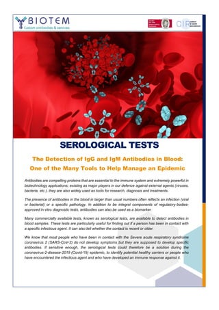
1. SEROLOGICAL TESTS
The Detection of IgG and IgM Antibodies in Blood:
One of the Many Tools to Help Manage an Epidemic
Antibodies are compelling proteins that are essential to the immune system and extremely powerful in
biotechnology applications; existing as major players in our defence against external agents (viruses,
bacteria, etc.), they are also widely used as tools for research, diagnosis and treatments.
The presence of antibodies in the blood in larger than usual numbers often reflects an infection (viral
or bacterial) or a specific pathology. In addition to be integral components of regulatory-bodies-
approved in vitro diagnostic tests, antibodies can also be used as a biomarker.
Many commercially available tests, known as serological tests, are available to detect antibodies in
blood samples. These tests are particularly useful for finding out if a person has been in contact with
a specific infectious agent. It can also tell whether the contact is recent or older.
We know that most people who have been in contact with the Severe acute respiratory syndrome
coronavirus 2 (SARS-CoV-2) do not develop symptoms but they are supposed to develop specific
antibodies. If sensitive enough, the serological tests could therefore be a solution during the
coronavirus-2-disease-2019 (Covid-19) epidemic, to identify potential healthy carriers or people who
have encountered the infectious agent and who have developed an immune response against it.
2. 2
ANTIBODY OVERVIEW
ntibodies, or immunoglobulins, are complex glycoproteins secreted by B lymphocytes (plasmocytes).
In general, these proteins are produced by the immune system in response to an infectious agent or a
pathogen. Antibodies will specifically recognize this agent, referred to as an antigen, in order to
neutralize and eliminate it. The production of antibodies and the antigen-antibody recognition is
therefore a completely natural defence system enabling the body to act against a pathogen.
The structure of antibodies (Abs) was first described in 1959 by Porter and Edelman. It is a protein of about 150
kDa. With a global structure similar to the letter Y (Figure 1), it consists of:
2 heavy chains (H for heavy – in blue) each with 1 variable and 3 or 4 constant domains
2 light chains (L for Light – in green) each with a variable and a constant domain
Heavy and light chains are connected to each
other by disulfide bridges (in yellow).
VH + VL = variable globular domain at the end
of the 2 branches of the « Y ». The antigen is
recognized by the N-terminal ends of the
variable domains.
CH1 + CL = first constant globular domain
between the variable domain and the hinge.
CH2 x 2 = second constant globular domain
under the hinge.
CH3 x 2 = third constant globular domain,
towards the C-terminus of the heavy chains.
FIGURE 1 ANTIBODY MODELLING. MOUSE IgG2a ISOTYPE. THE LIGHT CHAINS ARE GREEN, THE HEAVY CHAINS
ARE CYAN AND BLUE, THE GLYCAN IS IN ORANGE, AND THE INTERCHAIN DISULFIDES ARE YELLOW STICKS.
In humans and in mice, there are different categories of antibodies or isotypes in the blood stream (Figure 2):
IgG (normal values for IgG are ~70% of the
circulating Abs in humans)
IgG is the most present antibody isotype in normal
human serum. It consists of 4 subclasses (IgG1,
IgG2, IgG3 and IgG4) each containing a different
heavy chain type.
IgM (10%)
As a soluble globulin, IgM is most often present as a
pentamer, its basic structure held together by a J
chain. It can also exist as a hexameric form in the
serum and as a monomer on the surface of B-cells.
IgA (<20%)
IgA exists as a monomer or a dimer. In contrast to
the serum where this isotype is in minority, IgA is the
A
FIGURE 2 IMMUNOGLOBULIN ISOTYPES
3. 3
predominant antibody in mucous secretions such as saliva, tears, etc.
IgE (<1%)
IgE is very rare in the serum but is found on basophils and mast-cells. This class is known for its role in
allergies and to fight infection by parasites.
IgD (<1%)
IgD is very rare in human serum and more important on the surface of B-cells. Its role is still unknown.
IgM is the predominant antibody in the primary immune response. During the maturation of B lymphocytes, there
is an isotype switching: IgG will be expressed in larger quantities in serum, IgA in the mucosal tissues and IgE
for allergens or parasites.
FIGURE 3 AVERAGE ANTIBODY EXPRESSION AFTER EXPOSURE TO A GIVEN ANTIGEN THAT ELICATES IGGS
SEROLOGICAL TESTS OVERVIEW
erological tests allow to detect soluble proteins, hormones or biomarkers – antibodies in the case that
interests us today – in a blood serum sample. These tests can be qualitative or quantitative and are widely
used for screening, diagnosis, or patient follow-up, but also for epidemiological studies.
As all diagnostics tests these, tools are not 100% reliable. Indeed, due to the time of appearance of
immunoglobulins and also their expression in a wide range of quantities – starting from very low levels – the
tests must be very sensitive to avoid delivering false negatives (when the biomarker is present, but the test fails
to detect it). The question of the limit of detection (LOD) will be discussed below.
On the other hand, the specificity has to be good enough for the test to be meaningful; you do not want to have
too many false alerts that would not come from real infections (false positives), although in the case of Covid-
19, the risk for the patient is relatively low since the consequence will just be a quarantine, as far as no specific
treatment can be given (present situation). A true positive result means that the patient has been in contact with
a pathogen at some time. Unlike virological tests in which viral particles are detected, serological tests allow a
patient to be evaluated even if the virus is no longer present. In addition, long-term follow-up can provide
information, for example if the patient is in the course of its isotype switch.
S
4. 4
There are different types of serological tests:
Precipitation tests based on the precipitation that takes place when antibodies (especially IgMs) and
antigens are mixed together.
Neutralization tests, which depend on the capacity of antibodies to neutralize the infectious properties
of the infectious organisms.
Hemagglutinin-inhibition tests based on the agglutination of red blood cells. This agglutination will be
prevented by the presence of antibodies.
Enzyme Linked Immunosorbent Assay (ELISA) or Lateral Flow Immunoassay (LFIA) based on
antibody binding to the antigen on a solid surface (plate or strip).
We will focus only on these last two techniques which are the most common in routine (see BIOTEM’s
Immunoassay Solutions).
1. ELISA: Enzyme Linked Immunosorbent Assay
Principally used for research and for in vitro diagnostic purposes, ELISA tests are enzymatic immunoassays for
the detection and quantification of analytes in equipped laboratories. Several formats of ELISA are available
(Figure 5):
Indirect ELISA
This is a simple 2-step ELISA test. The antigen (or pathogen) of interest is coated on the plate. Serum
samples are then deposited and incubated. In the presence of antibodies (IgG / IgM) in the serum, these
will specifically bind to the antigen. Revelation is carried out with an anti-human isotype, secondary
antibody conjugated to a tracer, typically the enzyme HRP. This format is the most commonly used in
routine.
FIGURE 4 SEROLOGICAL TESTS. A) PRECIPITATION TEST (COMPLEMENT FIXATION TEST) ;
B) NEUTRALIZATION TEST ; C) HEMAGGLUTININ-INHIBITION TEST.
Sources: Online Microbiology Notes and Microbe Online
5. 5
Sandwich ELISA
The plate is coated with a capture antibody specific to the antigen / pathogen of interest. This primary
antibody could be commercially available or developed from scratch (see BIOTEM’s Antibody Solutions).
Secondly, the antigen in solution is deposed and incubated. Then serum samples are deposited and
incubated. Specific antibodies (IgG / IgM, but also other isotypes) that are present in the serum will bind
to the antigen. Revelation is carried out with an anti-human IgG or IgM, secondary antibody. The capture
antibody (the one coated on the plate) must be from another species than human in order to avoid non-
specific binding of the anti-human secondary antibody (the one used for revelation).
Competitive ELISA
The result is negatively proportional to the amount of biomarker to be measured. This format is rarely
used for serological tests.
2. LFIA: Lateral Flow Immunoassay or Rapid tests
Lateral Flow tests are based on the migration of nano or micro particles on strips for analytes detection. These
tests, strips or cassettes (example: the pregnancy test) are widely used as first-line emergency tests performed
at the patient’s bedside or directly in the field, thanks to its user-friendly design.
Several common names are used (although some are less adapted than others):
Lateral Flow ImmunoAssay (LFIA)
ImmunoChromatography assays or ImmunoChromatography Test (ICT)
Rapid Test or Quick Test
Point of Care (POC) Test
Strip Test or Dipstick test
As with the ELISA test, the Lateral Flow test can be used in different applications (Human health, Animal health,
Drug presence assessment, Industrial quality control, Agriculture, Biosecurity, Environment, etc.) and with
different types of samples (serum, plasma, urine, saliva, food, environmental media, water, etc.).
Although a Lateral Flow test is easy to use, it is based on a sophisticated device made up of several components
lying on a plastic support strip (Figure 6):
FIGURE 5 SCHEMATIC REPRESENTATION OF TWO TYPES OF ELISA TESTS
6. 6
● The Sample Pad is necessary for the proper distribution of the
sample towards the conjugate pad. The sample pad can also be
impregnated with different reactants (buffers, detergents, proteins,
etc.) to modify / adapt the chemical / physical characteristics of the
sample or to facilitate its migration and fixation on the test line. It
could also be useful as a filter. Typically, the sample pad is composed
of cellulose fiber filters.
● The function of the Conjugate Pad is to distribute the detector
particles onto the so-called ‘Membrane’ (the real support of the test)
in a consistent volume with respect to the sample. The analyte
present in the sample will therefore come in contact with the detector
particles and migrate together to the test line. The detector particles are typically antibodies conjugated to beads
(gold, coloured latex, etc.) that will allow the appearance of the test and control lines. The conjugate pad is
composed of fibers of cellulose, of glass or plastic, when the ‘membrane’ is usually made of nitrocellulose.
● The Test Line corresponds to the narrow space where the visualization of the target of interest will take place.
● The Control Line is used to check the correct migration of the sample. Whatever the result, this second line
must always be appearing.
● As the sample pad, the Absorbent Pad is a very important component for the good fluidics of the strip. It
facilitates the migration of the sample along the membrane by capillary effect and absorbs the overflow.
Several characteristics must be taken into account for the test and control lines in order to obtain good
performances:
Thickness
Weight
Tensile strength
Dimensions (length and width)
Material
As for ELISA tests, several formats are available (Figure 7):
Indirect or Sandwich LFIA
A positive result is represented by the presence of a coloured line at the test line position
Competitive LFIA
A positive result is represented by the absence of a coloured line at the test line position. This format is
rarely used for serological tests.
FIGURE 7 COMMON LATERAL FLOW FORMATS
FIGURE 6 LATERAL FLOW COMPONENTS
7. 7
3. Test Performances
The performances of a test are highly variable depending on the target, the type of samples (matrix effects) but
also depending on the limit of detection (LOD). Many parameters have to be taken into account in order to have
a test with optimal performance and avoid false negatives and/or false positives (Figure 8).
Limits of quantification
Limits of quantification are the highest and lowest concentrations of an analyte that have been
demonstrated to be measurable for a given device, with acceptable levels of precision and accuracy.
Sensitivity (Se)
The sensitivity of a test is the probability that the test will be positive if the person effectively has the
disease. It is the number of true positives divided by the total number of people with the disease (A /
A+C). The more sensitive a test is, the fewer false negatives it delivers.
Specificity (Sp)
The specificity of a test is the probability that the test will be negative if the person tested is effectively
free of the disease. It is the number of true negatives divided by the total number of people free of the
disease (D / B+D). The more specific a test is, the fewer false positives it delivers.
Positive Predictive Value (PPV)
The PPV of a test is the probability that the person is really sick if the test is positive. It is the number of
true positives divided by the total number of people with a positive test (A / A+B).
Negative Predictive Value (NPV)
The NPV of a test is the probability that the person will not have the disease if the test is negative. It is
the number of true negatives divided by the total number of people with a negative test (D / C+D).
Robustness:
The ability of a method to remain unaffected by small variations in method parameters.
Precision:
The closeness of agreement between independent test results obtained under specific conditions.
Reproducibility:
The precision between laboratories (standardization).
Repeatability:
The precision with the same operating conditions over a short interval of time (intra-assay precision).
Stability:
The capacity of an analyte (in a matrix) or a test to give the same results under specific conditions
(temperature, humidity, etc.) for given time intervals.
Non exhaustive list
FIGURE 8 DIAGRAM EXPLAINING THE INTRINSIC PERFORMANCE OF A TEST
8. 8
ELISA or LFIA? The choice is essentially based on the practicality of the test and therefore the situation in
which it will be used.
Often used as an emergency test, a rapid test is useful as a first-line test and for large scale testing that
would have to be easy to use. More accurate tests but that would demand scientific instrumentations, can
then be used for positive patient confirmations. ELISA testing is common but requires laboratory equipment
(Table 1).
Pros. Cons.
ELISA Quantitative assay
Optimal limit of detection (LOD)
Sensitivity
Reliable and Robust
Multiplex analysis
Laboratory equipment
Long turnaround time (> 30 min)
Cost
Refrigerated storage
LFIA/ Rapid Test Rapid test and user friendly
Short results (< 10 min)
Emergency test
Economical industrial production
Easy storage (room temperature)
Qualitative or semi-quantitative
Multiplex detection
Sensitivity
SEROLOGICAL TESTS & COVID-19 EPIDEMIC
lready today, more than 140 Covid-19 serological tests are available worldwide. Many of these tests
were available rapidly at the beginning of the epidemic but sometimes with performances that can be
further optimized. Other tests are still under development and should be available in the coming weeks.
Whereas virological tests allow detection of the viral gene (RNA) by PCR, serological tests allow the
detection of antibodies (IgG and IgM) specific to the virus even long time after its clearance from the patient’s
body. Therefore, these second test types are complementary to the first ones and allow the identification of
patients who have been in contact (recently or not) with the virus and who have developed an immune response.
Currently, no specific therapy or vaccine exists to treat or prevent Covid-19 infection. In order to limit the spread
of the virus and to discharge our hospital networks, emergency measures that have not been taken in over a
century have been adopted by many countries. One of the main measures is the confinement of the population.
This is an extremely urgent measure to contain the progression of the epidemic but it raises the question of what
will happen after this period. Several points are currently at steaks, including:
̶ How can we get out of confinement by reducing the number of new contaminations?
̶ Will a vaccine or treatment be available quickly?
Serological tests may be a partial short-term solution to assess the population and their immune response
against the virus, since a first contact might not be enough to fully immunize.
When can these tests be used? Are they effective enough? Do we have enough information about the virus and
its effect on the immune system? These are questions that doctors, specialists and researchers are working on
to identify the best solutions.
One thing is certain, virological testing remains for the moment the solution to identify infected and contagious
people. But serological tests are definitely valuable as part of the arsenal that we need to have against this
epidemic.
A
TABLE 1. COMPARISON ELISA VS. LFIA
9. 9
WE WOULD LIKE TO THANK ALL THE PEOPLE MOBILIZED TO FIGHT
THIS EPIDEMIC. THANK YOU TO THE MEDICAL STAFF FOR TAKING
CARE OF OUR PATIENTS. THANK YOU TO THE HOSPITAL STAFF, OFTEN
IN THE SHADOWS, FOR THEIR DAILY INVOLVEMENT. THANK YOU ALSO
TO ALL THE RESEARCHERS WHO ARE WORKING CONSTANTLY ON
FUTURE SCREENING AND THERAPY TOOLS.
THANK YOU TO OUR EMPLOYEES WHO ARE ALSO INVESTED IN
CARRYING OUT PROJECTS WITHRESEARCHERS. FINALLY, THANKYOU
TO OUR CUSTOMERS FOR THEIR TRUST.
TOGETHER WE WILL WIN AGAINST COVID-19!
SAVE LIVES, STAY HOME
10. 10
SOURCES
Antibody Structure and Function: The Basis for Engineering Therapeutics (PMID: 31816964)
Functional switching (Milestone 8 ; Nature https://www.nature.com/articles/ni.3607)
Atlas of immunology (Cruse, J.M., and Lewis, R.E. (2010). Boca Raton, FL: CRC Press/Taylor &
Francis).
Assay Validation Methods (https://www.fws.gov/aah/PDF/QI-Terms%20and%20Defs.pdf)
A practical guide to immunoassay method validation. (Front. Neurol., 19 August 2015 |
https://doi.org/10.3389/fneur.2015.00179)
Smart Servier Medical Art
11. 11
ABOUT BIOTEM
BIOTEM is a Contract Research Organization providing custom and high added value services in the field of
antibody and immunoassay development.
With 40 years of experience, BIOTEM has developed a large panel of improved strategies enabling it to
complete the most challenging projects. Our staff scientists have extensive experience in antigen development
(peptides, proteins), hybridoma & recombinant antibody generation and production. In addition, the team has
developed an exclusive know-how in antigen design / conjugation, tracer preparation (gold, latex, magnetic
particles, carbon, etc.) and selection, as well as in immunoassay production (ELISA plate, Lateral Flow strip
and cassette).
Our technology platforms allow us to choose among several production processes in order to perfectly match
each project specifications. Thanks to a high success rate (>96%), most of our contracts are proposed with
results commitments and are composed of Go / No-Go phases.
BIOTEM also complies with ISO 9001 and ISO 13485 (IVD) quality standards.
100% fee-for-service: BIOTEM does not claim any intellectual properties or any other rights on the
developed antibodies or immunoassays.
BIOTEM: YOUR PARTNER IN IMMUNOTECHNOLOGIES SINCE 1980!
WWW.BIOTEM-ANTIBODY.COM
Written by Jonathan MAYALI
jonathan.mayali@biotem.fr
+33 476 651 091