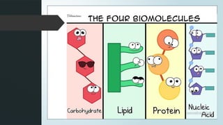
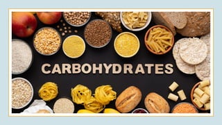
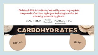
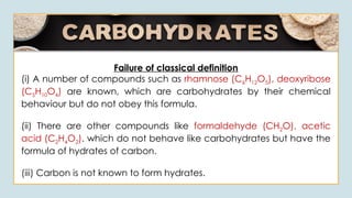
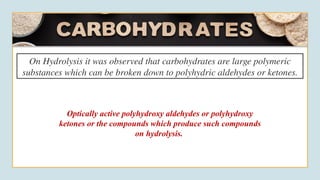
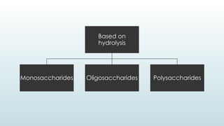
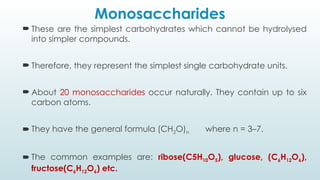
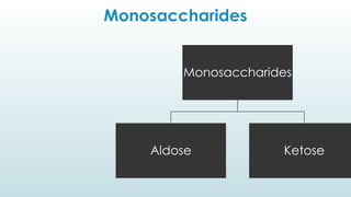
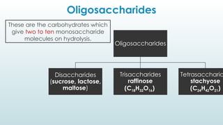
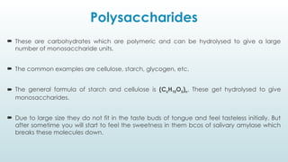
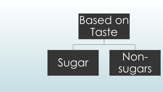
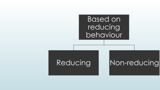
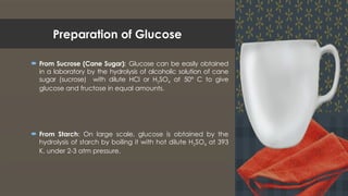
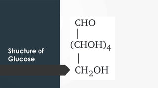
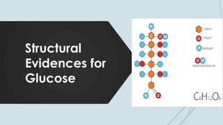
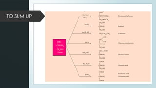
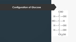
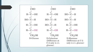
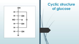
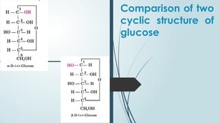
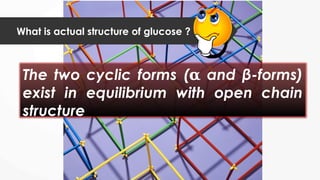
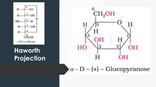
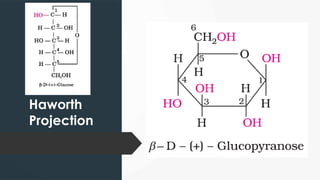
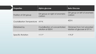
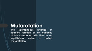
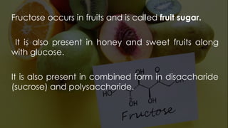
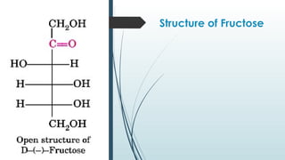
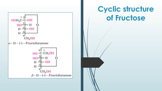
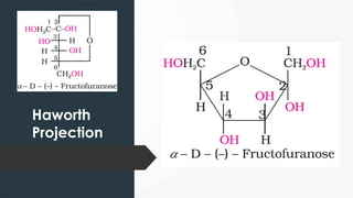
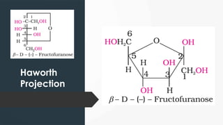
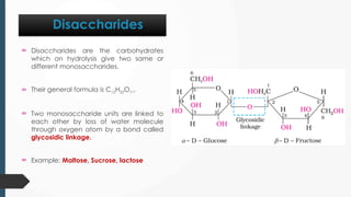
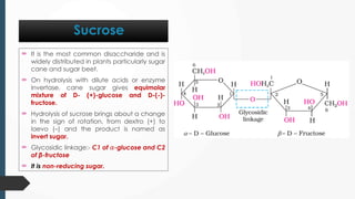
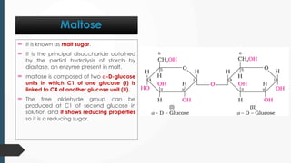
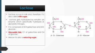
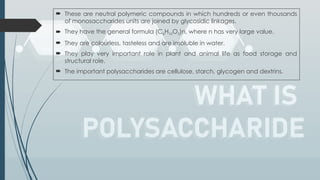
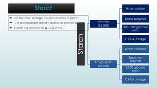
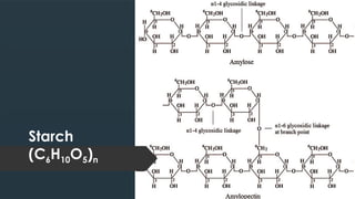
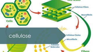
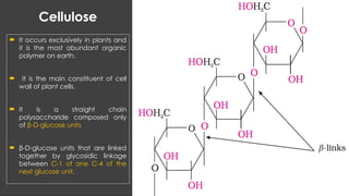
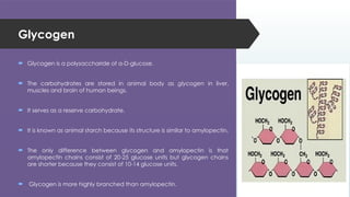
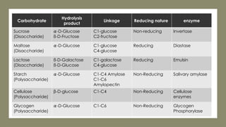
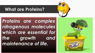
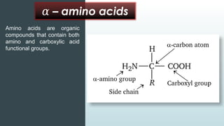
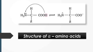
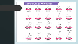
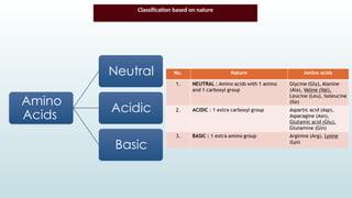
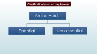
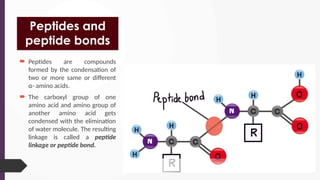
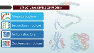
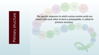
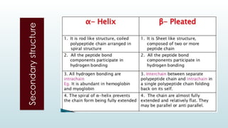
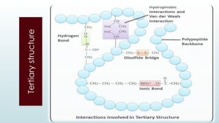
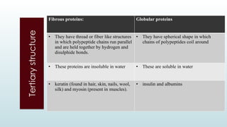
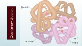
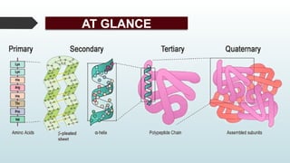
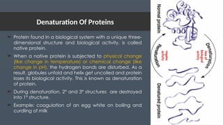
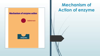
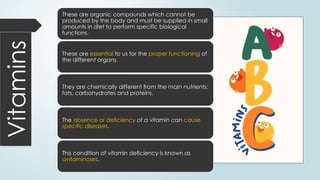
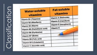
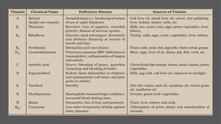
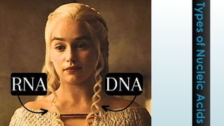
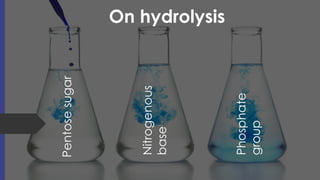
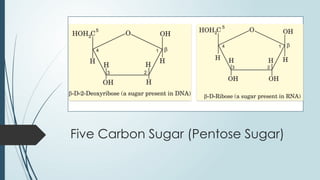
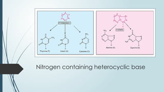
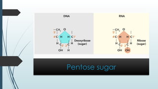
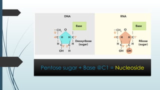
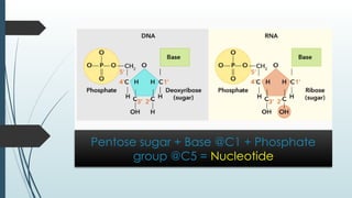
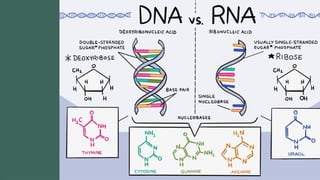
Chapter-10
Building block
of life
Biomolecules are the complex
lifeless molecules which build
up living organisms and are
required for their growth and
maintenance
Carbon Water
Carbohydrates are a class of naturally occurring organic
compounds of carbon, hydrogen and oxygen which are
primarily produced by plants.
Failure of classical definition
(i) A number of compounds such as rhamnose (C6H12O5), deoxyribose
(C5H10O4) are known, which are carbohydrates by their chemical
behaviour but do not obey this formula.
(ii) There are other compounds like formaldehyde (CH2O), acetic
acid (C2H4O2). which do not behave like carbohydrates but have the
formula of hydrates of carbon.
(iii) Carbon is not known to form hydrates.
On Hydrolysis it was observed that carbohydrates are large polymeric
substances which can be broken down to polyhydric aldehydes or ketones.
Optically active polyhydroxy aldehydes or polyhydroxy
ketones or the compounds which produce such compounds
on hydrolysis.
Classification of Carbohydrates
Based on
hydrolysis
Monosaccharides Oligosaccharides Polysaccharides
Monosaccharides
 These are the simplest carbohydrates which cannot be hydrolysed
into simpler compounds.
 Therefore, they represent the simplest single carbohydrate units.
 About 20 monosaccharides occur naturally. They contain up to six
carbon atoms.
 They have the general formula (CH2O)n where n = 3–7.
 The common examples are: ribose(C5H10O5), glucose, (C6H12O6),
fructose(C6H12O6) etc.
Monosaccharides
Monosaccharides
Aldose Ketose
Oligosaccharides
Oligosaccharides
Disaccharides
(sucrose, lactose,
maltose)
Trisaccharides
raffinose
(C18H32O16)
Tetrasaccharid
stachyose
(C24H42O21)
These are the carbohydrates which
give two to ten monosaccharide
molecules on hydrolysis.
Polysaccharides
 These are carbohydrates which are polymeric and can be hydrolysed to give a large
number of monosaccharide units.
 The common examples are cellulose, starch, glycogen, etc.
 The general formula of starch and cellulose is (C6H10O5)n. These get hydrolysed to give
monosaccharides.
 Due to large size they do not fit in the taste buds of tongue and feel tasteless initially. But
after sometime you will start to feel the sweetness in them bcos of salivary amylase which
breaks these molecules down.
Based on
Taste
Sugar
Non-
sugars
Based on
reducing
behaviour
Reducing Non-reducing
Glucose (C6H12O6)
Preparation of Glucose
 From Sucrose (Cane Sugar): Glucose can be easily obtained
in a laboratory by the hydrolysis of alcoholic solution of cane
sugar (sucrose) with dilute HCl or H2SO4 at 50° C to give
glucose and fructose in equal amounts.
 From Starch: On large scale, glucose is obtained by the
hydrolysis of starch by boiling it with hot dilute H2SO4 at 393
K, under 2-3 atm pressure.
Structure of
Glucose
Structural
Evidences for
Glucose
Reaction with
HI
(Presence of
straight chain)
Presence of
Carbonyl Group
Presence of
Aldehydic Group
Presence of
5 (-OH) Group
Presence of
1 (1O
-OH) Group
Reaction with
Na-Hg & H2
TO SUM UP
Configuration of Glucose
 Glucose do not restore the pink colour of Schiff 's reagent, despite having an
aldehydic group.
 It also fails to form the hydrogen sulphite addition product with NaHSO3.
 The pentaacetate of glucose does not react with hydroxylamine and 2,4-DNP
derivative which shows the absence of free —CHO group.
 Glucose exists in two different crystalline forms which are named as alpha ( )
𝛼
and beta form (β).
Limitations of straight chain structure
Cyclic structure of glucose
Cyclic structure
of glucose
Cyclic structure
of glucose
Comparison of two
cyclic structure of
glucose
What is actual structure of glucose ?
The two cyclic forms ( and β-forms)
𝛂
exist in equilibrium with open chain
structure
Haworth
Projection
Haworth
Projection
Properties Alpha glucose Beta Glucose
Position of OH group
OH group on right of anomeric
carbon
OH group on left of anomeric
carbon
Crystallization Temperature 419 K
423 K
Obtained by Crystallization of concentrated
solution at 303 K
Obtained from hot saturated
solution of glucose at 371 K
Specific Rotation +111° +19.2°
Mutarotation
The spontaneous change in
specific rotation of an optically
active compound with time to an
equilibrium value is called
mutarotation.
Fructose occurs in fruits and is called fruit sugar.
It is also present in honey and sweet fruits along
with glucose.
It is also present in combined form in disaccharide
(sucrose) and polysaccharide.
Structure of Fructose
Cyclic structure
of Fructose
Haworth
Projection
Haworth
Projection
Disaccharides
 Disaccharides are the carbohydrates
which on hydrolysis give two same or
different monosaccharides.
 Their general formula is C12H22O11.
 Two monosaccharide units are linked to
each other by loss of water molecule
through oxygen atom by a bond called
glycosidic linkage.
 Example: Maltose, Sucrose, lactose
 It is the most common disaccharide and is
widely distributed in plants particularly sugar
cane and sugar beet.
 On hydrolysis with dilute acids or enzyme
invertase, cane sugar gives equimolar
mixture of D- (+)-glucose and D-(-)-
fructose.
 Hydrolysis of sucrose brings about a change
in the sign of rotation, from dextro (+) to
laevo (–) and the product is named as
invert sugar.
 Glycosidic linkage:- C1 of 𝛼-glucose and C2
of β-fructose
 It is non-reducing sugar.
Sucrose
 It is known as malt sugar.
 It is the principal disaccharide obtained
by the partial hydrolysis of starch by
diastase, an enzyme present in malt.
 maltose is composed of two 𝛼-D-glucose
units in which C1 of one glucose (I) is
linked to C4 of another glucose unit (II).
 The free aldehyde group can be
produced at C1 of second glucose in
solution and it shows reducing properties
so it is a reducing sugar.
Maltose
 Lactose occurs in milk and, therefore, it is
also called milk sugar.
 Lactose gets hydrolyzed by emulsin, an
enzyme which specifically hydrolyses β-
glycosidic linkages.
 It is composed of β-D-galactose and β-D-
glucose.
 Glycosidic link: C1 of galactose and C4
of glucose.
 Hence it is also a reducing sugar.
Lactose
 These are neutral polymeric compounds in which hundreds or even thousands
of monosaccharides units are joined by glycosidic linkages.
 They have the general formula (C6H10O5)n, where n has very large value.
 They are colourless, tasteless and are insoluble in water.
 They play very important role in plant and animal life as food storage and
structural role.
 The important polysaccharides are cellulose, starch, glycogen and dextrins.
Starch
Starch
Amylose
(15-20%)
Water soluble
Linear polymer
200-1000 glucose
units
C1-C4 Linkage
Amylopectin
(80-85%)
Water Insoluble
Branched
polymer
25-30 glucose
units
C1-C6 Linkage
Starch
 It is the main storage polysaccharide of plants.
 It is an important dietary source for human beings
 Starch is a polymer of 𝛂-D-glucose.
Starch
(C6H10O5)n
cellulose
Cellulose
 It occurs exclusively in plants and
it is the most abundant organic
polymer on earth.
 It is the main constituent of cell
wall of plant cells.
 It is a straight chain
polysaccharide composed only
of β-D-glucose units
 β-D-glucose units that are linked
together by glycosidic linkage
between C-1 of one C-4 of the
next glucose unit.
Glycogen
 Glycogen is a polysaccharide of α-D-glucose.
 The carbohydrates are stored in animal body as glycogen in liver,
muscles and brain of human beings.
 It serves as a reserve carbohydrate.
 It is known as animal starch because its structure is similar to amylopectin.
 The only difference between glycogen and amylopectin is that
amylopectin chains consist of 20-25 glucose units but glycogen chains
are shorter because they consist of 10-14 glucose units.
 Glycogen is more highly branched than amylopectin.
Carbohydrate
Hydrolysis
product
Linkage Reducing nature enzyme
Sucrose
(Disaccharide)
𝛼-D-Glucose
ß-D-Fructose
C1-glucose
C2-fructose
Non-reducing Invertase
Maltose
(Disaccharide)
𝛼-D-Glucose C1-glucose
C4-glucose
Reducing Diastase
Lactose
(Disaccharide)
ß-D-Galactose
ß-D-Glucose
C1-galactose
C4-glucose
Reducing Emulsin
Starch
(Polysaccharide)
𝛼-D-Glucose C1-C4 Amylose
C1-C6
Amylopectin
Non-Reducing Salivary amylase
Cellulose
(Polysaccharide)
β-D-glucose C1-C4 Non-Reducing Cellulose
enzymes
Glycogen
(Polysaccharide)
𝛼-D-Glucose C1-C6 Non-Reducing Glycogen
Phosphorylase
PROTEIN
What are Proteins?
Proteins are complex
nitrogenous molecules
which are essential for
the growth and
maintenance of life.
On hydrolysis
Proteins are polymeric chain
of – amino acids
𝛼
–
𝛼 amino acids
Amino acids are organic
compounds that contain both
amino and carboxylic acid
functional groups.
Structure of – amino acids
𝛼
Effect of pH and Isoelectric point
Classification of Amino Acids
Classification based on nature
Amino
Acids
Neutral
Acidic
Basic
Amino Acids
Essential Non-essential
Classification based on requirement
Peptides and
peptide bonds
 Peptides are compounds
formed by the condensation of
two or more same or different
α- amino acids.
 The carboxyl group of one
amino acid and amino group of
another amino acid gets
condensed with the elimination
of water molecule. The resulting
linkage is called a peptide
linkage or peptide bond.
STRUCTURAL LEVELS OF PROTEIN
Primary structure
Secondary structure
Tertiary structure
Quaternary structure
Primary
structure The specific sequence in which various amino acids are
linked with each other to form a polypeptide, is called its
primary structure.
Secondary
structure
Secondary
structure
Tertiary
structure
Tertiary
structure
Fibrous proteins: Globular proteins
• They have thread or fiber like structures
in which polypeptide chains run parallel
and are held together by hydrogen and
disulphide bonds.
• They have spherical shape in which
chains of polypeptides coil around
• These proteins are insoluble in water • These are soluble in water
• keratin (found in hair, skin, nails, wool,
silk) and myosin (present in muscles).
• insulin and albumins
Quaternary
structure
AT GLANCE
Denaturation Of Proteins
 Protein found In a biological system with a unique three-
dimensional structure and biological activity, is called
native protein.
 When a native protein is subjected to physical change
(like change in temperature) or chemical change (like
change in pH), the hydrogen bonds are disturbed. As a
result, globules unfold and helix get uncoiled and protein
loses its biological activity. This is known as denaturation
of protein.
 During denaturation, 2° and 3° structures are destroyed
into 1° structure.
 Example: coagulation of an egg white on boiling and
curdling of milk
The enzymes are biological catalysts produced by living cells which catalyze the
biochemical reactions in living organisms.
Enzymes
 Chemically enzymes are naturally occurring
simple or globular proteins. Some enzymes
may be non-proteins also.
 The enzymes are generally named after the
compound or class of compound upon
which they work.
 Sometimes the enzymes are also named after
the reaction where they are used.
For example, the enzymes which catalyse the
oxidation of one substrate with simultaneous
reduction of another substrate are named as
oxide reductase enzymes.
 The ending of the name of an enzyme is –ase.
Some important Chemical reactions and their enzymes
1. High efficiency
2. Required in extremely small quantities
3. They are highly Specific in nature.
4. Work only at Optimum temperature and pH
Mechanism of
Action of enzyme
Vitamins These are organic compounds which cannot be
produced by the body and must be supplied in small
amounts in diet to perform specific biological
functions.
These are essential to us for the proper functioning of
the different organs.
They are chemically different from the main nutrients;
fats, carbohydrates and proteins.
The absence or deficiency of a vitamin can cause
specific diseases.
This condition of vitamin deficiency is known as
avitaminoses.
Classification
Nucleic Acids
Types
of
Nucleic
Acids
On hydrolysis
Pentose
sugar
Nitrogenous
base
Phosphate
group
Five Carbon Sugar (Pentose Sugar)
Nitrogen containing heterocyclic base
Phosphate Group
The formation
of Nucleic
Acids
Pentose sugar
Pentose sugar + Base @C1 = Nucleoside
Pentose sugar + Base @C1 + Phosphate
group @C5 = Nucleotide
Biomolecules_class12ncertwithrizwaninal.pptx

Biomolecules_class12ncertwithrizwaninal.pptx

  • 1.
  • 2.
  • 3.
    Biomolecules are thecomplex lifeless molecules which build up living organisms and are required for their growth and maintenance
  • 6.
    Carbon Water Carbohydrates area class of naturally occurring organic compounds of carbon, hydrogen and oxygen which are primarily produced by plants.
  • 7.
    Failure of classicaldefinition (i) A number of compounds such as rhamnose (C6H12O5), deoxyribose (C5H10O4) are known, which are carbohydrates by their chemical behaviour but do not obey this formula. (ii) There are other compounds like formaldehyde (CH2O), acetic acid (C2H4O2). which do not behave like carbohydrates but have the formula of hydrates of carbon. (iii) Carbon is not known to form hydrates.
  • 8.
    On Hydrolysis itwas observed that carbohydrates are large polymeric substances which can be broken down to polyhydric aldehydes or ketones. Optically active polyhydroxy aldehydes or polyhydroxy ketones or the compounds which produce such compounds on hydrolysis.
  • 9.
  • 10.
  • 11.
    Monosaccharides  These arethe simplest carbohydrates which cannot be hydrolysed into simpler compounds.  Therefore, they represent the simplest single carbohydrate units.  About 20 monosaccharides occur naturally. They contain up to six carbon atoms.  They have the general formula (CH2O)n where n = 3–7.  The common examples are: ribose(C5H10O5), glucose, (C6H12O6), fructose(C6H12O6) etc.
  • 12.
  • 13.
  • 14.
    Polysaccharides  These arecarbohydrates which are polymeric and can be hydrolysed to give a large number of monosaccharide units.  The common examples are cellulose, starch, glycogen, etc.  The general formula of starch and cellulose is (C6H10O5)n. These get hydrolysed to give monosaccharides.  Due to large size they do not fit in the taste buds of tongue and feel tasteless initially. But after sometime you will start to feel the sweetness in them bcos of salivary amylase which breaks these molecules down.
  • 15.
  • 16.
  • 18.
  • 19.
    Preparation of Glucose From Sucrose (Cane Sugar): Glucose can be easily obtained in a laboratory by the hydrolysis of alcoholic solution of cane sugar (sucrose) with dilute HCl or H2SO4 at 50° C to give glucose and fructose in equal amounts.  From Starch: On large scale, glucose is obtained by the hydrolysis of starch by boiling it with hot dilute H2SO4 at 393 K, under 2-3 atm pressure.
  • 20.
  • 21.
  • 22.
  • 23.
  • 24.
  • 25.
  • 26.
  • 27.
  • 28.
  • 29.
  • 31.
     Glucose donot restore the pink colour of Schiff 's reagent, despite having an aldehydic group.  It also fails to form the hydrogen sulphite addition product with NaHSO3.  The pentaacetate of glucose does not react with hydroxylamine and 2,4-DNP derivative which shows the absence of free —CHO group.  Glucose exists in two different crystalline forms which are named as alpha ( ) 𝛼 and beta form (β). Limitations of straight chain structure
  • 32.
  • 33.
  • 34.
  • 35.
    Comparison of two cyclicstructure of glucose
  • 36.
    What is actualstructure of glucose ? The two cyclic forms ( and β-forms) 𝛂 exist in equilibrium with open chain structure
  • 37.
  • 38.
  • 39.
    Properties Alpha glucoseBeta Glucose Position of OH group OH group on right of anomeric carbon OH group on left of anomeric carbon Crystallization Temperature 419 K 423 K Obtained by Crystallization of concentrated solution at 303 K Obtained from hot saturated solution of glucose at 371 K Specific Rotation +111° +19.2°
  • 40.
    Mutarotation The spontaneous changein specific rotation of an optically active compound with time to an equilibrium value is called mutarotation.
  • 41.
    Fructose occurs infruits and is called fruit sugar. It is also present in honey and sweet fruits along with glucose. It is also present in combined form in disaccharide (sucrose) and polysaccharide.
  • 42.
  • 43.
  • 44.
  • 45.
  • 47.
    Disaccharides  Disaccharides arethe carbohydrates which on hydrolysis give two same or different monosaccharides.  Their general formula is C12H22O11.  Two monosaccharide units are linked to each other by loss of water molecule through oxygen atom by a bond called glycosidic linkage.  Example: Maltose, Sucrose, lactose
  • 48.
     It isthe most common disaccharide and is widely distributed in plants particularly sugar cane and sugar beet.  On hydrolysis with dilute acids or enzyme invertase, cane sugar gives equimolar mixture of D- (+)-glucose and D-(-)- fructose.  Hydrolysis of sucrose brings about a change in the sign of rotation, from dextro (+) to laevo (–) and the product is named as invert sugar.  Glycosidic linkage:- C1 of 𝛼-glucose and C2 of β-fructose  It is non-reducing sugar. Sucrose
  • 49.
     It isknown as malt sugar.  It is the principal disaccharide obtained by the partial hydrolysis of starch by diastase, an enzyme present in malt.  maltose is composed of two 𝛼-D-glucose units in which C1 of one glucose (I) is linked to C4 of another glucose unit (II).  The free aldehyde group can be produced at C1 of second glucose in solution and it shows reducing properties so it is a reducing sugar. Maltose
  • 50.
     Lactose occursin milk and, therefore, it is also called milk sugar.  Lactose gets hydrolyzed by emulsin, an enzyme which specifically hydrolyses β- glycosidic linkages.  It is composed of β-D-galactose and β-D- glucose.  Glycosidic link: C1 of galactose and C4 of glucose.  Hence it is also a reducing sugar. Lactose
  • 52.
     These areneutral polymeric compounds in which hundreds or even thousands of monosaccharides units are joined by glycosidic linkages.  They have the general formula (C6H10O5)n, where n has very large value.  They are colourless, tasteless and are insoluble in water.  They play very important role in plant and animal life as food storage and structural role.  The important polysaccharides are cellulose, starch, glycogen and dextrins.
  • 53.
  • 54.
    Starch Amylose (15-20%) Water soluble Linear polymer 200-1000glucose units C1-C4 Linkage Amylopectin (80-85%) Water Insoluble Branched polymer 25-30 glucose units C1-C6 Linkage Starch  It is the main storage polysaccharide of plants.  It is an important dietary source for human beings  Starch is a polymer of 𝛂-D-glucose.
  • 55.
  • 56.
  • 57.
    Cellulose  It occursexclusively in plants and it is the most abundant organic polymer on earth.  It is the main constituent of cell wall of plant cells.  It is a straight chain polysaccharide composed only of β-D-glucose units  β-D-glucose units that are linked together by glycosidic linkage between C-1 of one C-4 of the next glucose unit.
  • 58.
    Glycogen  Glycogen isa polysaccharide of α-D-glucose.  The carbohydrates are stored in animal body as glycogen in liver, muscles and brain of human beings.  It serves as a reserve carbohydrate.  It is known as animal starch because its structure is similar to amylopectin.  The only difference between glycogen and amylopectin is that amylopectin chains consist of 20-25 glucose units but glycogen chains are shorter because they consist of 10-14 glucose units.  Glycogen is more highly branched than amylopectin.
  • 59.
    Carbohydrate Hydrolysis product Linkage Reducing natureenzyme Sucrose (Disaccharide) 𝛼-D-Glucose ß-D-Fructose C1-glucose C2-fructose Non-reducing Invertase Maltose (Disaccharide) 𝛼-D-Glucose C1-glucose C4-glucose Reducing Diastase Lactose (Disaccharide) ß-D-Galactose ß-D-Glucose C1-galactose C4-glucose Reducing Emulsin Starch (Polysaccharide) 𝛼-D-Glucose C1-C4 Amylose C1-C6 Amylopectin Non-Reducing Salivary amylase Cellulose (Polysaccharide) β-D-glucose C1-C4 Non-Reducing Cellulose enzymes Glycogen (Polysaccharide) 𝛼-D-Glucose C1-C6 Non-Reducing Glycogen Phosphorylase
  • 60.
  • 61.
    What are Proteins? Proteinsare complex nitrogenous molecules which are essential for the growth and maintenance of life.
  • 62.
    On hydrolysis Proteins arepolymeric chain of – amino acids 𝛼
  • 63.
    – 𝛼 amino acids Aminoacids are organic compounds that contain both amino and carboxylic acid functional groups.
  • 64.
    Structure of –amino acids 𝛼
  • 66.
    Effect of pHand Isoelectric point
  • 67.
  • 68.
    Classification based onnature Amino Acids Neutral Acidic Basic
  • 69.
  • 70.
    Peptides and peptide bonds Peptides are compounds formed by the condensation of two or more same or different α- amino acids.  The carboxyl group of one amino acid and amino group of another amino acid gets condensed with the elimination of water molecule. The resulting linkage is called a peptide linkage or peptide bond.
  • 71.
    STRUCTURAL LEVELS OFPROTEIN Primary structure Secondary structure Tertiary structure Quaternary structure
  • 72.
    Primary structure The specificsequence in which various amino acids are linked with each other to form a polypeptide, is called its primary structure.
  • 73.
  • 74.
  • 75.
  • 76.
    Tertiary structure Fibrous proteins: Globularproteins • They have thread or fiber like structures in which polypeptide chains run parallel and are held together by hydrogen and disulphide bonds. • They have spherical shape in which chains of polypeptides coil around • These proteins are insoluble in water • These are soluble in water • keratin (found in hair, skin, nails, wool, silk) and myosin (present in muscles). • insulin and albumins
  • 77.
  • 78.
  • 79.
    Denaturation Of Proteins Protein found In a biological system with a unique three- dimensional structure and biological activity, is called native protein.  When a native protein is subjected to physical change (like change in temperature) or chemical change (like change in pH), the hydrogen bonds are disturbed. As a result, globules unfold and helix get uncoiled and protein loses its biological activity. This is known as denaturation of protein.  During denaturation, 2° and 3° structures are destroyed into 1° structure.  Example: coagulation of an egg white on boiling and curdling of milk
  • 83.
    The enzymes arebiological catalysts produced by living cells which catalyze the biochemical reactions in living organisms.
  • 84.
    Enzymes  Chemically enzymesare naturally occurring simple or globular proteins. Some enzymes may be non-proteins also.  The enzymes are generally named after the compound or class of compound upon which they work.  Sometimes the enzymes are also named after the reaction where they are used. For example, the enzymes which catalyse the oxidation of one substrate with simultaneous reduction of another substrate are named as oxide reductase enzymes.  The ending of the name of an enzyme is –ase.
  • 85.
    Some important Chemicalreactions and their enzymes
  • 86.
    1. High efficiency 2.Required in extremely small quantities 3. They are highly Specific in nature. 4. Work only at Optimum temperature and pH
  • 87.
  • 88.
    Vitamins These areorganic compounds which cannot be produced by the body and must be supplied in small amounts in diet to perform specific biological functions. These are essential to us for the proper functioning of the different organs. They are chemically different from the main nutrients; fats, carbohydrates and proteins. The absence or deficiency of a vitamin can cause specific diseases. This condition of vitamin deficiency is known as avitaminoses.
  • 89.
  • 91.
  • 92.
  • 93.
  • 94.
    Five Carbon Sugar(Pentose Sugar)
  • 95.
  • 96.
  • 97.
  • 98.
  • 99.
    Pentose sugar +Base @C1 = Nucleoside
  • 100.
    Pentose sugar +Base @C1 + Phosphate group @C5 = Nucleotide

Editor's Notes

  • #30 Emil Fischer gave the exact spatial arrangement of different −OH groups in glucose after studying many other properties. D and L before the name of any compound commonly indicates the relative configuration of a particular stereoisomer If —OH on the lowest asymmetric carbon is on the right side, it is assigned D-configuration. If —OH on the lowest asymmetric carbon is on the left side, it is assigned L-configuration.
  • #43  It is also called laevulose.
  • #49 Since the laevorotation of fructose (–92.4°) is more than dextrorotation of glucose (+ 52.5°), the mixture is laevorotatory.
  • #58 Cellulose is not digested by the human digestive system because the system does not have the necessary enzymes to hydrolyse it. However, the grazing animals such as cows, buffaloes, etc., can digest cellulose. (Cellulolytic bacteria present in the stomach (rumen) of ruminant mammals (cattle, sheep, etc.) break down cellulose with the help of enzyme cellulase. It is then digested and converted into glucose. Cellulose is the major structural polysaccharide in higher plants. The bulk of cell walls of plants and vegetable tissues are made of cellulose. It is mainly present in wood (45-50%), cotton seeds (90-95%), flax (80-85%), hemp (75-80%), jute (60-65%) and cereal straws (40-45%). In fact, more than 50% of the total organic matter in the living world is cellulose.
  • #67 Proteins on complete hydrolysis yields Amino Acids There are 20 standard amino acids which are repeatedly found in the structure of proteins – animal, plant or microbial. Roughly 500 amino acids have been identified in nature, but just 20 amino acids make up the proteins found in the human body. 10 of the 20 amino acids found in proteins can be synthesized in the human body
  • #68 Amino acids contain both an acidic carboxyl (-COOH)) group and a basic amino (—NH2) group. Spectroscopic studies reveal that in neutral solution (pH = 7.0), both carboxyl and amino groups are in the ionised form. The —COOH group loses a proton to form a carboxylate (—COO) ion, whereas the amino group gains a proton to form —NH3 ion. Thus, in neutral aqueous solution, the amino acids form a dipolar ion structure called internal salt structure or zwitter ion structure.
  • #69 List of 20 amino acids of which proteins are made in the human body
  • #70 List of 20 amino acids of which proteins are made in the human body
  • #71 An amino acid exists as a positive ion in acidic solution and as a negative ion in the basic solution. Therefore, on passing electric current, it will migrate towards the cathode in acidic solution and towards the anode in basic solution. Hence, at a particular pH of the solution, the amino acid molecule should not migrate to either electrode and should exist as a neutral dipolar ion. This pH is known as isoelectric point. The isoelectric point depends on other functional groups present in the amino acid. The neutral amino acids have isoelectric points in the range of pH 5.5 to 6.3. At isoelectric point, the amino acids have least solubility in water and this property is exploited in the separation of different amino acids obtained from the hydrolysis of proteins.
  • #74 Histidine, isoleucine, leucine, lysine, methionine, phenylalanine, threonine, tryptophan, and valine
  • #76 If a large number of α-amino acids (hundreds to thousands) are joined by peptide bonds, the resulting compound is called polypeptide. By convention a peptide having molecular mass upto 10,000 u is called polypeptide, while a peptide having a molecular mass more than 10,000 u is called a protein. A polypeptide with a fewer amino acids may also be called a protein if it has a well- defined conformation of a protein such as insulin which contains 51 amino acids
  • #78 Proteins may have one or more polypeptide chains. Each polypeptide chain has a large number of α-amino acids linked to one another in a specific sequence. The change of just one amino acid in the sequence can alter its biological activity and can create a different protein. Sickel cell anameia leading because of defective amino acid sequence in HB. The first method was developed by Frederic Sanger in 1953, who succeeded in determining the amino acid sequence in insulin. Insulin has 51 amino acids arranged in two polypeptide chains which are cross-linked at two places by disulphide bonds. One chain contains 21 amino acids units and the other has 30 amino acids.
  • #79 The secondary structure of protein refers to the shape in which a long polypeptide chain can exist.
  • #80 The secondary structure of protein refers to the shape in which a long polypeptide chain can exist.
  • #81 The tertiary structure of proteins represents overall folding of the polypeptide chains i.e., further folding of the secondary structure. It gives rise to two major molecular shapes viz. fibrous and globular. The main forces which stabilise the 2° and 3° structures of proteins are hydrogen bonds, disulphide linkages, van der Waals and electrostatic forces of attraction.
  • #82 The tertiary structure of proteins represents overall folding of the polypeptide chains i.e., further folding of the secondary structure. It gives rise to two major molecular shapes viz. fibrous and globular. The main forces which stabilise the 2° and 3° structures of proteins are hydrogen bonds, disulphide linkages, van der Waals and electrostatic forces of attraction.
  • #83 Although many proteins exist as a single polypeptide chain, but some of the proteins are composed of two or more polypeptide chains called subunits. The spatial arrangement of these subunits with respect to each other is known as quaternary structure.
  • #98 As you know, every generation of each and every species resembles its ancestors in many ways. It has been established that the nucleus of a living cell is responsible for transmission of these characteristics (also called heredity) from one generation to the next. The particles present in the nucleus of the cell which are responsible for transmission of inherent characters are called chromosomes which are made up of proteins combined with biomolecules known as nucleic acids.
  • #103 These are responsible for the linkage in nucleic acid polymers The phosphate group is bonded to a hydroxyl group of sugar.
  • #105 Base + sugar = Nucleoside
  • #106 Base + sugar = Nucleoside
  • #107 Base + sugar = Nucleoside
  • #110 Base + sugar = Nucleoside