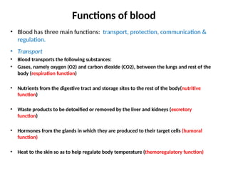
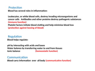
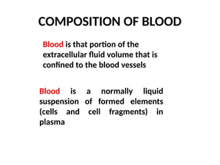
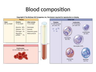
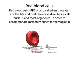
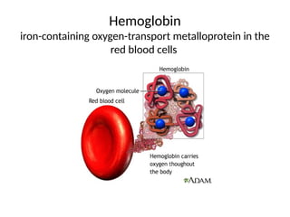
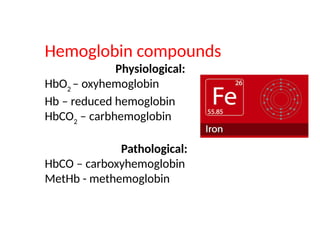
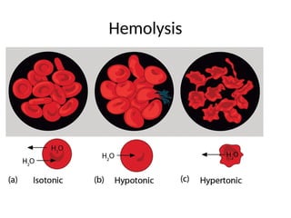
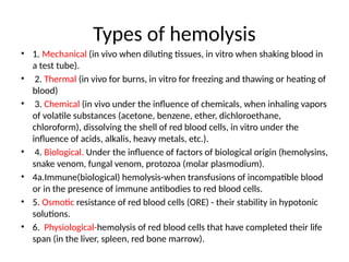
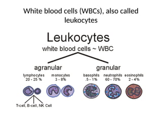
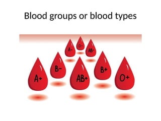
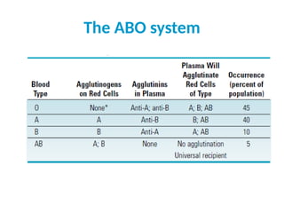
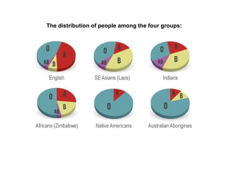
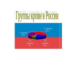
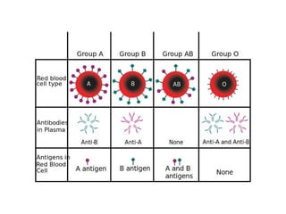
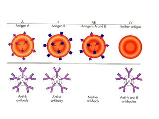
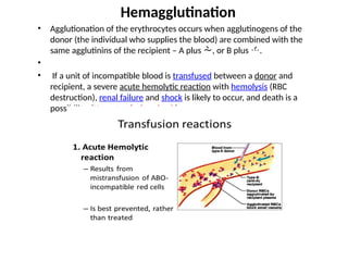
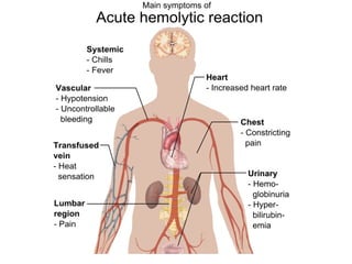
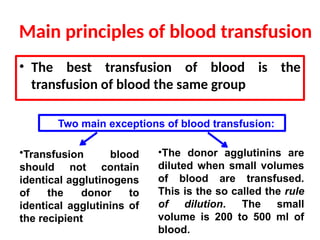
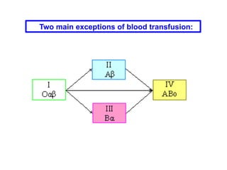
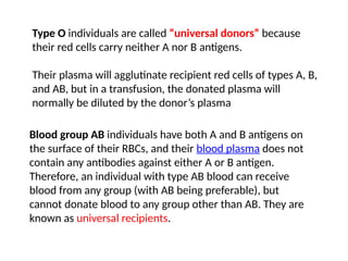
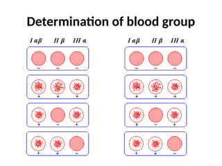
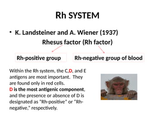
Physiology of blood
Functions of blood
• Blood has three main functions: transport, protection, communication &
regulation.
• Transport
• Blood transports the following substances:
• Gases, namely oxygen (O2) and carbon dioxide (CO2), between the lungs and rest of the
body (respiration function)
• Nutrients from the digestive tract and storage sites to the rest of the body(nutritive
function)
• Waste products to be detoxified or removed by the liver and kidneys (excretory
function)
• Hormones from the glands in which they are produced to their target cells (humoral
function)
• Heat to the skin so as to help regulate body temperature (themoregulatory function)
Protection
Blood has several roles in inflammation:
Leukocytes, or white blood cells, destroy invading microorganisms and
cancer cells Antibodies and other proteins destroy pathogenic substances
(immune function)
Platelet factors initiate blood clotting and help minimise blood loss
(protection against loosing of blood)
Regulation
Blood helps regulate:
pH by interacting with acids and bases
Water balance by transferring water to and from tissues
Ionic balance (homeostatic function)
Communication
Blood carry information over all body (communicative function)
COMPOSITION OF BLOOD
Blood is that portion of the
extracellular fluid volume that is
confined to the blood vessels
Blood is a normally liquid
suspension of formed elements
(cells and cell fragments) in
plasma
Blood composition
Red blood cells
Red blood cells (RBCs), also called erythrocytes
are flexible and oval biconcave disks lack a cell
nucleus and most organelles, in order to
accommodate maximum space for hemoglobin
Hemoglobin
iron-containing oxygen-transport metalloprotein in the
red blood cells
Hemoglobin compounds
Physiological:
HbO2
– oxyhemoglobin
Hb – reduced hemoglobin
HbCO2
– carbhemoglobin
Pathological:
HbCO – carboxyhemoglobin
MetHb - methemoglobin
Hemolysis
Types of hemolysis
• 1. Mechanical (in vivo when diluting tissues, in vitro when shaking blood in
a test tube).
• 2. Thermal (in vivo for burns, in vitro for freezing and thawing or heating of
blood)
• 3. Chemical (in vivo under the influence of chemicals, when inhaling vapors
of volatile substances (acetone, benzene, ether, dichloroethane,
chloroform), dissolving the shell of red blood cells, in vitro under the
influence of acids, alkalis, heavy metals, etc.).
• 4. Biological. Under the influence of factors of biological origin (hemolysins,
snake venom, fungal venom, protozoa (molar plasmodium).
• 4a.Immune(biological) hemolysis-when transfusions of incompatible blood
or in the presence of immune antibodies to red blood cells.
• 5. Osmotic resistance of red blood cells (ORE) - their stability in hypotonic
solutions.
• 6. Physiological-hemolysis of red blood cells that have completed their life
span (in the liver, spleen, red bone marrow).
Osmotic resistance of red blood cells (ORE) -
their stability in hypotonic solutions.
• Distinguish between:
• The minimum ORE is the concentration of the NaCl
solution at which hemolysis begins (0.48-0.46%). Less
stable ones are hemolysed.
• The maximum ORE. - the concentration of the NaCl
solution in which all red blood cells are hemolysed (0.34—
0.32%).
• Osmotic resistance of red blood cells depends on the
degree of their maturity and shape. Young forms of red
blood cells, coming from the bone marrow into the blood,
are most resistant to hypotension.
White blood cells (WBCs), also called
leukocytes
Blood groups or blood types
Immunologic properties
of blood
• Presence of blood group systems were discovered by
Karl Landsteiner during early experiments with blood
transfusion in 1901 .
• Blood of all people we can divide for different groups(or
types) according to presence or absence definite
agglutinogens.
• The most important are erythrocite blood groups.
• . A total of 35 human blood group systems are
now recognized by the
International Society of Blood Transfusion
(ISBT).
• The two most important ones are ABO and
the Rh(D) antigen; they determine someone's
blood type
Erythrocite blood group systems
Blood group
• When red cells from an individual are mixed with plasma
from another individual, an immune response (transfusion
reaction) will occur, in which the red cells will clump together
(agglutinate) and hemolysis, releasing their hemoglobin.
• The antigens are called agglutinogens and are carried on the
red cells in the blood as well as on cells in many other
tissues. In red cells, they are glycosphingolipids that differ by
only the last sugar in the carbohydrate chain that is attached
to a membrane sphingolipid.
• There are more than 400 different blood group antigens that
have been identified.
• There are more than 400 different blood
group antigens that have been identified.
• There are no two people with identical
fingerprints so there are no two persons with
identical antigenic set in blood.
System Антигены % распределение
групп крови
АВО A1, A2, A3, A4, A5, Az
, A0
, B1, B2, B3, BW
,
BX
, H, O
0 - 40 А - 39 В - 15 АВ - 6
MNSs M, M1, M2, Ma
, Mc
, Mk
, Mv
, Mg
, Mia
, Mur,
Mta
, N, N2, Nya
, S, Sta
, Sul, Sj, S2, s, U,
Tm, Hu, He, Hil, Vw
, Vr
, Ria
, Cla
M - 33 N - 19 MN - 48
P P1, P2, Pk
P1 - 79 P2 - 21 Pk
p
Rh C, Cw
, Cu
, Ces
, Ce, ce, CE, D, Du
, Dw
,
E, Eu
, Ew
, Et
, c, cE, d, e, e , LW
Rh+ 85 Rh- 15
Lu Lua
, Lub
Lua
-0,1 Lub
-92,4 Luab
-7,5
Le Lea
, Leb
, Lec
, Led
Lea
Leb
Lec
Led
K K, k, Kpa
, Kpb
, Jsa
, Jsb
Kk-0,2 Kk-9,8 kk-90
Fy Fya
, Fyb
Fya
-17 Fyb
-34 Fyab
-49
Jk Jka
,Jkb
Jka
-25 Jkb
-25 Jkab
-50
Di Dia
, Dib
Dia
Dib
Yt Yta
, Ytb
Yta
-91,9 Ytb
-0,2 Ytab
-7,9
Ii I, i I-99,98 i-0,02
Au Aua
, Aub
Aua
-82 Aub
-8
Bx Bx Bx+ 0,03 Bx-99,97
Xg Xga
Xga
+ 63M, 89F Xgа
-37M, 11F
Bua
Bua
Bua
+ 0,1 Bua
–99,9
Do Doa
Doa
+ 66 Doa
- 34
• K. Landsteiner and Y. Yansky (1901)
• The A and B antigens are the most important of the
different blood group antigens;
• They are inherited and are the basis for dividing
individuals into the four blood groups: O, A, B, and
AB.
• In neonatal life, we quickly develop antibodies against
the antigens that are not present on our red cells, and
these antibodies, called agglutinins ( and ), are
carried in plasma.
ABO antigens
Antigens of the ABO group
Antigens are glycosphingolipids on the surface of erythrocytes.
The H antigen that is present in individuals with type O blood.
The A antigen (type A blood) has a terminal N-acetylgalactosamine (NAG).
The B antigen (type B blood) has a terminal galactose (Gal).
Cer = ceramide; Fuc = fucose; Gal = galatose; Glu = glucose.
H antigen A antigen B antigen
The ABO system
The distribution of people among the four groups:
• Agglutionation of the erythrocytes occurs when agglutinogens of the
donor (the individual who supplies the blood) are combined with the
same agglutinins of the recipient – A plus , or B plus .
•
• If a unit of incompatible blood is transfused between a donor and
recipient, a severe acute hemolytic reaction with hemolysis (RBC
destruction), renal failure and shock is likely to occur, and death is a
possibility. (Hemotrasfusion shock)
Hemagglutination
• The best transfusion of blood is the
transfusion of blood the same group
Main principles of blood transfusion
•Transfusion blood
should not contain
identical agglutinogens
of the donor to
identical agglutinins of
the recipient
Two main exceptions of blood transfusion:
•The donor agglutinins are
diluted when small volumes
of blood are transfused.
This is the so called the rule
of dilution. The small
volume is 200 to 500 ml of
blood.
Two main exceptions of blood transfusion:
Type O individuals are called “universal donors” because
their red cells carry neither A nor B antigens.
Their plasma will agglutinate recipient red cells of types A, B,
and AB, but in a transfusion, the donated plasma will
normally be diluted by the donor’s plasma
Blood group AB individuals have both A and B antigens on
the surface of their RBCs, and their blood plasma does not
contain any antibodies against either A or B antigen.
Therefore, an individual with type AB blood can receive
blood from any group (with AB being preferable), but
cannot donate blood to any group other than AB. They are
known as universal recipients.
Determination of blood group
Rh SYSTEM
• K. Landsteiner and A. Wiener (1937)
Rhesus factor (Rh factor)
Rh-positive group Rh-negative group of blood
Within the Rh system, the C,D, and E
antigens are most important. They
are found only in red cells.
D is the most antigenic component,
and the presence or absence of D is
designated as “Rh-positive” or “Rh-
negative,” respectively.
Rhesus system
• Under normal circumstances, human plasma
does not contain anti-Rh antibodies(agglutinins).
• However, if an Rh- person received Rh+ blood
the body starts to make anti-Rh anti-bodies that
will remain in the blood.
• If a second transfusion of Rh+ blood is given
later, the previously formed antibodies will react
against the donated blood and a severe reaction
may occur (rhesus conflict).
• This ( rhesus conflict) can occur also when Rh+
fetal blood mixes with the circulation of an
Rh– mother.
• When an Rh- mother carries an Rh+ fetus and
small amounts of fetal blood mix with
maternal blood during delivery, the mother
may develop significant levels of anti-Rh
antibodies in her plasma. During the next
pregnancy, the mother’s Rh agglutinins cross
the placenta into the fetus and can cause
severe hemolytic disease in the fetus.
Hemotransfusion
Rules for hemotransfusion
• 1. Determination blood group belonging
(donor & recepient) ABO & Rh systems
• 2. Cross test
• 3. Biological test
15ml of donors blood(streamly) (wait 3 min), 3 times
• 4. Hemotransfution
Good bye!

Biochemistry. Topic name: Blood.

  • 1.
  • 2.
    Functions of blood •Blood has three main functions: transport, protection, communication & regulation. • Transport • Blood transports the following substances: • Gases, namely oxygen (O2) and carbon dioxide (CO2), between the lungs and rest of the body (respiration function) • Nutrients from the digestive tract and storage sites to the rest of the body(nutritive function) • Waste products to be detoxified or removed by the liver and kidneys (excretory function) • Hormones from the glands in which they are produced to their target cells (humoral function) • Heat to the skin so as to help regulate body temperature (themoregulatory function)
  • 3.
    Protection Blood has severalroles in inflammation: Leukocytes, or white blood cells, destroy invading microorganisms and cancer cells Antibodies and other proteins destroy pathogenic substances (immune function) Platelet factors initiate blood clotting and help minimise blood loss (protection against loosing of blood) Regulation Blood helps regulate: pH by interacting with acids and bases Water balance by transferring water to and from tissues Ionic balance (homeostatic function) Communication Blood carry information over all body (communicative function)
  • 4.
    COMPOSITION OF BLOOD Bloodis that portion of the extracellular fluid volume that is confined to the blood vessels Blood is a normally liquid suspension of formed elements (cells and cell fragments) in plasma
  • 5.
  • 6.
    Red blood cells Redblood cells (RBCs), also called erythrocytes are flexible and oval biconcave disks lack a cell nucleus and most organelles, in order to accommodate maximum space for hemoglobin
  • 7.
  • 8.
    Hemoglobin compounds Physiological: HbO2 – oxyhemoglobin Hb– reduced hemoglobin HbCO2 – carbhemoglobin Pathological: HbCO – carboxyhemoglobin MetHb - methemoglobin
  • 9.
  • 10.
    Types of hemolysis •1. Mechanical (in vivo when diluting tissues, in vitro when shaking blood in a test tube). • 2. Thermal (in vivo for burns, in vitro for freezing and thawing or heating of blood) • 3. Chemical (in vivo under the influence of chemicals, when inhaling vapors of volatile substances (acetone, benzene, ether, dichloroethane, chloroform), dissolving the shell of red blood cells, in vitro under the influence of acids, alkalis, heavy metals, etc.). • 4. Biological. Under the influence of factors of biological origin (hemolysins, snake venom, fungal venom, protozoa (molar plasmodium). • 4a.Immune(biological) hemolysis-when transfusions of incompatible blood or in the presence of immune antibodies to red blood cells. • 5. Osmotic resistance of red blood cells (ORE) - their stability in hypotonic solutions. • 6. Physiological-hemolysis of red blood cells that have completed their life span (in the liver, spleen, red bone marrow).
  • 11.
    Osmotic resistance ofred blood cells (ORE) - their stability in hypotonic solutions. • Distinguish between: • The minimum ORE is the concentration of the NaCl solution at which hemolysis begins (0.48-0.46%). Less stable ones are hemolysed. • The maximum ORE. - the concentration of the NaCl solution in which all red blood cells are hemolysed (0.34— 0.32%). • Osmotic resistance of red blood cells depends on the degree of their maturity and shape. Young forms of red blood cells, coming from the bone marrow into the blood, are most resistant to hypotension.
  • 12.
    White blood cells(WBCs), also called leukocytes
  • 13.
    Blood groups orblood types
  • 14.
    Immunologic properties of blood •Presence of blood group systems were discovered by Karl Landsteiner during early experiments with blood transfusion in 1901 . • Blood of all people we can divide for different groups(or types) according to presence or absence definite agglutinogens. • The most important are erythrocite blood groups.
  • 15.
    • . Atotal of 35 human blood group systems are now recognized by the International Society of Blood Transfusion (ISBT). • The two most important ones are ABO and the Rh(D) antigen; they determine someone's blood type Erythrocite blood group systems
  • 16.
    Blood group • Whenred cells from an individual are mixed with plasma from another individual, an immune response (transfusion reaction) will occur, in which the red cells will clump together (agglutinate) and hemolysis, releasing their hemoglobin. • The antigens are called agglutinogens and are carried on the red cells in the blood as well as on cells in many other tissues. In red cells, they are glycosphingolipids that differ by only the last sugar in the carbohydrate chain that is attached to a membrane sphingolipid. • There are more than 400 different blood group antigens that have been identified.
  • 17.
    • There aremore than 400 different blood group antigens that have been identified. • There are no two people with identical fingerprints so there are no two persons with identical antigenic set in blood.
  • 18.
    System Антигены %распределение групп крови АВО A1, A2, A3, A4, A5, Az , A0 , B1, B2, B3, BW , BX , H, O 0 - 40 А - 39 В - 15 АВ - 6 MNSs M, M1, M2, Ma , Mc , Mk , Mv , Mg , Mia , Mur, Mta , N, N2, Nya , S, Sta , Sul, Sj, S2, s, U, Tm, Hu, He, Hil, Vw , Vr , Ria , Cla M - 33 N - 19 MN - 48 P P1, P2, Pk P1 - 79 P2 - 21 Pk p Rh C, Cw , Cu , Ces , Ce, ce, CE, D, Du , Dw , E, Eu , Ew , Et , c, cE, d, e, e , LW Rh+ 85 Rh- 15 Lu Lua , Lub Lua -0,1 Lub -92,4 Luab -7,5 Le Lea , Leb , Lec , Led Lea Leb Lec Led K K, k, Kpa , Kpb , Jsa , Jsb Kk-0,2 Kk-9,8 kk-90 Fy Fya , Fyb Fya -17 Fyb -34 Fyab -49 Jk Jka ,Jkb Jka -25 Jkb -25 Jkab -50 Di Dia , Dib Dia Dib Yt Yta , Ytb Yta -91,9 Ytb -0,2 Ytab -7,9 Ii I, i I-99,98 i-0,02 Au Aua , Aub Aua -82 Aub -8 Bx Bx Bx+ 0,03 Bx-99,97 Xg Xga Xga + 63M, 89F Xgа -37M, 11F Bua Bua Bua + 0,1 Bua –99,9 Do Doa Doa + 66 Doa - 34
  • 19.
    • K. Landsteinerand Y. Yansky (1901) • The A and B antigens are the most important of the different blood group antigens; • They are inherited and are the basis for dividing individuals into the four blood groups: O, A, B, and AB. • In neonatal life, we quickly develop antibodies against the antigens that are not present on our red cells, and these antibodies, called agglutinins ( and ), are carried in plasma. ABO antigens
  • 20.
    Antigens of theABO group Antigens are glycosphingolipids on the surface of erythrocytes. The H antigen that is present in individuals with type O blood. The A antigen (type A blood) has a terminal N-acetylgalactosamine (NAG). The B antigen (type B blood) has a terminal galactose (Gal). Cer = ceramide; Fuc = fucose; Gal = galatose; Glu = glucose. H antigen A antigen B antigen
  • 21.
  • 22.
    The distribution ofpeople among the four groups:
  • 27.
    • Agglutionation ofthe erythrocytes occurs when agglutinogens of the donor (the individual who supplies the blood) are combined with the same agglutinins of the recipient – A plus , or B plus . • • If a unit of incompatible blood is transfused between a donor and recipient, a severe acute hemolytic reaction with hemolysis (RBC destruction), renal failure and shock is likely to occur, and death is a possibility. (Hemotrasfusion shock) Hemagglutination
  • 29.
    • The besttransfusion of blood is the transfusion of blood the same group Main principles of blood transfusion •Transfusion blood should not contain identical agglutinogens of the donor to identical agglutinins of the recipient Two main exceptions of blood transfusion: •The donor agglutinins are diluted when small volumes of blood are transfused. This is the so called the rule of dilution. The small volume is 200 to 500 ml of blood.
  • 30.
    Two main exceptionsof blood transfusion:
  • 31.
    Type O individualsare called “universal donors” because their red cells carry neither A nor B antigens. Their plasma will agglutinate recipient red cells of types A, B, and AB, but in a transfusion, the donated plasma will normally be diluted by the donor’s plasma Blood group AB individuals have both A and B antigens on the surface of their RBCs, and their blood plasma does not contain any antibodies against either A or B antigen. Therefore, an individual with type AB blood can receive blood from any group (with AB being preferable), but cannot donate blood to any group other than AB. They are known as universal recipients.
  • 32.
  • 34.
    Rh SYSTEM • K.Landsteiner and A. Wiener (1937) Rhesus factor (Rh factor) Rh-positive group Rh-negative group of blood Within the Rh system, the C,D, and E antigens are most important. They are found only in red cells. D is the most antigenic component, and the presence or absence of D is designated as “Rh-positive” or “Rh- negative,” respectively.
  • 35.
    Rhesus system • Undernormal circumstances, human plasma does not contain anti-Rh antibodies(agglutinins). • However, if an Rh- person received Rh+ blood the body starts to make anti-Rh anti-bodies that will remain in the blood. • If a second transfusion of Rh+ blood is given later, the previously formed antibodies will react against the donated blood and a severe reaction may occur (rhesus conflict).
  • 36.
    • This (rhesus conflict) can occur also when Rh+ fetal blood mixes with the circulation of an Rh– mother. • When an Rh- mother carries an Rh+ fetus and small amounts of fetal blood mix with maternal blood during delivery, the mother may develop significant levels of anti-Rh antibodies in her plasma. During the next pregnancy, the mother’s Rh agglutinins cross the placenta into the fetus and can cause severe hemolytic disease in the fetus.
  • 38.
  • 39.
    Rules for hemotransfusion •1. Determination blood group belonging (donor & recepient) ABO & Rh systems • 2. Cross test • 3. Biological test 15ml of donors blood(streamly) (wait 3 min), 3 times • 4. Hemotransfution
  • 40.