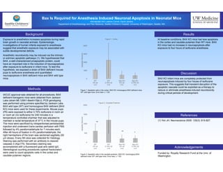
This study investigated the role of the BAX protein in isoflurane-induced neuronal apoptosis in neonatal mice. The researchers found that BAX knockout mice had less baseline apoptosis in the cortex and caudate-putamen compared to wild type mice. Exposure to isoflurane anesthesia for 4 hours did not increase neuroapoptosis in BAX knockout mice, but did increase it in wild type mice. This suggests that blocking the apoptotic cascade by inhibiting BAX may reduce or eliminate anesthesia-induced neurotoxicity in developing brains.
1. Bax Is Required for Anesthesia Induced Neuronal Apoptosis in Neonatal Mice
Hikmatullah Arif, Leanne Cornel, David Jardine
Department of Anesthesiology and Pain Medicine, Seattle Children’s Hospital, University of Washington, Seattle, WA
Background
Exposure to anesthetics increases apoptosis during rapid
brain growth in neonatal animals. Epidemiologic
investigations of human infants exposed to anesthesia
suggest that anesthetic exposure may be associated with
subtle developmental deficits.
Anesthetic neurotoxicity may be induced via the intrinsic
or extrinsic apoptotic pathways (1). We hypothesized that
BAX, a well-characterized proapoptotic protein, could
have an important role in the induction of neuroapoptosis
after exposure to isoflurane in infant mice. To test this
hypothesis, we exposed a strain of BAX deficient mouse
pups to isoflurane anesthesia and quantitated
neuroapoptosis in BAX deficient mice and BAX wild type
mice.
IACUC approval was obtained for all procedures. BAX
deficient transgenic mice were obtained from Jackson
Labs (strain B6.129X1-Baxtm1Sjk/J). PCR genotyping
was performed using primers specified by Jackson Labs.
BAX wild type (WT) and homozygous BAX deficient (BAX
KO) mice were used for these experiments. Mouse pups
(P6) were exposed to either 0.75% isoflurane in room air
or room air (no isoflurane) for 240 minutes in a
temperature controlled chamber that was adjusted to
maintain a rectal temperature of 37°C in the mouse pups.
The mice were sacrificed by intraperitoneal pentobarbital
injection and underwent trans-cardiac perfusion with PBS
followed by 4% paraformaldehyde for 7 minutes each.
After 48 hours of fixation in 4% paraformaldehyde, the
right hemisphere of the brain was sectioned sagittally (50
µm slices). Every 5th slice was collected for histology.
Staining was performed with an antibody to cleaved
caspase-3 (Asp175). Secondary staining was
accomplished with a fluorescent goat anti-rabbit IgG.
Stereological counting procedures (optical fractionator)
were used to count apoptotic cells in the cortex and
caudate-putamen regions.
At baseline conditions, BAX KO mice had less apoptosis
in the cortex and caudate-putamen than WT mice. BAX
KO mice had no increase in neuroapoptosis after
exposure to four hours of isoflurane anesthesia.
Funded by: Royalty Research Fund at the Univ. of
Washington.
Methods
Discussion
Acknowledgements
BAX KO infant mice are completely protected from
neuroapoptosis induced by four house of isoflurane
exposure. This suggests that transient disruption of the
apoptotic cascade could be exploited as a therapy to
reduce or eliminate anesthesia induced neurotoxicity
during critical periods of development.
UW Medicine
SCHOOL OF MEDICINE
Results
References
(1) Yon JH. Neuroscience 2005: 135(3): 815-827.
Figure 1: Apoptotic cells in the cortex. BAX KO: homozygous BAX deficient mice;
WT: wild type mice. Error bars: ± 1 SD.
Figure 2: Apoptotic cells in the caudate-putamen. BAX KO: homozygous BAX
deficient mice; WT: wild type mice. Error bars: ± 1 SD.
0
1000
2000
3000
4000
WT
0% Isf
n=3
WT
0.75% Isf
n=5
Bax KO
0% Isf
n=4
Bax KO
0.75% Isf
n=3
ApoptoticNeurons
Figure 1: Cortex
0
500
1000
1500
2000
WT
0% Isf
n=3
WT
0.75% Isf
n=5
Bax KO
0% Isf
n=4
Bax KO
0.75% Isf
n=3
ApoptoticNeurons
Figure 2: Caudate-Putamen
*
*
NS
NS