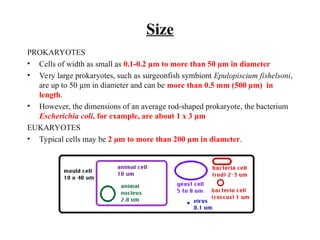
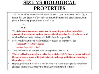
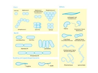
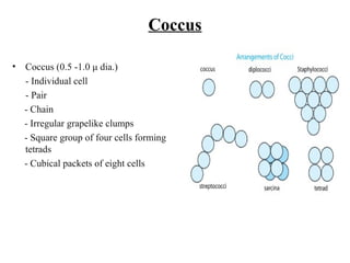
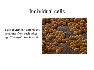
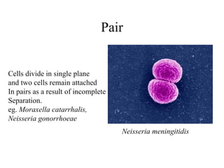
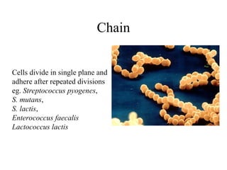
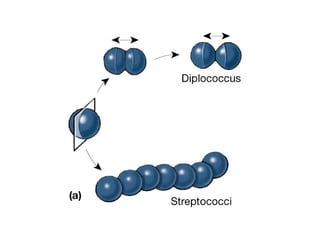
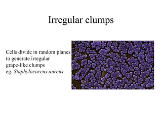
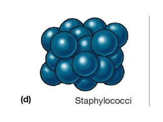
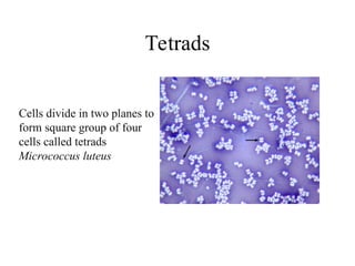
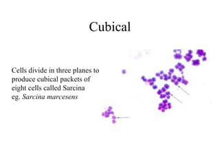
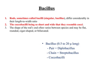
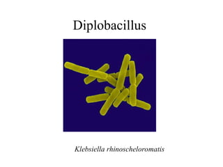
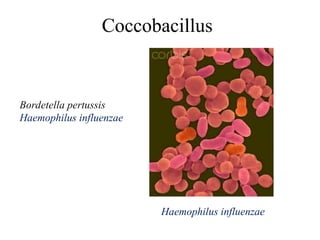
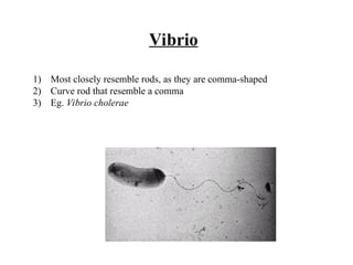
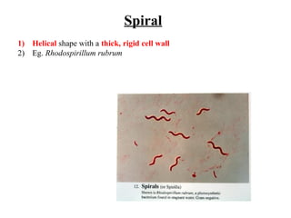
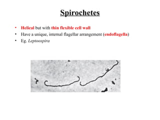
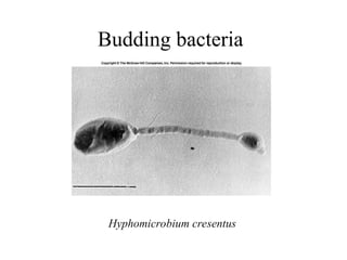
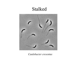
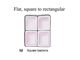
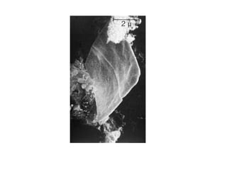
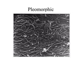
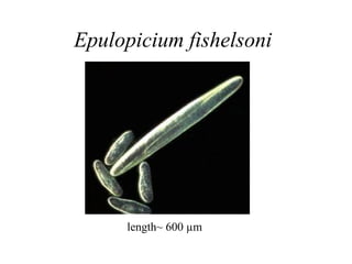
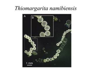
Morphology of Bacteria
Size
PROKARYOTES
• Cells of width as small as 0.1-0.2 µm to more than 50 µm in diameter
• Very large prokaryotes, such as surgeonfish symbiont Epulopiscium fishelsoni,
are up to 50 µm in diameter and can be more than 0.5 mm (500 µm) in
length.
• However, the dimensions of an average rod-shaped prokaryote, the bacterium
Escherichia coli, for example, are about 1 x 3 µm
EUKARYOTES
• Typical cells may be 2 µm to more than 200 µm in diameter.
SIZE VS BIOLOGICAL
PROPERTIES
• The rate at which nutrients and waste products pass into and out of a cell, a
factor that can greatly affect cellular metabolic rates and growth rates, is in
general inversely proportional to cell size.
WHY
• This is because transport rates are to some degree a function of the
amount of membrane surface area available relative to cell volume, and
small cells have more surface available than do large cells.
• Most readily explained mathematically in case of a spherical cell:
– Volume (V) = 4/3πr3
whereas
– surface Area (SA) = 4πr2
• Thus surface are to volume ratio in a spherical cell is 3/r.
• Thus a cell with a smaller r value has a higher SA/V than a larger cell and
thus can have a more efficient nutrient exchange with its surroundings
than a larger cell.
• Higher growth and metabolic rate in turn can cause major physicochemical
changes in an ecosystem over a relatively short period of time.
Coccus
• Coccus (0.5 -1.0 μ dia.)
- Individual cell
- Pair
- Chain
- Irregular grapelike clumps
- Square group of four cells forming
tetrads
- Cubical packets of eight cells
Individual cells
Cells divide and completely
separates from each other
eg. Chlamydia trachomatis
Pair
Cells divide in single plane
and two cells remain attached
In pairs as a result of incomplete
Separation.
eg. Moraxella catarrhalis,
Neisseria gonorrhoeae
Neisseria meningitidis
Chain
Cells divide in single plane and
adhere after repeated divisions
eg. Streptococcus pyogenes,
S. mutans,
S. lactis,
Enterococcus faecalis
Lactococcus lactis
Irregular clumps
Cells divide in random planes
to generate irregular
grape-like clumps
eg. Staphylococcus aureus
Tetrads
Cells divide in two planes to
form square group of four
cells called tetrads
Micrococcus luteus
Cubical
Cells divide in three planes to
produce cubical packets of
eight cells called Sarcina
eg. Sarcina marcesens
Bacillus
• Bacillus (0.5 to 20 μ long)
- Pair = Diplobacillus
- Chain = Streptobacillus
- Coccobacilli
1. Rods, sometimes called bacilli (singular, bacillus), differ considerably in
their length-to-width ratio
2. The coccobacilli being so short and wide that they resemble cocci.
3. The shape of the rod’s end often varies between species and may be flat,
rounded, cigar-shaped, or bifurcated.
Diplobacillus
Klebsiella rhinoscheloromatis
Streptobacillus
Streptobacillus moniliformis
Bacillus megaterium
Coccobacillus
Haemophilus influenzae
Bordetella pertussis
Haemophilus influenzae
Vibrio
1) Most closely resemble rods, as they are comma-shaped
2) Curve rod that resemble a comma
3) Eg. Vibrio cholerae
Spiral
1) Helical shape with a thick, rigid cell wall
2) Eg. Rhodospirillum rubrum
Spirochetes
• Helical but with thin flexible cell wall
• Have a unique, internal flagellar arrangement (endoflagella)
• Eg. Leptoospira
Actinomycetes
• Actinomycetes typically form long filaments called hyphae that may
branch to produce a network called a mycelium.
• In this sense, they are similar to filamentous fungi, a group of
eucaryotic microbes.
Budding bacteria
Hyphomicrobium cresentus
Stalked
Caulobacter cresentus
Flat, square to rectangular
Pleomorphic
Epulopicium fishelsoni
length~ 600 µm
Thiomargarita namibiensis
End
Endoflagella
Borrelia burgdorferi
1) Endoflagella run along length in the corkscrew shaped spirochetes.
2) Although these bacteria are gram negative (with a double membrane envelope
structure), they possess only 2, to 4 rings (the S, M and presumably the C rings
and sometimes an extra pair) in the basal complex - they lack the P and L
rings.
3) The result is that the basal complex does not cross the outer membrane and
the filaments lie between the peptidoglycan and outer membrane (in the
periplasm) wrapped around the cell (giving it a spiral appearance) as shown
above.
4) These internal flagella form a bundle or axial filament.

Bacterial morphology (SIZE SHAPE ARRANGEMENT) (1).ppt

  • 1.
  • 2.
    Size PROKARYOTES • Cells ofwidth as small as 0.1-0.2 µm to more than 50 µm in diameter • Very large prokaryotes, such as surgeonfish symbiont Epulopiscium fishelsoni, are up to 50 µm in diameter and can be more than 0.5 mm (500 µm) in length. • However, the dimensions of an average rod-shaped prokaryote, the bacterium Escherichia coli, for example, are about 1 x 3 µm EUKARYOTES • Typical cells may be 2 µm to more than 200 µm in diameter.
  • 3.
    SIZE VS BIOLOGICAL PROPERTIES •The rate at which nutrients and waste products pass into and out of a cell, a factor that can greatly affect cellular metabolic rates and growth rates, is in general inversely proportional to cell size. WHY • This is because transport rates are to some degree a function of the amount of membrane surface area available relative to cell volume, and small cells have more surface available than do large cells. • Most readily explained mathematically in case of a spherical cell: – Volume (V) = 4/3πr3 whereas – surface Area (SA) = 4πr2 • Thus surface are to volume ratio in a spherical cell is 3/r. • Thus a cell with a smaller r value has a higher SA/V than a larger cell and thus can have a more efficient nutrient exchange with its surroundings than a larger cell. • Higher growth and metabolic rate in turn can cause major physicochemical changes in an ecosystem over a relatively short period of time.
  • 5.
    Coccus • Coccus (0.5-1.0 μ dia.) - Individual cell - Pair - Chain - Irregular grapelike clumps - Square group of four cells forming tetrads - Cubical packets of eight cells
  • 6.
    Individual cells Cells divideand completely separates from each other eg. Chlamydia trachomatis
  • 7.
    Pair Cells divide insingle plane and two cells remain attached In pairs as a result of incomplete Separation. eg. Moraxella catarrhalis, Neisseria gonorrhoeae Neisseria meningitidis
  • 8.
    Chain Cells divide insingle plane and adhere after repeated divisions eg. Streptococcus pyogenes, S. mutans, S. lactis, Enterococcus faecalis Lactococcus lactis
  • 10.
    Irregular clumps Cells dividein random planes to generate irregular grape-like clumps eg. Staphylococcus aureus
  • 12.
    Tetrads Cells divide intwo planes to form square group of four cells called tetrads Micrococcus luteus
  • 14.
    Cubical Cells divide inthree planes to produce cubical packets of eight cells called Sarcina eg. Sarcina marcesens
  • 15.
    Bacillus • Bacillus (0.5to 20 μ long) - Pair = Diplobacillus - Chain = Streptobacillus - Coccobacilli 1. Rods, sometimes called bacilli (singular, bacillus), differ considerably in their length-to-width ratio 2. The coccobacilli being so short and wide that they resemble cocci. 3. The shape of the rod’s end often varies between species and may be flat, rounded, cigar-shaped, or bifurcated.
  • 16.
  • 17.
  • 18.
  • 19.
    Vibrio 1) Most closelyresemble rods, as they are comma-shaped 2) Curve rod that resemble a comma 3) Eg. Vibrio cholerae
  • 20.
    Spiral 1) Helical shapewith a thick, rigid cell wall 2) Eg. Rhodospirillum rubrum
  • 21.
    Spirochetes • Helical butwith thin flexible cell wall • Have a unique, internal flagellar arrangement (endoflagella) • Eg. Leptoospira
  • 22.
    Actinomycetes • Actinomycetes typicallyform long filaments called hyphae that may branch to produce a network called a mycelium. • In this sense, they are similar to filamentous fungi, a group of eucaryotic microbes.
  • 23.
  • 24.
  • 25.
    Flat, square torectangular
  • 27.
  • 28.
  • 29.
  • 30.
  • 31.
    Endoflagella Borrelia burgdorferi 1) Endoflagellarun along length in the corkscrew shaped spirochetes. 2) Although these bacteria are gram negative (with a double membrane envelope structure), they possess only 2, to 4 rings (the S, M and presumably the C rings and sometimes an extra pair) in the basal complex - they lack the P and L rings. 3) The result is that the basal complex does not cross the outer membrane and the filaments lie between the peptidoglycan and outer membrane (in the periplasm) wrapped around the cell (giving it a spiral appearance) as shown above. 4) These internal flagella form a bundle or axial filament.