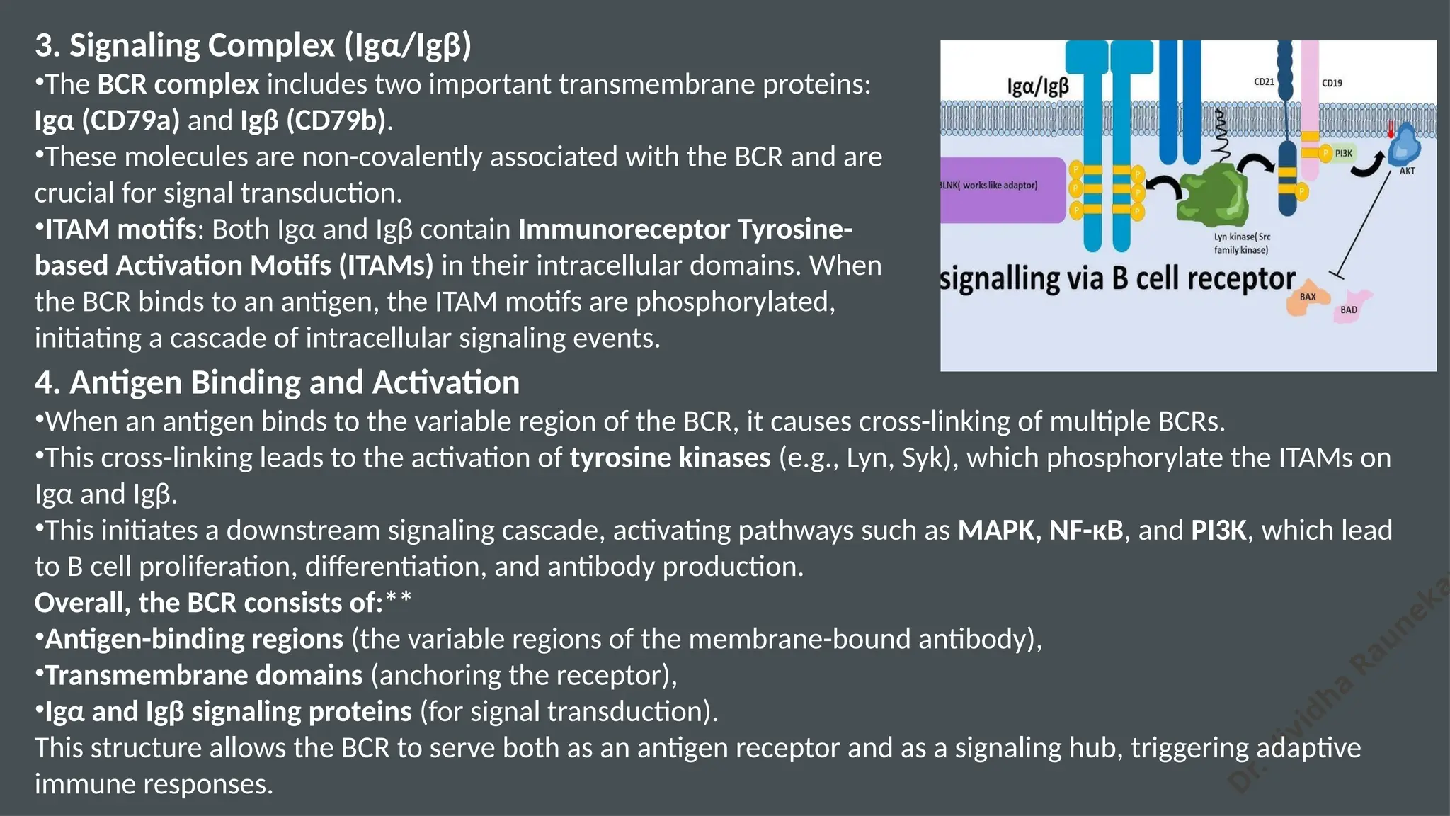
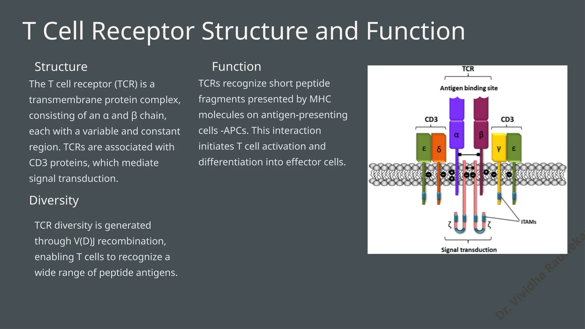
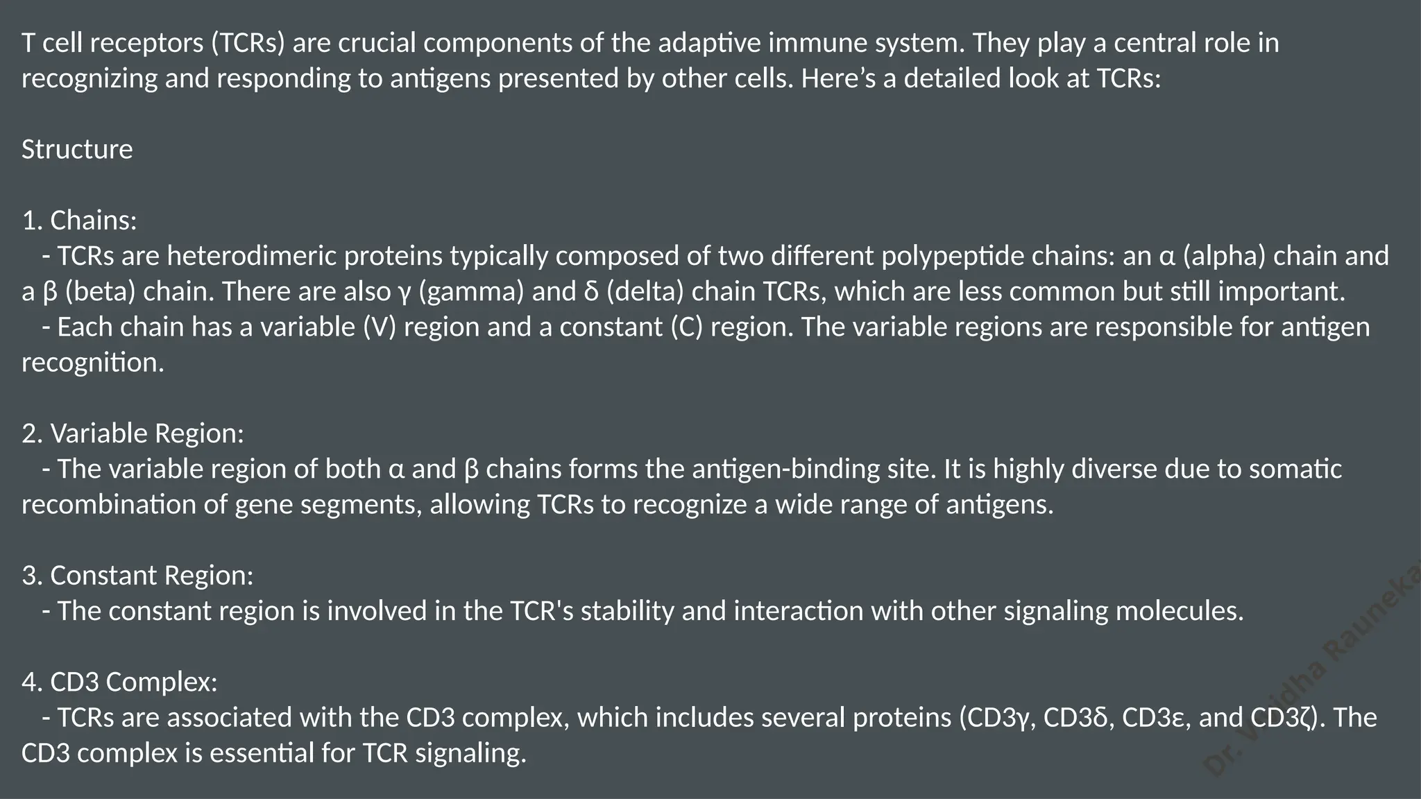
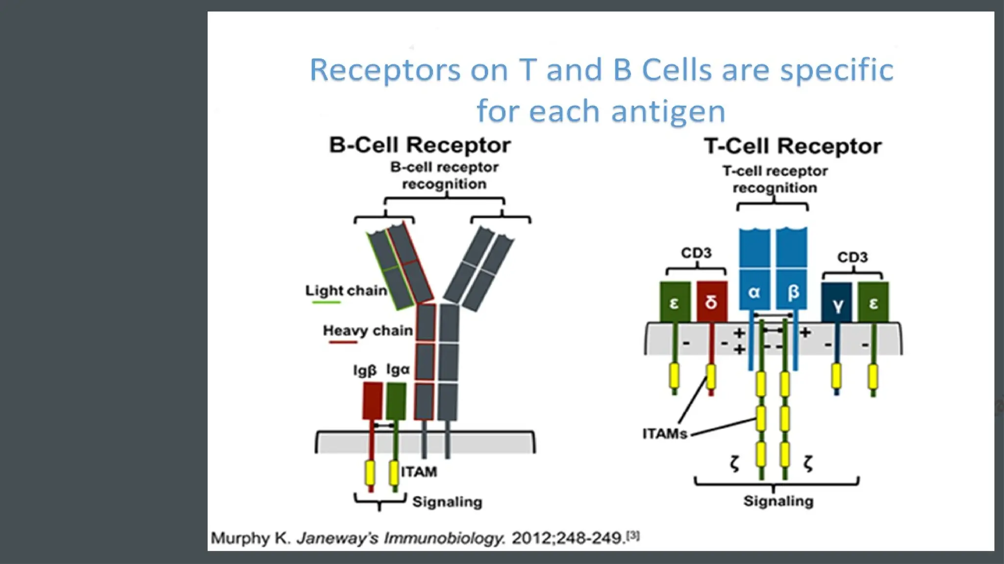
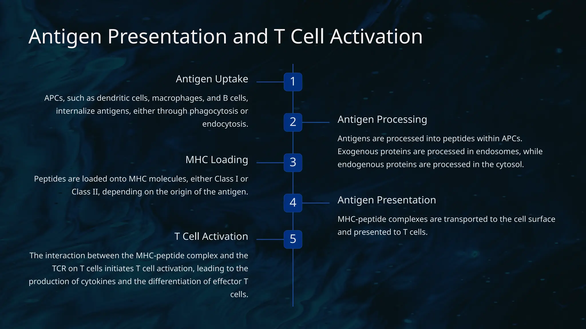
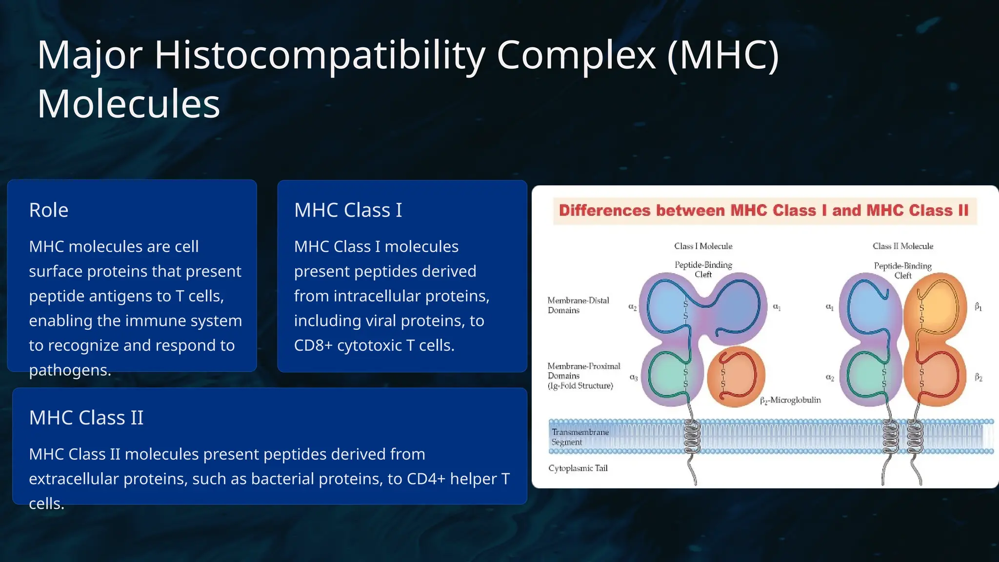
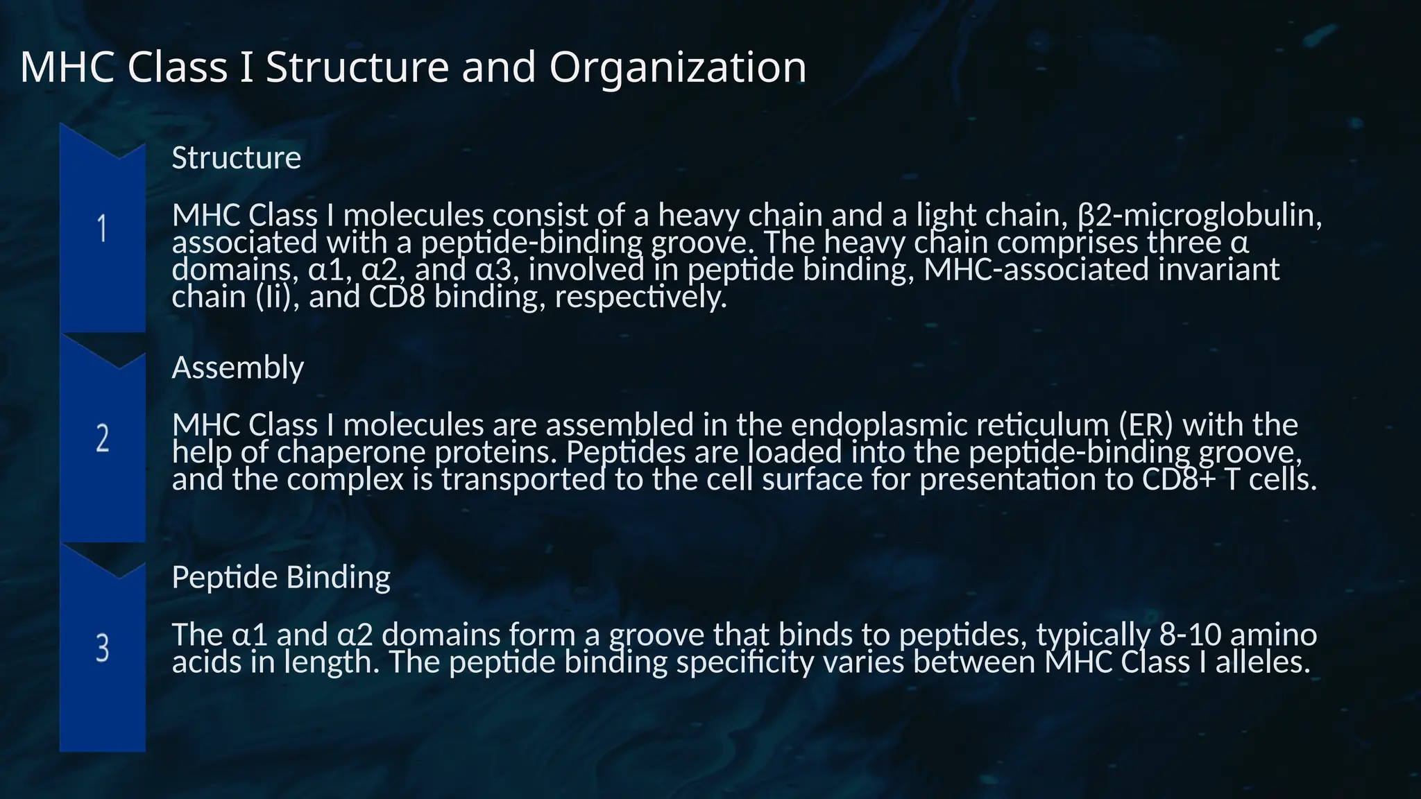
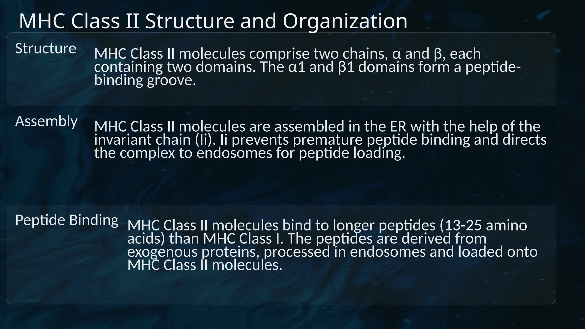
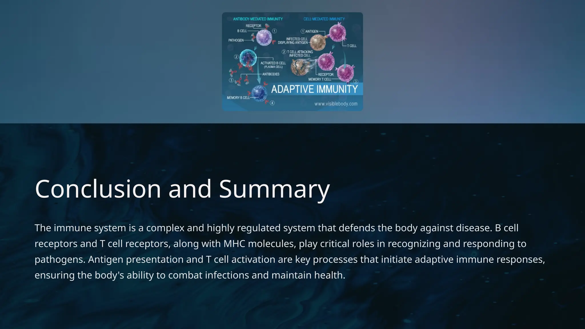
D
r. Vividha
Rauneka
D
r. Vividha
Raunekar
B cell and T cell receptors:
structure and function
Dr. Vividha Raunekar
D
r. Vividha
Rauneka
B Cell Receptor Structure and
Function
1 Structure
The B cell receptor (BCR) is a
membrane-bound antibody,
composed of two heavy
chains and two light chains
connected by disulfide
bonds. Each chain contains a
variable region that
recognizes specific antigens
and a constant region
involved in effector
functions.
2 Function
BCRs are responsible for
recognizing and binding to
specific antigens. This
interaction triggers a
cascade of signaling events
that lead to B cell activation
and antibody production.
3 Diversity
The diversity of BCRs is
generated through genetic
recombination, allowing B
cells to recognize a vast array
of antigens.
4 Effector Functions
Following activation, B cells
differentiate into plasma
cells that secrete antibodies,
which neutralize pathogens
and activate other immune
cells.
D
r. Vividha
Rauneka
The B-cell receptor (BCR) is a membrane-bound immunoglobulin (Ig) on the surface of B cells that is essential for
recognizing antigens and initiating B cell activation. Here are the detailed structural components of the BCR:
1. Immunoglobulin (Ig) Component
•Structure: The BCR is essentially a membrane-bound antibody, which has a similar structure to soluble antibodies.
• Heavy Chains (H-chains): Each BCR contains two identical heavy chains.
• Light Chains (L-chains): It also contains two identical light chains.
• Variable Region: The variable regions of the heavy and light chains are located at the tips of the receptor and
are responsible for antigen recognition. These regions are highly diverse, allowing the BCR to bind a wide
variety of antigens.
• Constant Region: The constant regions of both heavy and light chains are located closer to the B cell
membrane and contribute to the structural integrity of the receptor.
•Domains:
• Fab region (Fragment, antigen-binding): Consists of one constant and one variable domain from each of the
heavy and light chains. This is the part responsible for binding to specific antigens.
• Fc region (Fragment, crystallizable): Located at the base of the BCR and consists of the constant regions of
the heavy chains. It plays a role in receptor stability and interacting with cell signaling molecules.
2. Membrane-Bound Component
•Transmembrane Region: The constant region of the heavy chain extends into the membrane, anchoring the BCR to
the B cell’s surface.
•Intracellular Tail: The BCR itself has a short intracellular tail, which is insufficient for signaling. Instead, it relies on
associated signaling molecules.
D
r. Vividha
Rauneka
3. Signaling Complex (Igα/Igβ)
•The BCR complex includes two important transmembrane proteins:
Igα (CD79a) and Igβ (CD79b).
•These molecules are non-covalently associated with the BCR and are
crucial for signal transduction.
•ITAM motifs: Both Igα and Igβ contain Immunoreceptor Tyrosine-
based Activation Motifs (ITAMs) in their intracellular domains. When
the BCR binds to an antigen, the ITAM motifs are phosphorylated,
initiating a cascade of intracellular signaling events.
4. Antigen Binding and Activation
•When an antigen binds to the variable region of the BCR, it causes cross-linking of multiple BCRs.
•This cross-linking leads to the activation of tyrosine kinases (e.g., Lyn, Syk), which phosphorylate the ITAMs on
Igα and Igβ.
•This initiates a downstream signaling cascade, activating pathways such as MAPK, NF-κB, and PI3K, which lead
to B cell proliferation, differentiation, and antibody production.
Overall, the BCR consists of:**
•Antigen-binding regions (the variable regions of the membrane-bound antibody),
•Transmembrane domains (anchoring the receptor),
•Igα and Igβ signaling proteins (for signal transduction).
This structure allows the BCR to serve both as an antigen receptor and as a signaling hub, triggering adaptive
immune responses.
D
r. Vividha
Rauneka
T Cell Receptor Structure and Function
Structure
The T cell receptor (TCR) is a
transmembrane protein complex,
consisting of an α and β chain,
each with a variable and constant
region. TCRs are associated with
CD3 proteins, which mediate
signal transduction.
Function
TCRs recognize short peptide
fragments presented by MHC
molecules on antigen-presenting
cells -APCs. This interaction
initiates T cell activation and
differentiation into effector cells.
Diversity
TCR diversity is generated
through V(D)J recombination,
enabling T cells to recognize a
wide range of peptide antigens.
D
r. Vividha
Rauneka
T cell receptors (TCRs) are crucial components of the adaptive immune system. They play a central role in
recognizing and responding to antigens presented by other cells. Here’s a detailed look at TCRs:
Structure
1. Chains:
- TCRs are heterodimeric proteins typically composed of two different polypeptide chains: an α (alpha) chain and
a β (beta) chain. There are also γ (gamma) and δ (delta) chain TCRs, which are less common but still important.
- Each chain has a variable (V) region and a constant (C) region. The variable regions are responsible for antigen
recognition.
2. Variable Region:
- The variable region of both α and β chains forms the antigen-binding site. It is highly diverse due to somatic
recombination of gene segments, allowing TCRs to recognize a wide range of antigens.
3. Constant Region:
- The constant region is involved in the TCR's stability and interaction with other signaling molecules.
4. CD3 Complex:
- TCRs are associated with the CD3 complex, which includes several proteins (CD3γ, CD3δ, CD3ε, and CD3ζ). The
CD3 complex is essential for TCR signaling.
D
r. Vividha
Rauneka
Function
1. Antigen Recognition:
- TCRs recognize peptide antigens that are presented by Major Histocompatibility Complex (MHC) molecules on the surface of antigen-
presenting cells.
- Class I MHC molecules present peptides to CD8+ T cells (cytotoxic T cells), while **Class II MHC** molecules present to CD4+ T cells
(helper T cells).
2. Signaling:
- Upon antigen recognition, TCRs transmit signals into the T cell through the CD3 complex. This signaling triggers a cascade of intracellular
events that leads to T cell activation, proliferation, and differentiation.
3. T Cell Activation:
- Besides TCR signaling, full T cell activation often requires additional signals from co-stimulatory molecules (e.g., CD28) and cytokines.
4. Antigen Specificity:
- Each T cell has a unique TCR that recognizes a specific peptide-MHC complex, allowing the immune system to target a diverse array of
pathogens.
Diversity
- Somatic Recombination:
- TCR diversity is generated through somatic recombination of V, D (diversity), and J (joining) gene segments in developing T cells. This
process allows for the production of a vast array of TCRs, each with unique antigen specificity.
- Receptor Editing:
- In some cases, T cells undergo receptor editing to refine their antigen specificity and avoid self-reactivity.
D
r. Vividha
Rauneka
D
r. Vividha
Rauneka
Antigen Presentation and T Cell Activation
1
Antigen Uptake
APCs, such as dendritic cells, macrophages, and B cells,
internalize antigens, either through phagocytosis or
endocytosis. 2 Antigen Processing
Antigens are processed into peptides within APCs.
Exogenous proteins are processed in endosomes, while
endogenous proteins are processed in the cytosol.
3
MHC Loading
Peptides are loaded onto MHC molecules, either Class I or
Class II, depending on the origin of the antigen.
4 Antigen Presentation
MHC-peptide complexes are transported to the cell surface
and presented to T cells.
5
T Cell Activation
The interaction between the MHC-peptide complex and the
TCR on T cells initiates T cell activation, leading to the
production of cytokines and the differentiation of effector T
cells.
D
r. Vividha
Rauneka
Major Histocompatibility Complex (MHC)
Molecules
Role
MHC molecules are cell
surface proteins that present
peptide antigens to T cells,
enabling the immune system
to recognize and respond to
pathogens.
MHC Class I
MHC Class I molecules
present peptides derived
from intracellular proteins,
including viral proteins, to
CD8+ cytotoxic T cells.
MHC Class II
MHC Class II molecules present peptides derived from
extracellular proteins, such as bacterial proteins, to CD4+ helper T
cells.
D
r. Vividha
Rauneka
MHC Class I Structure and Organization
Structure
MHC Class I molecules consist of a heavy chain and a light chain, β2-microglobulin,
associated with a peptide-binding groove. The heavy chain comprises three α
domains, α1, α2, and α3, involved in peptide binding, MHC-associated invariant
chain (Ii), and CD8 binding, respectively.
Assembly
MHC Class I molecules are assembled in the endoplasmic reticulum (ER) with the
help of chaperone proteins. Peptides are loaded into the peptide-binding groove,
and the complex is transported to the cell surface for presentation to CD8+ T cells.
Peptide Binding
The α1 and α2 domains form a groove that binds to peptides, typically 8-10 amino
acids in length. The peptide binding specificity varies between MHC Class I alleles.
D
r. Vividha
Rauneka
Key Features:
• The peptide-binding groove is closed, restricting the length of the peptides that
can be presented.
• MHC Class I molecules are expressed on all nucleated cells.
Function:
• Presentation of intracellular antigens (e.g., viral or tumor peptides) to CD8+
cytotoxic T cells.
• Helps the immune system target and destroy cells that are infected with viruses or
have become cancerous.
• MHC Class I molecules display fragments of proteins synthesized within the cell,
allowing for the recognition of "self" versus "non-self."
D
r. Vividha
Rauneka
MHC Class II Structure and Organization
Structure MHC Class II molecules comprise two chains, α and β, each
containing two domains. The α1 and β1 domains form a peptide-
binding groove.
Assembly MHC Class II molecules are assembled in the ER with the help of the
invariant chain (Ii). Ii prevents premature peptide binding and directs
the complex to endosomes for peptide loading.
Peptide Binding MHC Class II molecules bind to longer peptides (13-25 amino
acids) than MHC Class I. The peptides are derived from
exogenous proteins, processed in endosomes and loaded onto
MHC Class II molecules.
D
r. Vividha
Rauneka
Key Features:
• The peptide-binding groove can accommodate larger peptides than MHC Class
I.
• MHC Class II molecules are expressed primarily on antigen-presenting cells
(APCs) such as dendritic cells, macrophages, and B cells.
Function:
• Presentation of extracellular antigens (e.g., bacteria, toxins) to CD4+ helper T
cells.
• Plays a critical role in activating immune responses against pathogens that are
taken up from the extracellular environment.
• MHC Class II molecules bind peptides derived from proteins that have been
processed in the endocytic pathway.
D
r. Vividha
Rauneka
Common Features of MHC Molecules:
• Polymorphism: MHC molecules are highly polymorphic, meaning there are many different
alleles in the population, enhancing the immune system’s ability to recognize a vast array of
antigens.
• Co-dominant Expression: Both maternal and paternal alleles of MHC genes are expressed,
increasing the diversity of antigens that can be presented.
Overall Function in Immune Response:
• MHC molecules play a critical role in antigen presentation and the activation of T cells.
• MHC Class I molecules present antigens to CD8+ cytotoxic T cells, leading to the destruction of
infected or abnormal cells.
• MHC Class II molecules present antigens to CD4+ helper T cells, which then help to coordinate
the broader immune response, including the activation of B cells and macrophages.
• These molecules are crucial for immune surveillance and play a major role in self-tolerance,
preventing the immune system from attacking normal, healthy tissues.
D
r. Vividha
Rauneka
Polymorphism of MHC Molecules:
The polymorphism of MHC (Major Histocompatibility Complex) molecules is one of the key features of the
immune system that allows it to recognize a wide array of pathogens. This polymorphism refers to the existence
of multiple alleles for each MHC gene within a population, contributing to individual and population-level
immune diversity.
1. Characteristics of MHC Polymorphism:
• Highly Polymorphic Genes: MHC genes are among the most polymorphic in the human genome, meaning
there are many different alleles (variants) of each gene in the population.
• MHC Class I Polymorphism: Found in HLA-A, HLA-B, and HLA-C loci in humans (HLA stands for Human
Leukocyte Antigen). Each locus can have hundreds of alleles.
• MHC Class II Polymorphism: Found primarily in HLA-DR, HLA-DQ, and HLA-DP loci. These also exhibit high
allelic diversity.
• Example: The HLA-B locus has over 5,000 known alleles, making it one of the most polymorphic loci in the
human genome.
2. Location of Polymorphisms:
• MHC polymorphisms are concentrated in specific regions that are functionally important:
• Peptide-binding groove: The majority of polymorphisms occur in the regions that form the peptide-binding
groove. This allows different MHC alleles to bind a diverse range of peptides (antigens).
D
r. Vividha
Rauneka
• α1 and α2 domains (Class I): In MHC Class I molecules, most of the polymorphisms are found in the α1 and
α2 domains, which form the antigen-binding cleft.
• α1 and β1 domains (Class II): In MHC Class II molecules, polymorphisms are found primarily in the α1 and β1
domains, where the peptide is presented.
3. Functional Implications of MHC Polymorphism:
• Diverse Antigen Presentation: MHC polymorphism ensures that different individuals in a population can
present a wide variety of peptides from pathogens, increasing the likelihood that some individuals will have
an immune response capable of combating a given pathogen.
• Survival of Species: This diversity is important for species survival because it allows the population to
collectively recognize and respond to a broad range of pathogens. If all individuals had the same MHC alleles,
they might all be susceptible to the same pathogens.
• Heterozygote Advantage: Individuals who are heterozygous (carry different alleles at a given MHC locus) can
present a wider variety of antigens than homozygotes (who carry two identical alleles), providing a selective
advantage in immune responses.
• Example: In some studies, heterozygous individuals at HLA loci have been shown to have better survival rates
when infected with certain pathogens, like HIV.
D
r. Vividha
Rauneka
Mechanism of MHC Polymorphism:
• Gene Duplication and Divergence: The MHC genes are thought to have evolved through a process of gene duplication followed by
divergent evolution, leading to multiple similar genes within the MHC region.
• Balancing Selection: This refers to a form of natural selection that maintains genetic diversity within a population. Pathogens are
constantly evolving, and balancing selection helps maintain a diverse set of MHC alleles in the population, allowing better
adaptability to new or mutating pathogens.
• Frequency-dependent Selection: When rare MHC alleles provide an advantage because pathogens have not yet evolved to evade
them, these alleles may increase in frequency.
5. Consequences of MHC Polymorphism:
• Transplant Rejection: Because MHC molecules are so polymorphic, individuals often have different MHC alleles, making it difficult to
find matching MHC types for organ transplantation. Mismatches between donor and recipient MHC molecules can lead to transplant
rejection.
• Autoimmune Diseases: Some MHC alleles are associated with an increased risk of autoimmune diseases, where the immune system
mistakenly targets self-antigens. For example, HLA-B27 is associated with an increased risk of ankylosing spondylitis, and HLA-DR4 is
linked to rheumatoid arthritis.
• Pathogen Evasion: Some pathogens, such as HIV and certain cancers, have evolved mechanisms to evade detection by specific MHC
alleles, creating a continuous evolutionary arms race between host immune defenses and pathogens.
6. HLA Typing and Its Importance:
• HLA typing refers to the identification of an individual’s specific MHC alleles, particularly important for:
• Transplant Matching: Helps in matching organ or bone marrow donors with recipients to avoid rejection.
• Disease Susceptibility Research: Understanding the link between certain HLA types and susceptibility to diseases like autoimmune
disorders, infectious diseases, and even cancer.
• Population Studies: Provides insights into the evolutionary pressures and migration patterns of human populations.
D
r. Vividha
Rauneka
7. Examples of MHC Allelic Diversity:
• HLA-A: Over 3,000 different alleles.
• HLA-B: More than 5,000 known alleles.
• HLA-DRB1 (Class II): Over 1,000 different alleles.
• These allelic variants differ primarily in the amino acids that line the peptide-binding groove, resulting in
differences in the repertoire of peptides that each MHC molecule can present.
Evolutionary Role of MHC Polymorphism:
• The extreme polymorphism in MHC genes has been maintained throughout evolution because it provides a
population-level defense mechanism against emerging diseases. Pathogens that escape detection by certain
MHC molecules will still be recognized by others, preventing widespread infection.
• MHC polymorphism exemplifies the concept of immunogenetic diversity, allowing a balance between immune
defense and self-tolerance, and shaping the immune response in both health and disease.
• This genetic diversity is crucial for a well-functioning immune system, both at the individual and population
level, enhancing our ability to respond to the ever-changing landscape of pathogens.
D
r. Vividha
Rauneka
Conclusion and Summary
The immune system is a complex and highly regulated system that defends the body against disease. B cell
receptors and T cell receptors, along with MHC molecules, play critical roles in recognizing and responding to
pathogens. Antigen presentation and T cell activation are key processes that initiate adaptive immune responses,
ensuring the body's ability to combat infections and maintain health.

B cell and T cell receptors: structure and function

  • 1.
    D r. Vividha Rauneka D r. Vividha Raunekar Bcell and T cell receptors: structure and function Dr. Vividha Raunekar
  • 2.
    D r. Vividha Rauneka B CellReceptor Structure and Function 1 Structure The B cell receptor (BCR) is a membrane-bound antibody, composed of two heavy chains and two light chains connected by disulfide bonds. Each chain contains a variable region that recognizes specific antigens and a constant region involved in effector functions. 2 Function BCRs are responsible for recognizing and binding to specific antigens. This interaction triggers a cascade of signaling events that lead to B cell activation and antibody production. 3 Diversity The diversity of BCRs is generated through genetic recombination, allowing B cells to recognize a vast array of antigens. 4 Effector Functions Following activation, B cells differentiate into plasma cells that secrete antibodies, which neutralize pathogens and activate other immune cells.
  • 3.
    D r. Vividha Rauneka The B-cellreceptor (BCR) is a membrane-bound immunoglobulin (Ig) on the surface of B cells that is essential for recognizing antigens and initiating B cell activation. Here are the detailed structural components of the BCR: 1. Immunoglobulin (Ig) Component •Structure: The BCR is essentially a membrane-bound antibody, which has a similar structure to soluble antibodies. • Heavy Chains (H-chains): Each BCR contains two identical heavy chains. • Light Chains (L-chains): It also contains two identical light chains. • Variable Region: The variable regions of the heavy and light chains are located at the tips of the receptor and are responsible for antigen recognition. These regions are highly diverse, allowing the BCR to bind a wide variety of antigens. • Constant Region: The constant regions of both heavy and light chains are located closer to the B cell membrane and contribute to the structural integrity of the receptor. •Domains: • Fab region (Fragment, antigen-binding): Consists of one constant and one variable domain from each of the heavy and light chains. This is the part responsible for binding to specific antigens. • Fc region (Fragment, crystallizable): Located at the base of the BCR and consists of the constant regions of the heavy chains. It plays a role in receptor stability and interacting with cell signaling molecules. 2. Membrane-Bound Component •Transmembrane Region: The constant region of the heavy chain extends into the membrane, anchoring the BCR to the B cell’s surface. •Intracellular Tail: The BCR itself has a short intracellular tail, which is insufficient for signaling. Instead, it relies on associated signaling molecules.
  • 4.
    D r. Vividha Rauneka 3. SignalingComplex (Igα/Igβ) •The BCR complex includes two important transmembrane proteins: Igα (CD79a) and Igβ (CD79b). •These molecules are non-covalently associated with the BCR and are crucial for signal transduction. •ITAM motifs: Both Igα and Igβ contain Immunoreceptor Tyrosine- based Activation Motifs (ITAMs) in their intracellular domains. When the BCR binds to an antigen, the ITAM motifs are phosphorylated, initiating a cascade of intracellular signaling events. 4. Antigen Binding and Activation •When an antigen binds to the variable region of the BCR, it causes cross-linking of multiple BCRs. •This cross-linking leads to the activation of tyrosine kinases (e.g., Lyn, Syk), which phosphorylate the ITAMs on Igα and Igβ. •This initiates a downstream signaling cascade, activating pathways such as MAPK, NF-κB, and PI3K, which lead to B cell proliferation, differentiation, and antibody production. Overall, the BCR consists of:** •Antigen-binding regions (the variable regions of the membrane-bound antibody), •Transmembrane domains (anchoring the receptor), •Igα and Igβ signaling proteins (for signal transduction). This structure allows the BCR to serve both as an antigen receptor and as a signaling hub, triggering adaptive immune responses.
  • 5.
    D r. Vividha Rauneka T CellReceptor Structure and Function Structure The T cell receptor (TCR) is a transmembrane protein complex, consisting of an α and β chain, each with a variable and constant region. TCRs are associated with CD3 proteins, which mediate signal transduction. Function TCRs recognize short peptide fragments presented by MHC molecules on antigen-presenting cells -APCs. This interaction initiates T cell activation and differentiation into effector cells. Diversity TCR diversity is generated through V(D)J recombination, enabling T cells to recognize a wide range of peptide antigens.
  • 6.
    D r. Vividha Rauneka T cellreceptors (TCRs) are crucial components of the adaptive immune system. They play a central role in recognizing and responding to antigens presented by other cells. Here’s a detailed look at TCRs: Structure 1. Chains: - TCRs are heterodimeric proteins typically composed of two different polypeptide chains: an α (alpha) chain and a β (beta) chain. There are also γ (gamma) and δ (delta) chain TCRs, which are less common but still important. - Each chain has a variable (V) region and a constant (C) region. The variable regions are responsible for antigen recognition. 2. Variable Region: - The variable region of both α and β chains forms the antigen-binding site. It is highly diverse due to somatic recombination of gene segments, allowing TCRs to recognize a wide range of antigens. 3. Constant Region: - The constant region is involved in the TCR's stability and interaction with other signaling molecules. 4. CD3 Complex: - TCRs are associated with the CD3 complex, which includes several proteins (CD3γ, CD3δ, CD3ε, and CD3ζ). The CD3 complex is essential for TCR signaling.
  • 7.
    D r. Vividha Rauneka Function 1. AntigenRecognition: - TCRs recognize peptide antigens that are presented by Major Histocompatibility Complex (MHC) molecules on the surface of antigen- presenting cells. - Class I MHC molecules present peptides to CD8+ T cells (cytotoxic T cells), while **Class II MHC** molecules present to CD4+ T cells (helper T cells). 2. Signaling: - Upon antigen recognition, TCRs transmit signals into the T cell through the CD3 complex. This signaling triggers a cascade of intracellular events that leads to T cell activation, proliferation, and differentiation. 3. T Cell Activation: - Besides TCR signaling, full T cell activation often requires additional signals from co-stimulatory molecules (e.g., CD28) and cytokines. 4. Antigen Specificity: - Each T cell has a unique TCR that recognizes a specific peptide-MHC complex, allowing the immune system to target a diverse array of pathogens. Diversity - Somatic Recombination: - TCR diversity is generated through somatic recombination of V, D (diversity), and J (joining) gene segments in developing T cells. This process allows for the production of a vast array of TCRs, each with unique antigen specificity. - Receptor Editing: - In some cases, T cells undergo receptor editing to refine their antigen specificity and avoid self-reactivity.
  • 8.
  • 9.
    D r. Vividha Rauneka Antigen Presentationand T Cell Activation 1 Antigen Uptake APCs, such as dendritic cells, macrophages, and B cells, internalize antigens, either through phagocytosis or endocytosis. 2 Antigen Processing Antigens are processed into peptides within APCs. Exogenous proteins are processed in endosomes, while endogenous proteins are processed in the cytosol. 3 MHC Loading Peptides are loaded onto MHC molecules, either Class I or Class II, depending on the origin of the antigen. 4 Antigen Presentation MHC-peptide complexes are transported to the cell surface and presented to T cells. 5 T Cell Activation The interaction between the MHC-peptide complex and the TCR on T cells initiates T cell activation, leading to the production of cytokines and the differentiation of effector T cells.
  • 10.
    D r. Vividha Rauneka Major HistocompatibilityComplex (MHC) Molecules Role MHC molecules are cell surface proteins that present peptide antigens to T cells, enabling the immune system to recognize and respond to pathogens. MHC Class I MHC Class I molecules present peptides derived from intracellular proteins, including viral proteins, to CD8+ cytotoxic T cells. MHC Class II MHC Class II molecules present peptides derived from extracellular proteins, such as bacterial proteins, to CD4+ helper T cells.
  • 11.
    D r. Vividha Rauneka MHC ClassI Structure and Organization Structure MHC Class I molecules consist of a heavy chain and a light chain, β2-microglobulin, associated with a peptide-binding groove. The heavy chain comprises three α domains, α1, α2, and α3, involved in peptide binding, MHC-associated invariant chain (Ii), and CD8 binding, respectively. Assembly MHC Class I molecules are assembled in the endoplasmic reticulum (ER) with the help of chaperone proteins. Peptides are loaded into the peptide-binding groove, and the complex is transported to the cell surface for presentation to CD8+ T cells. Peptide Binding The α1 and α2 domains form a groove that binds to peptides, typically 8-10 amino acids in length. The peptide binding specificity varies between MHC Class I alleles.
  • 12.
    D r. Vividha Rauneka Key Features: •The peptide-binding groove is closed, restricting the length of the peptides that can be presented. • MHC Class I molecules are expressed on all nucleated cells. Function: • Presentation of intracellular antigens (e.g., viral or tumor peptides) to CD8+ cytotoxic T cells. • Helps the immune system target and destroy cells that are infected with viruses or have become cancerous. • MHC Class I molecules display fragments of proteins synthesized within the cell, allowing for the recognition of "self" versus "non-self."
  • 13.
    D r. Vividha Rauneka MHC ClassII Structure and Organization Structure MHC Class II molecules comprise two chains, α and β, each containing two domains. The α1 and β1 domains form a peptide- binding groove. Assembly MHC Class II molecules are assembled in the ER with the help of the invariant chain (Ii). Ii prevents premature peptide binding and directs the complex to endosomes for peptide loading. Peptide Binding MHC Class II molecules bind to longer peptides (13-25 amino acids) than MHC Class I. The peptides are derived from exogenous proteins, processed in endosomes and loaded onto MHC Class II molecules.
  • 14.
    D r. Vividha Rauneka Key Features: •The peptide-binding groove can accommodate larger peptides than MHC Class I. • MHC Class II molecules are expressed primarily on antigen-presenting cells (APCs) such as dendritic cells, macrophages, and B cells. Function: • Presentation of extracellular antigens (e.g., bacteria, toxins) to CD4+ helper T cells. • Plays a critical role in activating immune responses against pathogens that are taken up from the extracellular environment. • MHC Class II molecules bind peptides derived from proteins that have been processed in the endocytic pathway.
  • 15.
    D r. Vividha Rauneka Common Featuresof MHC Molecules: • Polymorphism: MHC molecules are highly polymorphic, meaning there are many different alleles in the population, enhancing the immune system’s ability to recognize a vast array of antigens. • Co-dominant Expression: Both maternal and paternal alleles of MHC genes are expressed, increasing the diversity of antigens that can be presented. Overall Function in Immune Response: • MHC molecules play a critical role in antigen presentation and the activation of T cells. • MHC Class I molecules present antigens to CD8+ cytotoxic T cells, leading to the destruction of infected or abnormal cells. • MHC Class II molecules present antigens to CD4+ helper T cells, which then help to coordinate the broader immune response, including the activation of B cells and macrophages. • These molecules are crucial for immune surveillance and play a major role in self-tolerance, preventing the immune system from attacking normal, healthy tissues.
  • 16.
    D r. Vividha Rauneka Polymorphism ofMHC Molecules: The polymorphism of MHC (Major Histocompatibility Complex) molecules is one of the key features of the immune system that allows it to recognize a wide array of pathogens. This polymorphism refers to the existence of multiple alleles for each MHC gene within a population, contributing to individual and population-level immune diversity. 1. Characteristics of MHC Polymorphism: • Highly Polymorphic Genes: MHC genes are among the most polymorphic in the human genome, meaning there are many different alleles (variants) of each gene in the population. • MHC Class I Polymorphism: Found in HLA-A, HLA-B, and HLA-C loci in humans (HLA stands for Human Leukocyte Antigen). Each locus can have hundreds of alleles. • MHC Class II Polymorphism: Found primarily in HLA-DR, HLA-DQ, and HLA-DP loci. These also exhibit high allelic diversity. • Example: The HLA-B locus has over 5,000 known alleles, making it one of the most polymorphic loci in the human genome. 2. Location of Polymorphisms: • MHC polymorphisms are concentrated in specific regions that are functionally important: • Peptide-binding groove: The majority of polymorphisms occur in the regions that form the peptide-binding groove. This allows different MHC alleles to bind a diverse range of peptides (antigens).
  • 17.
    D r. Vividha Rauneka • α1and α2 domains (Class I): In MHC Class I molecules, most of the polymorphisms are found in the α1 and α2 domains, which form the antigen-binding cleft. • α1 and β1 domains (Class II): In MHC Class II molecules, polymorphisms are found primarily in the α1 and β1 domains, where the peptide is presented. 3. Functional Implications of MHC Polymorphism: • Diverse Antigen Presentation: MHC polymorphism ensures that different individuals in a population can present a wide variety of peptides from pathogens, increasing the likelihood that some individuals will have an immune response capable of combating a given pathogen. • Survival of Species: This diversity is important for species survival because it allows the population to collectively recognize and respond to a broad range of pathogens. If all individuals had the same MHC alleles, they might all be susceptible to the same pathogens. • Heterozygote Advantage: Individuals who are heterozygous (carry different alleles at a given MHC locus) can present a wider variety of antigens than homozygotes (who carry two identical alleles), providing a selective advantage in immune responses. • Example: In some studies, heterozygous individuals at HLA loci have been shown to have better survival rates when infected with certain pathogens, like HIV.
  • 18.
    D r. Vividha Rauneka Mechanism ofMHC Polymorphism: • Gene Duplication and Divergence: The MHC genes are thought to have evolved through a process of gene duplication followed by divergent evolution, leading to multiple similar genes within the MHC region. • Balancing Selection: This refers to a form of natural selection that maintains genetic diversity within a population. Pathogens are constantly evolving, and balancing selection helps maintain a diverse set of MHC alleles in the population, allowing better adaptability to new or mutating pathogens. • Frequency-dependent Selection: When rare MHC alleles provide an advantage because pathogens have not yet evolved to evade them, these alleles may increase in frequency. 5. Consequences of MHC Polymorphism: • Transplant Rejection: Because MHC molecules are so polymorphic, individuals often have different MHC alleles, making it difficult to find matching MHC types for organ transplantation. Mismatches between donor and recipient MHC molecules can lead to transplant rejection. • Autoimmune Diseases: Some MHC alleles are associated with an increased risk of autoimmune diseases, where the immune system mistakenly targets self-antigens. For example, HLA-B27 is associated with an increased risk of ankylosing spondylitis, and HLA-DR4 is linked to rheumatoid arthritis. • Pathogen Evasion: Some pathogens, such as HIV and certain cancers, have evolved mechanisms to evade detection by specific MHC alleles, creating a continuous evolutionary arms race between host immune defenses and pathogens. 6. HLA Typing and Its Importance: • HLA typing refers to the identification of an individual’s specific MHC alleles, particularly important for: • Transplant Matching: Helps in matching organ or bone marrow donors with recipients to avoid rejection. • Disease Susceptibility Research: Understanding the link between certain HLA types and susceptibility to diseases like autoimmune disorders, infectious diseases, and even cancer. • Population Studies: Provides insights into the evolutionary pressures and migration patterns of human populations.
  • 19.
    D r. Vividha Rauneka 7. Examplesof MHC Allelic Diversity: • HLA-A: Over 3,000 different alleles. • HLA-B: More than 5,000 known alleles. • HLA-DRB1 (Class II): Over 1,000 different alleles. • These allelic variants differ primarily in the amino acids that line the peptide-binding groove, resulting in differences in the repertoire of peptides that each MHC molecule can present. Evolutionary Role of MHC Polymorphism: • The extreme polymorphism in MHC genes has been maintained throughout evolution because it provides a population-level defense mechanism against emerging diseases. Pathogens that escape detection by certain MHC molecules will still be recognized by others, preventing widespread infection. • MHC polymorphism exemplifies the concept of immunogenetic diversity, allowing a balance between immune defense and self-tolerance, and shaping the immune response in both health and disease. • This genetic diversity is crucial for a well-functioning immune system, both at the individual and population level, enhancing our ability to respond to the ever-changing landscape of pathogens.
  • 20.
    D r. Vividha Rauneka Conclusion andSummary The immune system is a complex and highly regulated system that defends the body against disease. B cell receptors and T cell receptors, along with MHC molecules, play critical roles in recognizing and responding to pathogens. Antigen presentation and T cell activation are key processes that initiate adaptive immune responses, ensuring the body's ability to combat infections and maintain health.