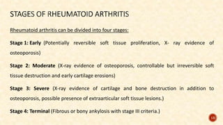
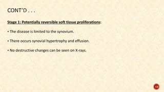
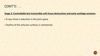
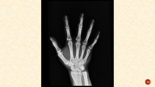
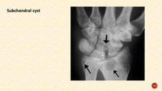
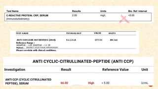
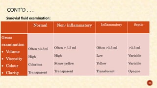
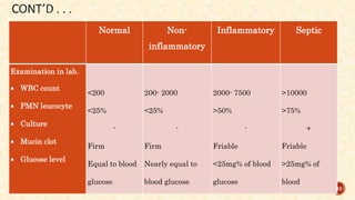
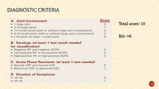
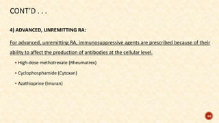
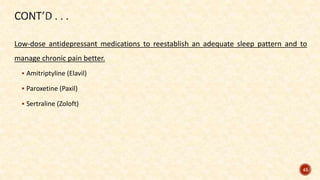
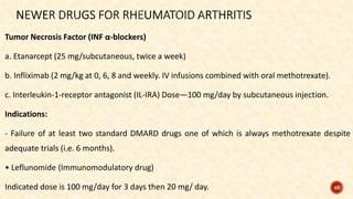
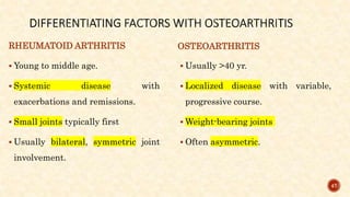
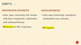
Mrs. Sunita presents to her primary care physician complaining of joint pain, stiffness, and
swelling, primarily affecting her finger, palm and wrists. She reports that the symptoms
have been progressively worsening over the past several months and are interfering with
her daily activities, including typing on the computer and grasping objects.
The physician orders blood investigations and X-rays to rule out the disease. After which
he prescribes Diclofenac Sodium and initiates exercises for relieving stiffness.,
1
ARTHIRITIS
PRESENTED BY:
DAKSH
M.Sc NURSING 2ND YEAR
MSN- ORTHOPAEDICS
2
 Human body is composed of various organs and organ systems that functions together
to make body work.
 Among all systems, musculoskeletal system allows the body for movement and provides
protection to the internal organs as well.
 There are various structures associated that combines together and makes joints that
makes movements possible.
 There can be conditions when deformities can occur due to external or internal factors
like trauma/ injuries or autoimmune response respectively which can erode the
structures of musculoskeletal system making it difficult to perform functions effectively.
3
RHEUMATOID
ARTHRITIS
Rheumatoid arthritis a systemic disease of young and middle-aged adults characterized by
proliferative and destructive changes in synovial membrane, periarticular structures,
skeletal muscles and perineural sheaths.
It is characterized by pain, swelling and limitation of joint movement.
Eventually, joints are destroyed, fibrosed or ankylosed.
5
Prevalence to be around 0.5-1% of the adult population in India.
Sex: Eighty percent affected are women; Male: Female ratio is 1:3.
6
Monoarthritis
– Pyogenic arthritis
– Tubercular arthritis
– Haemophilic arthritis
– Secondary osteoarthritis
– Gout – sometimes
Polyarthritis
– Rheumatoid arthritis
– Rheumatic fever
– Juvenile chronic polyarthritis
– Primary osteoarthritis
– Seronegative spondarthritis
7
The exact cause is unknown, but malfunction of the cellular and humoral arms of the
immune system are cited as the probable cause.
Current Hypothesis: An initiating antigen triggers an aberrant response, which becomes
self-perpetuating long after the offending antigen has been cleared.
Antigenic Agents: Antigenic agents, which probably act as predisposing factors, are
viruses: rubella, Epstein-Barr, etc. genetic (common in people with HLA DR4 60%),
psychological stress, allergic factors, endocrine factors and metabolic factors.
Genetic predisposition: It is strongly suspected because of certain histocompatibility
markers associated with it (HLA-drw4/HLA-DR1).
8
9
Pain
Swelling
Warmth
Erythema
Lack of function
Stiffness
Immobilization
Rheumatoid nodules
10
Hitchhiker’s thumb deformity Claw toe deformity
Swan neck deformity Boutonniere deformity 11
Extra-articular features
 Fever
 Weight loss
 Fatigue
 Anaemia
 Neuropathy
 Scleritis
 Pericarditis
 Lymph node enlargement
 Raynaud’s phenomenon (cold- and stress-
induced vasospasm causing episodes of
digital blanching or cyanosis)
 Arteritis
 Splenomegaly
 Sjögren’s syndrome
12
Rheumatoid arthritis can be divided into four stages:
Stage 1: Early (Potentially reversible soft tissue proliferation, X- ray evidence of
osteoporosis)
Stage 2: Moderate (X-ray evidence of osteoporosis, controllable but irreversible soft
tissue destruction and early cartilage erosions)
Stage 3: Severe (X-ray evidence of cartilage and bone destruction in addition to
osteoporosis, possible presence of extraarticular soft tissue lesions.)
Stage 4: Terminal (Fibrous or bony ankylosis with stage III criteria.)
13
Stage 1: Potentially reversible soft tissue proliferations:
 The disease is limited to the synovium.
 There occurs synovial hypertrophy and effusion.
 No destructive changes can be seen on X-rays.
14
Stage 2: Controllable but irreversible soft tissue destruction and early cartilage erosions:
 X-rays show a reduction in the joint space
 Outline of the articular surfaces is maintained.
15
Stage 3: Irreversible soft tissue and bony changes:
 The pannus ultimately destroys the articular cartilage and erodes the subchondral bone.
 The joint becomes ankylosed usually in a deformed position (fibrous ankylosis).
 Subluxation or dislocation.
16
Assessment of the condition is done by:
• History collection
• Physical examination
• Diagnostic criteria
• X- ray
• Blood profile
• Synovial fluid examination
17
History collection:
The patients complains of joint symptoms, duration of symptoms, morning stiffness.
Past medical history of Rheumatoid arthritis or any other Rheumatic disease.
Family history of the patient with Rheumatoid arthritis.
18
Physical examination:
Pain and stiffness in multiple joints (at least four, metacarpophalangeal joints)
swollen boggy joints
19
Physical examination:
synovial hypertrophy
20
Physical examination:
Edema of the periarticular structures
21
Physical examination:
Deformity
Limited range of motion of the joints
Subluxation or dislocation of joints
Signs of infective arthritis
22
X-rays of both hands and of the affected joints:
Reduced joint space
Erosion of articular margins
Subchondral cysts
Juxta-articular rarefaction
Soft tissue shadow at the level of the joint because of joint effusion or synovial
hypertrophy
Deformities of the hand and fingers
23
Reduced joint space
24
25
Erosion of articular margins
26
27
Subchondral cyst
Blood investigations:
 Elevated ESR
 Rheumatoid factor (RF)
 CRP
 Antinuclear antibody
 Anti CCP (Anti Citrullinated Peptide)
28
29
30
Synovial fluid examination:
Normal Non- inflammatory Inflammatory Septic
Gross
examination
 Volume
 Viscosity
 Colour
 Clarity
Often <3.5ml
High
Colorless
Transparent
Often > 3.5 ml
High
Straw yellow
Transparent
Often >3.5 ml
Low
Yellow
Translucent
>3.5 ml
Variable
Variable
Opaque
31
Normal Non-
inflammatory
Inflammatory Septic
Examination in lab.
 WBC count
 PMN leucocyte
 Culture
 Mucin clot
 Glucose level
<200
<25%
-
Firm
Equal to blood
glucose
200- 2000
<25%
-
Firm
Nearly equal to
blood glucose
2000- 7500
>50%
-
Friable
<25mg% of blood
glucose
>10000
>75%
+
Friable
>25mg% of
blood
32
Total score: 10
RA: >6
33
First Line of Drugs: NSAIDs
These are aspirin/ibuprofen/ketoprofen/diclofenac sodium/ naproxen/piroxicam, etc.
34
 Second Line of Drugs: Disease-modifying antirheumatic drugs (DMARD)
 Penicillamine/ Sulphasalazine/ Antimalarial drugs (e.g. chloroquine)/ Dapsone and
Levamisole.
 Active synovitis with generalized inflammation in many joints.
 Used only if first line fails.
 Symptomatic NSAIDs need to be continued in parallel.
35
Third line of drugs:
Azathioprim/ Cyclophosphamide/ Chlorambucil
Immunosuppressant
Cyclosporine
36
1) EARLY-STAGE RA:
 Education, a balance of rest and exercise.
 NSAIDS
 Disease modifying antirheumatic agents
 Methotrexate
 Biologic response modifiers
 Opioid analgesics are avoided because of the potential for continuing need for pain
relief. 37
2) MODERATE, EROSIVE RA:
 Occupational and physical therapy.
 Cyclosporine
38
3) SEVERE, EROSIVE RA:
Systemic corticosteroids:
Used in unremitting inflammation and pain or needs a “bridging” medication while
waiting for the slower disease-modifying antirheumatic agent (e.g., methotrexate) to
begin working.
Low-dose corticosteroid therapy is prescribed for the shortest time necessary to minimize
side effects.
39
4) TERMINAL, EROSIVE RA:
Reconstructive surgery
Reconstructive surgery is indicated when pain cannot be relieved by conservative
measures.
• Synovectomy (excision of the synovial membrane)
• Tenorrhaphy (suturing a tendon)
• Arthrodesis (surgical fusion of the joint)
• Arthroplasty (surgical repair and replacement of the joint).
40
Synovectomy
41
Arthrodesis surgery
42
Arthroplasty surgery
43
4) ADVANCED, UNREMITTING RA:
For advanced, unremitting RA, immunosuppressive agents are prescribed because of their
ability to affect the production of antibodies at the cellular level.
 High-dose methotrexate (Rheumatrex)
 Cyclophosphamide (Cytoxan)
 Azathioprine (Imuran)
44
Low-dose antidepressant medications to reestablish an adequate sleep pattern and to
manage chronic pain better.
 Amitriptyline (Elavil)
 Paroxetine (Paxil)
 Sertraline (Zoloft)
45
Tumor Necrosis Factor (INF α-blockers)
a. Etanarcept (25 mg/subcutaneous, twice a week)
b. Infliximab (2 mg/kg at 0, 6, 8 and weekly. IV infusions combined with oral methotrexate).
c. Interleukin-1-receptor antagonist (IL-IRA) Dose—100 mg/day by subcutaneous injection.
Indications:
- Failure of at least two standard DMARD drugs one of which is always methotrexate despite
adequate trials (i.e. 6 months).
• Leflunomide (Immunomodulatory drug)
Indicated dose is 100 mg/day for 3 days then 20 mg/ day. 46
RHEUMATOID ARTHRITIS
 Young to middle age.
 Systemic disease with
exacerbations and remissions.
 Small joints typically first
 Usually bilateral, symmetric joint
involvement.
OSTEOARTHRITIS
 Usually >40 yr.
 Localized disease with variable,
progressive course.
 Weight-bearing joints
 Often asymmetric.
47
RHEUMATOID ARTHRITIS
 Stiffness lasts 1 hour to all day and
may decrease with use.
 Pain is variable, may disrupt sleep.
 Effusions are common.
OSTEOARTHRITIS
 Stiffness occurs on arising but
usually subsides after 30 min.
 Pain gradually worsens with joint
use and disease progression,
relieved with rest.
 Effusions are uncommon.
48
RHEUMATOID ARTHRITIS
 Joint space narrowing and erosion
with bony overgrowths, subluxation
with advanced disease.
 RF positive in 80% of patients.
OSTEOARTHRITIS
 Joint space narrowing, osteophytes,
subchondral cysts, sclerosis.
 RF negative.
49
OSTEOARTHRITIS
Osteoarthritis is defined as a degenerative, non-inflammatory joint disease characterized
by destruction of articular cartilage and the formation of new bone at the joint surfaces
and margins.
52
Osteoarthritis is the second most common rheumatologic problem and it is the most
frequent joint disease with a prevalence of 22% to 39% in India.
53
Primary osteoarthritis:
 Idiopathic
 This occurs in a joint de novo.
 It occurs in old age, mainly in the
weight-bearing joints (knee and hip).
 Primary OA is commoner than
secondary OA.
Secondary osteoarthritis:
 Congenital maldevelopment of a joint.
 Trauma
 Previous disease producing a damaged
articular surface
 Internal derangement of the knee, such as a
loose body
 Mal-alignment (bow legs etc.)
 Obesity and excessive weight
54
55
Etiological factors
(Idiopathic, congenital maldevelopment of a joint, trauma,
previous disease producing a damaged articular surface etc.)
cartilage damage at the level of the chondrocytes
affected cartilage gradually becomes softer, less elastic, and less able to
resist wear with heavy use
56
fissuring and erosion of the articular surfaces
development of osteophytes
reduction in motion
Inflammatory changes
 Pain
 Stiffness
 Functional impairment
 Crepitus
 Swelling
 Feeling of 'instability' of the joint,
and 'locking’
 Bouchard nodes
 Heberden nodes
57
58
History collection:
History will be collected for the following:
 Family history
 Pain
 Activities increasing pain
59
Physical examination:
 Tenderness on the joint line
 Irregular and enlarged-looking joint due to the formation of peripheral osteophytes
 Deformity – varus of the knee, flexion-adduction external rotation of the hip
60
Physical examination:
 Tenderness on the joint line
 Crepitus on moving the joint
61
Physical examination:
 Irregular and enlarged-looking joint due to the formation of peripheral osteophytes
 Deformity – varus of the knee, flexion-adduction external rotation of the hip
62
 Effusion
 Terminal limitation of joint movement
 Subluxation
63
Radiological examination:
 Deformity of the joint
64
Radiological examination:
 Narrowing of joint space
 Osteophyte formation
65
 Subchondral sclerosis
66
 Loose bodies
67
Goal of management is to:
a) To delay the occurrence of the disease.
b) To stall progress of the disease and relieve symptoms.
c) To rehabilitate the patient, with or without surgery
68
SUPPORTIVE MANAGEMENT
 Weight reduction, in an obese patient.
 Avoidance of stress and strain
 Rest and joint protection
 Local heat
 Exercises
 Local application of counter-irritants and liniments
69
MEDICAL MANAGEMENT
Analgesics:
 Initial analgesic therapy is acetaminophen.
 NSAIDs
 Topical analgesics such as Capsaicin and Methylsalicylate
 Opioids
70
Viscosupplementation:
 Intra-articular injection of hyaluronic acids
Chondroprotective agents/ Disease-Modifying Osteoarthritis Drugs:
 Glucosamine and Chondroitin sulphate
71
Intra-articular corticosteroid injections:
Corticosteroid injections directly into the affected joint can provide temporary pain relief
and reduce inflammation in OA. (e.g. Triamcinolone acetonide, Methylprednisolone etc.)
72
SURGICAL MANAGEMENT:
Joint debridement: The affected joint is opened, degenerated cartilage smoothened, and
osteophytes and the hypertrophied synovium excised.
73
Osteotomy: Osteotomy is a surgical procedure in which a bone is cut, reshaped, or
realigned to correct deformities or improve its function.
74
Joint replacement: Damaged or dysfunctional joint is replaced with an artificial joint,
called a prosthesis. For cases crippled with advanced damage to the joint, total joint
replacement operation.
75
GOUT
 Gout is defined as a heterogeneous group of conditions related to a genetic defect of
purine metabolism resulting in hyperuricemia.
 Over secretion of uric acid or a renal defect resulting in decreased excretion of uric acid,
or a combination of both, occurs.
 Gout is also called monosodium urate arthropathy (gout).
77
78
 It is about 0.3/1000.
 It occurs more commonly in males than females.
79
 It is usually monoarticular and the first metatarsophalangeal joint is the most common
site of involvement (75%).
 Ankle, knee, wrist, fingers and elbow are other joints affected.
 Distal and lower extremity joints are involved more often.
80
Gout is a disease of:
 Affluence
 Alcoholism
 Obesity
 Old age
 Diuretic treatment
81
 Age
 Heredity
 Renal defect
 Severe dieting or starvation
 Excessive intake of foods that are high in purines (shellfish, organ meats)
 Drugs
82
Etiological factors
(age, hereditary, obesity etc.)
Hyperuricemia
Increased uric acid production
Decreased uric acid excretion
83
Urate crystal deposition
Inflammatory response
Acute gouty attack
Chronic gout
84
Tophi
Renal urate lithiasis (kidney stones) with chronic renal disease
85
86
 Pain
 Swelling
 Tenderness
 Increased temperature
 Sleep disturbance
 Tophi
 Gouty nephropathy
 Symptom-free period until the next
attack
87
88
History collection
History will be collected regarding following:
 Onset of pain
 Duration of symptoms
 Food intake
 Elimination
 Genetic history
89
oJoint Swelling and Redness
oTenderness
oWarmth
oLimited Range of Motion
oMetatarsophalangeal joint involvement
oTophi
oSkin changes (shiny and stretched skin)
90
Physical examination
91
oESR
oSynovial fluid study
oRaised SUA level
92
BLOOD INVESTIGATIONS
Attacks (4R’s)
 Rapid onset
 Recurrent
 Rarely seen > 10 days
 Remissions
93
Joints (FRAME)
 First MTP joint involved
 Red hot joint
 Articular involvement
 Urate crystals
 Extreme pain
Extra-articular features
 Hyperuricemia
 Tophi
94
95
MEDICAL MANAGEMENT
Analgesics/ anti-inflammatory drugs:
 Colchicine
 Indomethacin
Uricosuric agents:
 Probenecid
96
Xanthine oxidase inhibitors:
 Allopurinol
Corticosteroids
97
SURGICAL INTERVENTIONS
Arthrocentesis
Joint debridement
Tophi excision
Athritis (RA, OA, Gout) MSN.pptx

Athritis (RA, OA, Gout) MSN.pptx

  • 1.
    Mrs. Sunita presentsto her primary care physician complaining of joint pain, stiffness, and swelling, primarily affecting her finger, palm and wrists. She reports that the symptoms have been progressively worsening over the past several months and are interfering with her daily activities, including typing on the computer and grasping objects. The physician orders blood investigations and X-rays to rule out the disease. After which he prescribes Diclofenac Sodium and initiates exercises for relieving stiffness., 1
  • 2.
    ARTHIRITIS PRESENTED BY: DAKSH M.Sc NURSING2ND YEAR MSN- ORTHOPAEDICS 2
  • 3.
     Human bodyis composed of various organs and organ systems that functions together to make body work.  Among all systems, musculoskeletal system allows the body for movement and provides protection to the internal organs as well.  There are various structures associated that combines together and makes joints that makes movements possible.  There can be conditions when deformities can occur due to external or internal factors like trauma/ injuries or autoimmune response respectively which can erode the structures of musculoskeletal system making it difficult to perform functions effectively. 3
  • 4.
  • 5.
    Rheumatoid arthritis asystemic disease of young and middle-aged adults characterized by proliferative and destructive changes in synovial membrane, periarticular structures, skeletal muscles and perineural sheaths. It is characterized by pain, swelling and limitation of joint movement. Eventually, joints are destroyed, fibrosed or ankylosed. 5
  • 6.
    Prevalence to bearound 0.5-1% of the adult population in India. Sex: Eighty percent affected are women; Male: Female ratio is 1:3. 6
  • 7.
    Monoarthritis – Pyogenic arthritis –Tubercular arthritis – Haemophilic arthritis – Secondary osteoarthritis – Gout – sometimes Polyarthritis – Rheumatoid arthritis – Rheumatic fever – Juvenile chronic polyarthritis – Primary osteoarthritis – Seronegative spondarthritis 7
  • 8.
    The exact causeis unknown, but malfunction of the cellular and humoral arms of the immune system are cited as the probable cause. Current Hypothesis: An initiating antigen triggers an aberrant response, which becomes self-perpetuating long after the offending antigen has been cleared. Antigenic Agents: Antigenic agents, which probably act as predisposing factors, are viruses: rubella, Epstein-Barr, etc. genetic (common in people with HLA DR4 60%), psychological stress, allergic factors, endocrine factors and metabolic factors. Genetic predisposition: It is strongly suspected because of certain histocompatibility markers associated with it (HLA-drw4/HLA-DR1). 8
  • 9.
  • 10.
  • 11.
    Hitchhiker’s thumb deformityClaw toe deformity Swan neck deformity Boutonniere deformity 11
  • 12.
    Extra-articular features  Fever Weight loss  Fatigue  Anaemia  Neuropathy  Scleritis  Pericarditis  Lymph node enlargement  Raynaud’s phenomenon (cold- and stress- induced vasospasm causing episodes of digital blanching or cyanosis)  Arteritis  Splenomegaly  Sjögren’s syndrome 12
  • 13.
    Rheumatoid arthritis canbe divided into four stages: Stage 1: Early (Potentially reversible soft tissue proliferation, X- ray evidence of osteoporosis) Stage 2: Moderate (X-ray evidence of osteoporosis, controllable but irreversible soft tissue destruction and early cartilage erosions) Stage 3: Severe (X-ray evidence of cartilage and bone destruction in addition to osteoporosis, possible presence of extraarticular soft tissue lesions.) Stage 4: Terminal (Fibrous or bony ankylosis with stage III criteria.) 13
  • 14.
    Stage 1: Potentiallyreversible soft tissue proliferations:  The disease is limited to the synovium.  There occurs synovial hypertrophy and effusion.  No destructive changes can be seen on X-rays. 14
  • 15.
    Stage 2: Controllablebut irreversible soft tissue destruction and early cartilage erosions:  X-rays show a reduction in the joint space  Outline of the articular surfaces is maintained. 15
  • 16.
    Stage 3: Irreversiblesoft tissue and bony changes:  The pannus ultimately destroys the articular cartilage and erodes the subchondral bone.  The joint becomes ankylosed usually in a deformed position (fibrous ankylosis).  Subluxation or dislocation. 16
  • 17.
    Assessment of thecondition is done by: • History collection • Physical examination • Diagnostic criteria • X- ray • Blood profile • Synovial fluid examination 17
  • 18.
    History collection: The patientscomplains of joint symptoms, duration of symptoms, morning stiffness. Past medical history of Rheumatoid arthritis or any other Rheumatic disease. Family history of the patient with Rheumatoid arthritis. 18
  • 19.
    Physical examination: Pain andstiffness in multiple joints (at least four, metacarpophalangeal joints) swollen boggy joints 19
  • 20.
  • 21.
    Physical examination: Edema ofthe periarticular structures 21
  • 22.
    Physical examination: Deformity Limited rangeof motion of the joints Subluxation or dislocation of joints Signs of infective arthritis 22
  • 23.
    X-rays of bothhands and of the affected joints: Reduced joint space Erosion of articular margins Subchondral cysts Juxta-articular rarefaction Soft tissue shadow at the level of the joint because of joint effusion or synovial hypertrophy Deformities of the hand and fingers 23
  • 24.
  • 25.
  • 26.
  • 27.
  • 28.
    Blood investigations:  ElevatedESR  Rheumatoid factor (RF)  CRP  Antinuclear antibody  Anti CCP (Anti Citrullinated Peptide) 28
  • 29.
  • 30.
  • 31.
    Synovial fluid examination: NormalNon- inflammatory Inflammatory Septic Gross examination  Volume  Viscosity  Colour  Clarity Often <3.5ml High Colorless Transparent Often > 3.5 ml High Straw yellow Transparent Often >3.5 ml Low Yellow Translucent >3.5 ml Variable Variable Opaque 31
  • 32.
    Normal Non- inflammatory Inflammatory Septic Examinationin lab.  WBC count  PMN leucocyte  Culture  Mucin clot  Glucose level <200 <25% - Firm Equal to blood glucose 200- 2000 <25% - Firm Nearly equal to blood glucose 2000- 7500 >50% - Friable <25mg% of blood glucose >10000 >75% + Friable >25mg% of blood 32
  • 33.
  • 34.
    First Line ofDrugs: NSAIDs These are aspirin/ibuprofen/ketoprofen/diclofenac sodium/ naproxen/piroxicam, etc. 34
  • 35.
     Second Lineof Drugs: Disease-modifying antirheumatic drugs (DMARD)  Penicillamine/ Sulphasalazine/ Antimalarial drugs (e.g. chloroquine)/ Dapsone and Levamisole.  Active synovitis with generalized inflammation in many joints.  Used only if first line fails.  Symptomatic NSAIDs need to be continued in parallel. 35
  • 36.
    Third line ofdrugs: Azathioprim/ Cyclophosphamide/ Chlorambucil Immunosuppressant Cyclosporine 36
  • 37.
    1) EARLY-STAGE RA: Education, a balance of rest and exercise.  NSAIDS  Disease modifying antirheumatic agents  Methotrexate  Biologic response modifiers  Opioid analgesics are avoided because of the potential for continuing need for pain relief. 37
  • 38.
    2) MODERATE, EROSIVERA:  Occupational and physical therapy.  Cyclosporine 38
  • 39.
    3) SEVERE, EROSIVERA: Systemic corticosteroids: Used in unremitting inflammation and pain or needs a “bridging” medication while waiting for the slower disease-modifying antirheumatic agent (e.g., methotrexate) to begin working. Low-dose corticosteroid therapy is prescribed for the shortest time necessary to minimize side effects. 39
  • 40.
    4) TERMINAL, EROSIVERA: Reconstructive surgery Reconstructive surgery is indicated when pain cannot be relieved by conservative measures. • Synovectomy (excision of the synovial membrane) • Tenorrhaphy (suturing a tendon) • Arthrodesis (surgical fusion of the joint) • Arthroplasty (surgical repair and replacement of the joint). 40
  • 41.
  • 42.
  • 43.
  • 44.
    4) ADVANCED, UNREMITTINGRA: For advanced, unremitting RA, immunosuppressive agents are prescribed because of their ability to affect the production of antibodies at the cellular level.  High-dose methotrexate (Rheumatrex)  Cyclophosphamide (Cytoxan)  Azathioprine (Imuran) 44
  • 45.
    Low-dose antidepressant medicationsto reestablish an adequate sleep pattern and to manage chronic pain better.  Amitriptyline (Elavil)  Paroxetine (Paxil)  Sertraline (Zoloft) 45
  • 46.
    Tumor Necrosis Factor(INF α-blockers) a. Etanarcept (25 mg/subcutaneous, twice a week) b. Infliximab (2 mg/kg at 0, 6, 8 and weekly. IV infusions combined with oral methotrexate). c. Interleukin-1-receptor antagonist (IL-IRA) Dose—100 mg/day by subcutaneous injection. Indications: - Failure of at least two standard DMARD drugs one of which is always methotrexate despite adequate trials (i.e. 6 months). • Leflunomide (Immunomodulatory drug) Indicated dose is 100 mg/day for 3 days then 20 mg/ day. 46
  • 47.
    RHEUMATOID ARTHRITIS  Youngto middle age.  Systemic disease with exacerbations and remissions.  Small joints typically first  Usually bilateral, symmetric joint involvement. OSTEOARTHRITIS  Usually >40 yr.  Localized disease with variable, progressive course.  Weight-bearing joints  Often asymmetric. 47
  • 48.
    RHEUMATOID ARTHRITIS  Stiffnesslasts 1 hour to all day and may decrease with use.  Pain is variable, may disrupt sleep.  Effusions are common. OSTEOARTHRITIS  Stiffness occurs on arising but usually subsides after 30 min.  Pain gradually worsens with joint use and disease progression, relieved with rest.  Effusions are uncommon. 48
  • 49.
    RHEUMATOID ARTHRITIS  Jointspace narrowing and erosion with bony overgrowths, subluxation with advanced disease.  RF positive in 80% of patients. OSTEOARTHRITIS  Joint space narrowing, osteophytes, subchondral cysts, sclerosis.  RF negative. 49
  • 51.
  • 52.
    Osteoarthritis is definedas a degenerative, non-inflammatory joint disease characterized by destruction of articular cartilage and the formation of new bone at the joint surfaces and margins. 52
  • 53.
    Osteoarthritis is thesecond most common rheumatologic problem and it is the most frequent joint disease with a prevalence of 22% to 39% in India. 53
  • 54.
    Primary osteoarthritis:  Idiopathic This occurs in a joint de novo.  It occurs in old age, mainly in the weight-bearing joints (knee and hip).  Primary OA is commoner than secondary OA. Secondary osteoarthritis:  Congenital maldevelopment of a joint.  Trauma  Previous disease producing a damaged articular surface  Internal derangement of the knee, such as a loose body  Mal-alignment (bow legs etc.)  Obesity and excessive weight 54
  • 55.
    55 Etiological factors (Idiopathic, congenitalmaldevelopment of a joint, trauma, previous disease producing a damaged articular surface etc.) cartilage damage at the level of the chondrocytes affected cartilage gradually becomes softer, less elastic, and less able to resist wear with heavy use
  • 56.
    56 fissuring and erosionof the articular surfaces development of osteophytes reduction in motion Inflammatory changes
  • 57.
     Pain  Stiffness Functional impairment  Crepitus  Swelling  Feeling of 'instability' of the joint, and 'locking’  Bouchard nodes  Heberden nodes 57
  • 58.
  • 59.
    History collection: History willbe collected for the following:  Family history  Pain  Activities increasing pain 59
  • 60.
    Physical examination:  Tendernesson the joint line  Irregular and enlarged-looking joint due to the formation of peripheral osteophytes  Deformity – varus of the knee, flexion-adduction external rotation of the hip 60
  • 61.
    Physical examination:  Tendernesson the joint line  Crepitus on moving the joint 61
  • 62.
    Physical examination:  Irregularand enlarged-looking joint due to the formation of peripheral osteophytes  Deformity – varus of the knee, flexion-adduction external rotation of the hip 62
  • 63.
     Effusion  Terminallimitation of joint movement  Subluxation 63
  • 64.
  • 65.
    Radiological examination:  Narrowingof joint space  Osteophyte formation 65
  • 66.
  • 67.
  • 68.
    Goal of managementis to: a) To delay the occurrence of the disease. b) To stall progress of the disease and relieve symptoms. c) To rehabilitate the patient, with or without surgery 68
  • 69.
    SUPPORTIVE MANAGEMENT  Weightreduction, in an obese patient.  Avoidance of stress and strain  Rest and joint protection  Local heat  Exercises  Local application of counter-irritants and liniments 69
  • 70.
    MEDICAL MANAGEMENT Analgesics:  Initialanalgesic therapy is acetaminophen.  NSAIDs  Topical analgesics such as Capsaicin and Methylsalicylate  Opioids 70
  • 71.
    Viscosupplementation:  Intra-articular injectionof hyaluronic acids Chondroprotective agents/ Disease-Modifying Osteoarthritis Drugs:  Glucosamine and Chondroitin sulphate 71
  • 72.
    Intra-articular corticosteroid injections: Corticosteroidinjections directly into the affected joint can provide temporary pain relief and reduce inflammation in OA. (e.g. Triamcinolone acetonide, Methylprednisolone etc.) 72
  • 73.
    SURGICAL MANAGEMENT: Joint debridement:The affected joint is opened, degenerated cartilage smoothened, and osteophytes and the hypertrophied synovium excised. 73
  • 74.
    Osteotomy: Osteotomy isa surgical procedure in which a bone is cut, reshaped, or realigned to correct deformities or improve its function. 74
  • 75.
    Joint replacement: Damagedor dysfunctional joint is replaced with an artificial joint, called a prosthesis. For cases crippled with advanced damage to the joint, total joint replacement operation. 75
  • 76.
  • 77.
     Gout isdefined as a heterogeneous group of conditions related to a genetic defect of purine metabolism resulting in hyperuricemia.  Over secretion of uric acid or a renal defect resulting in decreased excretion of uric acid, or a combination of both, occurs.  Gout is also called monosodium urate arthropathy (gout). 77
  • 78.
  • 79.
     It isabout 0.3/1000.  It occurs more commonly in males than females. 79
  • 80.
     It isusually monoarticular and the first metatarsophalangeal joint is the most common site of involvement (75%).  Ankle, knee, wrist, fingers and elbow are other joints affected.  Distal and lower extremity joints are involved more often. 80
  • 81.
    Gout is adisease of:  Affluence  Alcoholism  Obesity  Old age  Diuretic treatment 81
  • 82.
     Age  Heredity Renal defect  Severe dieting or starvation  Excessive intake of foods that are high in purines (shellfish, organ meats)  Drugs 82
  • 83.
    Etiological factors (age, hereditary,obesity etc.) Hyperuricemia Increased uric acid production Decreased uric acid excretion 83
  • 84.
    Urate crystal deposition Inflammatoryresponse Acute gouty attack Chronic gout 84
  • 85.
    Tophi Renal urate lithiasis(kidney stones) with chronic renal disease 85
  • 86.
  • 87.
     Pain  Swelling Tenderness  Increased temperature  Sleep disturbance  Tophi  Gouty nephropathy  Symptom-free period until the next attack 87
  • 88.
  • 89.
    History collection History willbe collected regarding following:  Onset of pain  Duration of symptoms  Food intake  Elimination  Genetic history 89
  • 90.
    oJoint Swelling andRedness oTenderness oWarmth oLimited Range of Motion oMetatarsophalangeal joint involvement oTophi oSkin changes (shiny and stretched skin) 90 Physical examination
  • 91.
  • 92.
    oESR oSynovial fluid study oRaisedSUA level 92 BLOOD INVESTIGATIONS
  • 93.
    Attacks (4R’s)  Rapidonset  Recurrent  Rarely seen > 10 days  Remissions 93 Joints (FRAME)  First MTP joint involved  Red hot joint  Articular involvement  Urate crystals  Extreme pain Extra-articular features  Hyperuricemia  Tophi
  • 94.
  • 95.
    95 MEDICAL MANAGEMENT Analgesics/ anti-inflammatorydrugs:  Colchicine  Indomethacin Uricosuric agents:  Probenecid
  • 96.
    96 Xanthine oxidase inhibitors: Allopurinol Corticosteroids
  • 97.