AVN Bone Death
•Download as PPTX, PDF•
21 likes•4,769 views

This document discusses avascular necrosis (AVN), also called osteonecrosis. It defines AVN as bone death caused by sudden obstruction of blood supply to bone. Common causes include trauma, corticosteroid use, and decompression sickness. Sites often affected are the femoral head, scaphoid, and talus. The goal of treatment is to improve joint function, stop further bone damage, and preserve the bone and joint. Methods include nonsurgical options to delay progression and surgical interventions like core decompression or grafts, with joint replacement usually performed later.

Recommended
More Related Content
What's hot
What's hot (20)
Viewers also liked
Viewers also liked (14)
Similar to AVN Bone Death
Similar to AVN Bone Death (20)
Recently uploaded
Recently uploaded (20)
AVN Bone Death
- 2. Definition AVN OF BONE/OSTEONECROSIS means “BONE DEATH” Sudden Obstruction in arterial blood supply to a part of bone O S T E O N E C R O S I S
- 3. Causes Traumatic: Fracture -Femur Neck, Talus, Scaphoid. Dislocations – hip Non-Traumatic : Caisson Disease Sickle Cell Disease Gaucher’s Disease Coagulation Disorders Cortisone Administration Organ /Bone transplantation Metal corrosion Alcoholism Exposure to xrays and radioactive substances
- 4. Common sites of predilection -Femoral head -Scaphoid(Preiser’s Disease) -Talus -Segmental fracture -Others – capitellum , radial head , lateral femoral condyle
- 5. Pathogenesis BONE ISCHAEMIA is due to - Interruption of arterial inflow - Occlusion of Venous Outflow - Intravascular Blockage of arterioles & capillaries - Increase in marrow pressure
- 6. Repair process in a cancellous bone • Proliferating capillaries and fibrous stroma penetrate marrow space of dead bone • Phagocytes remove the marrow debris • Osteoblasts lay down immature woven bone • All these process further increases the radiodensity of necrotic bone
- 7. • Later osteoclastic resorption removes the woven bone as well as the old trabaeculae • This is replaced by well organised lamellar bone • This process of apposition of new bone on some surface and osteoclastic resorption on other surface is called CREEPING SUBSTITUTION
- 8. Repair process in cortical bone • Excavation of haversian canal by osteoclastic resorption proceeds • This enlarges the canal which multiple in number and uniform in size • Later osteoblasts lay down concentric rings of new bone
- 9. Pathology • Microscopically 4 stages are recognised -Stage of marrow necrosis and cell death. -Reactive vascularisation and infiltration. -Distortion of shape by collapse and compression of trabeculae -Subchondral collapse Deformation of articular cartilage
- 10. Dysbaric osteonecrosis Caisson disease /Decompressionsickness/ Aeroembolism. Seen in deep sea divers , tunnel workers, working in unpressurized aircrafts. Nitrogen gas bubbles liberated in a concentration that cannot be readily absorbed by blood stream or excreted by lungs. As a result gas bubbles accumulate in tissues causing local ischemia or intravascular occlusion
- 11. Corticosteroid induced AVN 1. Fat embolism theory : • Fat accumulates in liver in patients treated with steroid , and serum lipid concentration also increases • It gives rise to fat embolism and AVN 2. Subchondral osteoporotic fracture : Steroids induce protein catabolism Resulting in generalised osteoporosis Produces subchondral fractures and aseptic necrosis
- 12. Clinical Features Early stages: Pain-near joint Later stages: Stiffness, Limitation of movements Advanced stage: Fixed Deformities
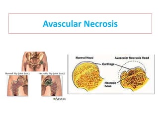
- 13. Radiological features • Initially necrotic bone appears radiodense • Surrounding vascular bone shows relative osteoporosis • In Early stages- Articular cartilage is not affected - so joint space is normal • In later stages -Partial collapse,flattening of head,joint space narrowing,osteoarthotic changes
- 14. AVN of femoral head • Occurs mainly due to femur neck fracture and hip dislocation • Due to disruption of vascular channel in femoral neck
- 15. Vascular supply around femoral neck
- 17. Radiography of femur head - AVN
- 18. Treatment Core Decompression. Vasularised Fibular Graft. Hemiarthroplasty . Total hip arthroplasty.
- 19. Legg-calve perthe’s disease AVN of ossification centre of capital epiphysis of femoral head. 3-12 age group. c/f: Limp, antalgic gait, limited motion HIP DEFORMITY xray: Early findings include Medial joint space widening irregularity of femoral head ossification cresent sign (represents a subchondral fracture)
- 20. Treatment: Aim- preserve the femoral head, acetabular congruity, eliminate or reduce weight bearing. Nonoperative -observation, activity restriction, partial weight bearing, traction, and physical therapy. .Ambulation-Abduction Brace Operative- children > 8 years of age. -Femoral Osteotomy proximal femoral varus osteotomy -Pelvic Osteotomy.
- 21. AVN of scaphoid
- 22. Vascular supply of scaphoid
- 23. Vascular disruption in # scaphoid
- 24. Radiography of scaphoid # AVN
- 25. AVN of talus
- 26. Vascular supply of talus
- 28. Other bones susceptible for AVN Lateral femoral condyle Capitellum
- 29. The goal in treating avascular necrosis Is to improve the patient's use of the affected joint, Stop further damage to the bone and Ensure bone and joint survival.
- 30. • Various methods for delaying disease progression • Non surgical – • Bisphosphonate • Anticoagulants • Vasodilators • Biophysical modalities • Surgical – • Core decompression • Vascularised bone graft/muscle pedicle graft Usually arthroplasty is awaited without any big surgical intervention