The document reports on a study investigating the role of the p38 mitogen-activated protein kinase p38γ in Alzheimer's disease (AD) pathogenesis. The study found that depletion of p38γ exacerbated neuronal excitotoxicity, cognitive deficits, neuronal circuit abnormalities, and premature mortality in an AD mouse model overexpressing amyloid-β (Aβ). In contrast, increasing p38γ activity abolished these Aβ-induced deficits. Furthermore, mimicking site-specific tau phosphorylation by p38γ alleviated Aβ-induced neuronal death and excitotoxicity. The findings suggest p38γ phosphorylation of tau at specific sites inhibits Aβ toxicity in early AD, challenging the view that tau phosphorylation is purely pathogenic.
![APP23.p38g−/−
with tau−/−
mice. The exacer-
bating effects of p38g loss on reduced survival,
memory deficits, and neuronal network dys-
function of APP23 mice were virtually abolished
in APP23.p38g−/−
.tau−/−
mice (Fig. 2, A to C, and
fig. S11). These data also showed that, com-
pared with APP23 mice, APP23.p38g−/−
animals
had aggravated memory deficits that persisted
with aging. In contrast, increasing tau levels in
p38g−/−
mice [brought about by crossing with
nonmutant tau-expressing Alz17 mice (23)] sig-
nificantly enhanced PTZ-induced seizures in
Alz17.p38g−/−
mice (Fig. 2, D to F). Conversely,
when compared to tau−/−
.p38g+/+
mice, tau−/−
.
p38g−/−
animals showed similar protection from
PTZ-induced seizures (Fig. 2, G to I). Taken
together, the effects of p38g on excitotoxicity
and Ab toxicity were tau-dependent.
We have previously shown that postsynaptic
PSD-95/tau/Fyn complexes mediate Ab-induced
excitotoxicity (9). PSD-95/tau/Fyn interaction was
enhanced in Alz17.p38g−/−
animals versus Alz17.
p38g+/+
mice (Fig. 3A and fig. S12). Conversely, no
PSD-95/tau/Fyn complexes were isolated from
tau−/−
and tau−/−
.p38g−/−
brains (fig. S12). In-
creasing p38g levels compromised PSD-95/tau/
Fyn interaction in cells, and expression of a con-
stitutively active p38g variant (p38gCA
) com-
pletely abolished this interaction (Fig. 3B and
fig. S13). Pan-p38 inhibition stopped p38g/p38gCA
-
induced disruption of PSD-95/tau/Fyn complexes
(Fig. 3B). PSD-95 copurified more tau and Fyn
from p38g−/−
versus p38g+/+
brains, and even
more from APP23.p38g−/−
compared with APP23.
p38g+/+
and p38g−/−
brains (Fig. 3C). Conversely,
PSD-95/tau/Fyn interaction was reduced in trans-
genic mice with neuronal expression of p38gCA
(Fig. 3D and fig. S14). PTZ transiently increased
PSD-95/tau/Fyn complex formation in p38g+/+
animals; this effect was even more noticeable
in p38g−/−
mice (fig. S12). Fyn-mediated NR2B
phosphorylation at Tyr1472
(Y1472) facilitates
PSD-95/NR2B interaction (24). Consistent with
increased PSD-95/tau/Fyn complex formation,
NR2B phosphorylation at Y1472 was increased
in p38g−/−
brains (fig. S15). Conversely, cellular
expression of p38g and p38gCA
—but not p38aCA
,
p38bCA
, or p38dCA
—reduced Y1472 phosphoryl-
ation of NR2B (fig. S15). Hence, p38g regulated
PSD-95/tau/Fyn complexes, likely at the level of
PSD-95/tau interaction (fig. S16).
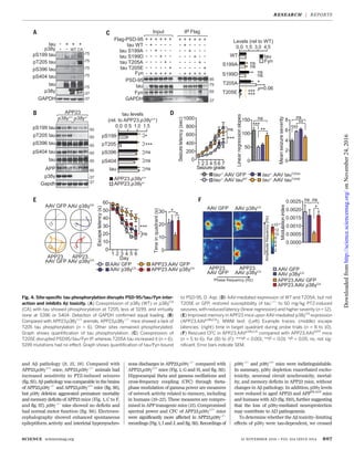
Although p38g hyperphosphorylates tau dur-
ing long-term in vitro kinase assays (25), the
temporal profile of p38g-induced tau phospho-
rylation in acute signaling remains unknown.
Short-term in vitro kinase reactions using phos-
phorylation site–specific tau antibodies revealed
phosphorylation at Ser199
(S199), Thr205
(T205),
S396, and S404 (fig. S17). Mass spectrometric
analysis confirmed these and 14 additional,
though low-abundant, sites (figs. S17C and S18
and table S4). Coexpression of p38g or p38gCA
and tau in cells revealed tau phosphorylation
(p) at T205, less at S199, and hardly any at S396
or S404 (Fig. 4A). Similarly, T205 (and, less so,
S199 and S396) were phosphorylated in p38gCA
transgenic mice (fig. S19). pT205 increased after
PTZ in p38g+/+
animals but was virtually abolished
in p38g−/−
mice, whereas pS199, pS396, and pS404
were induced in both p38g+/+
and p38g−/−
mice
(fig. S19). Similarly, pT205 was markedly reduced
in APP23.p38g−/−
animals compared with APP23.
p38g+/+
mice (Fig. 4B). In primary neurons,
pT205 (but not p199) was markedly reduced by
pan-p38 inhibition (fig. S20). Taken together,
these findings indicate that pT205 was a primary
p38g site in tau.
Next, we showed that a phosphorylation-
mimicking Thr205
→Glu205
(T205E) tau variant
coprecipitated significantly less with PSD-95 as
compared with nonmutant and T205A (A, Ala) tau
(Fig. 4C and fig. S21). In contrast, phosphorylation-
mimicking mutants of all other identified sites
had no effect on PSD-95/tau/Fyn interaction (fig.
S18). Microscale thermophoresis and glutathione
S-transferase–pulldown in vitro and fluorescence-
lifetime imaging microscopy (FLIM)–fluorescence
resonance energy transfer (FRET) analysis in
live cells confirmed the markedly compromised
interaction of T205E tau with PSD-95 (fig. S22).
The T205E mutation did not hinder tau/Fyn
interaction (fig. S21). Phosphorylation of T205 by
p38gCA
was required for disrupting PSD-95/tau/
Fyn complexes (fig. S21). Hence, p38g regulated
PSD-95/tau/Fyn complexes via phosphorylating
tau at T205.
The disruption of NR/PSD-95/tau/Fyn com-
plexes prevents excitotoxicity and Ab toxicity (9).
Hence, phosphorylation of tau at T205 should
similarly mitigate neurotoxicity. Ab caused cell
death in WT and T205A neurons but signifi-
cantly less in T205E tau-expressing neurons
(fig. S23). Similarly, neurons expressing p38g
and, more so, p38gCA
were significantly more
resistant to Ab-induced cell death than controls
(fig. S24). PTZ-induced seizures are reduced in
tau−/−
mice (8, 9). Adeno-associated virus (AAV)–
mediated expression of WT and T205A neurons,
but not T205E tau or green fluorescent protein
(GFP), in the forebrains of tau−/−
mice enhanced
PTZ-induced seizures (Fig. 4D and fig. S25). In
contrast, expression of p38gCA
in WT mice using
AAV or in Thy1.2-p38gCA
transgenic mice de-
creased PTZ-induced seizures (fig. S25). AAV-
mediated p38gCA
expression in APP23 mice
rescued memory deficits and network aberra-
tions; the same was true for crossing APP23
with Thy1.2-p38gCA
mice (Fig. 4, E and F, and
figs. S26 and S27). In summary, the levels of
active p38g kinase and tau phosphorylation at
T205 determined susceptibility to excitotoxicity
and Ab toxicity.
Here we have shown that T205 phosphoryl-
ation of tau is part of an Ab toxicity–inhibiting
response. This is contrary to the current view
that tau phosphorylation downstream of Ab tox-
icity is a pathological response (3). However, this
finding is in line with the idea that tau is in-
volved in normal physiologic NR signaling events
in neurons (12). Finally, we found that tau-
dependent Ab toxicity was modulated by site-
specific tau phosphorylation, which inhibited
postsynaptic PSD-95/tau/Fyn complexes, reveal-
ing an Ab toxicity–limiting role of p38g in AD
that is distinct and opposite to the effects of
p38a and p38b (11, 13, 14).
REFERENCES AND NOTES
1. C. Ballatore, V. M. Lee, J. Q. Trojanowski, Nat. Rev. Neurosci. 8,
663–672 (2007).
2. C. Haass, D. J. Selkoe, Nat. Rev. Mol. Cell Biol. 8, 101–112
(2007).
3. L. M. Ittner, J. Götz, Nat. Rev. Neurosci. 12, 67–72
(2011).
4. K. Iqbal, F. Liu, C.-X. Gong, A del C. Alonso,
I. Grundke-Iqbal, Acta Neuropathol. 118, 53–69
(2009).
5. E. M. Mandelkow, E. Mandelkow, Cold Spring Harb. Perspect.
Med. 2, a006247 (2012).
6. E. S. Musiek, D. M. Holtzman, Nat. Neurosci. 18, 800–806
(2015).
7. M. Rapoport, H. N. Dawson, L. I. Binder, M. P. Vitek,
A. Ferreira, Proc. Natl. Acad. Sci. U.S.A. 99, 6364–6369
(2002).
8. E. D. Roberson et al., Science 316, 750–754 (2007).
9. L. M. Ittner et al., Cell 142, 387–397 (2010).
10. J. J. Palop, L. Mucke, Nat. Neurosci. 13, 812–818
(2010).
11. G. E. Hardingham, H. Bading, Nat. Rev. Neurosci. 11, 682–696
(2010).
12. L. Mucke, D. J. Selkoe, Cold Spring Harb. Perspect. Med. 2,
a006338 (2012).
13. Q. Wang, D. M. Walsh, M. J. Rowan, D. J. Selkoe,
R. Anwyl, J. Neurosci. 24, 3370–3378 (2004).
14. S. Li et al., J. Neurosci. 31, 6627–6638 (2011).
15. A. A. Ittner, A. Gladbach, J. Bertz, L. S. Suh, L. M. Ittner, Acta
Neuropathol. Commun. 2, 149 (2014).
16. M. A. Fabian et al., Nat. Biotechnol. 23, 329–336 (2005).
17. M. B. Menon, S. Dhamija, A. Kotlyarov, M. Gaestel, Autophagy
11, 1425–1427 (2015).
18. C. Sturchler-Pierrat et al., Proc. Natl. Acad. Sci. U.S.A. 94,
13287–13292 (1997).
19. G. Buzsáki, E. I. Moser, Nat. Neurosci. 16, 130–138
(2013).
20. R. Goutagny, J. Jackson, S. Williams, Nat. Neurosci. 12,
1491–1493 (2009).
21. R. T. Canolty et al., Science 313, 1626–1628 (2006).
22. A. B. Tort, R. W. Komorowski, J. R. Manns, N. J. Kopell,
H. Eichenbaum, Proc. Natl. Acad. Sci. U.S.A. 106,
20942–20947 (2009).
23. A. Probst et al., Acta Neuropathol. 99, 469–481
(2000).
24. M. Aarts et al., Science 298, 846–850 (2002).
25. M. Goedert et al., FEBS Lett. 409, 57–62 (1997).
ACKNOWLEDGMENTS
We thank the staff of the Biological Resources
Centre of UNSW for animal care, E. Hinde for help
with FLIM-FRET experiments, D. Sullivan and T. Foo
for APOE genotyping of human samples, and T. Saito
and T. C. Saido for APPNL-G-F
mice. The data
from mass spectrometry experiments can be found
in the supplementary materials. This work was
funded by the National Health and Medical Research
Council (grants 1081916, 1037746, and 1003083),
the Australian Research Council (grants DP130102027
and DE130101591), Alzheimer’s Association (grant
NIRG000070035), Alzheimer’s Australia (grants
DGP14-39 and DGP14-95), the NIH (grant R28AA012725),
and UNSW Australia. A.I. and L.M.I. are inventors
on Australian patent application number APO/2016/900764,
submitted by UNSW, which covers increasing p38g
activity to prevent neuronal toxicity.
SUPPLEMENTARY MATERIALS
www.sciencemag.org/content/354/6314/904/suppl/DC1
Materials and Methods
Figs. S1 to S27
Tables S1 to S5
References (26–53)
22 July 2016; resubmitted 9 October 2016
Accepted 19 October 2016
10.1126/science.aah6205
908 18 NOVEMBER 2016 • VOL 354 ISSUE 6314 sciencemag.org SCIENCE
RESEARCH | REPORTS
onNovember24,2016http://science.sciencemag.org/Downloadedfrom](https://image.slidesharecdn.com/artculoalzheimer-161124185754/85/Articulo-alzheimer-5-320.jpg)
![(6314), 904-908. [doi: 10.1126/science.aah6205]354Science
Ke and Lars M. Ittner (November 17, 2016)
Ana P. G. Silva, Joel Mackay, Anne Poljak, Fabien Delerue, Yazi D.
Lisa S. Suh, Alexander Macmillan, Greg Sutherland, Jillian J. Kril,
Mian Bi, Annika van Hummel, Claire H. Stevens, Stefania Ippati,
Julia van der Hoven, Amadeus Gladbach, Magdalena Przybyla,
Arne Ittner, Sook Wern Chua, Josefine Bertz, Alexander Volkerling,
in Alzheimer's mice
toxicityβSite-specific phosphorylation of tau inhibits amyloid-
Editor's Summary
, this issue p. 904Science
phosphorylation only mediates toxic processes.
the postsynapse. A protective role of phosphorylated tau in disease challenges the dogma that tau
in early Alzheimer's disease. This protection involves specific tau phosphorylation at threonine 205 at
found evidence for a protective role of tauet al.Working in Alzheimer's disease mouse models, Ittner
induces phosphorylation of tau, which in turn mediates neuronal dysfunction.βthe field is that A
) plaques and tau tangles. The prevailing idea inβ(AβAlzheimer's disease presents with amyloid-
not all bad−−Tau phosphorylation
This copy is for your personal, non-commercial use only.
Article Tools
http://science.sciencemag.org/content/354/6314/904
article tools:
Visit the online version of this article to access the personalization and
Permissions
http://www.sciencemag.org/about/permissions.dtl
Obtain information about reproducing this article:
is a registered trademark of AAAS.ScienceAdvancement of Science; all rights reserved. The title
Avenue NW, Washington, DC 20005. Copyright 2016 by the American Association for the
in December, by the American Association for the Advancement of Science, 1200 New York
(print ISSN 0036-8075; online ISSN 1095-9203) is published weekly, except the last weekScience
onNovember24,2016http://science.sciencemag.org/Downloadedfrom](https://image.slidesharecdn.com/artculoalzheimer-161124185754/85/Articulo-alzheimer-6-320.jpg)