2. Saleh RO et al.
agents (4).
Although E. faecalis is typically a gram-positive
bacterium found in the gut as a commensal, it is
commonly associated with various significant human
infections. These infections include UTIs, endocarditis,
bacteremia, and wound infections. Urinary tract
infections caused by E. faecalis are highly prevalent,
accounting for approximately 110 000 cases annually.
Many of these infections are acquired in health care
settings (nosocomial). Enterococcus faecalis has been
identified as the third-most significant uropathogen in
terms of causing intermittent and chronic UTIs among
patients in intensive care units (ICUs) (5).
In recent years, there has been a renewed focus on
exploring plants as potential sources of antimicrobial
agents. This interest stems from their traditional
medicinal use and the reliance of a significant portion
of the global population, particularly in developing
countries, on plants for the treatment of both infectious
and non-infectious diseases. The World Health
Organization (WHO) estimates that over 80% of people
worldwide utilize various plant extracts in folk medicine
(6). One of the key advantages of plant extracts is their
perceived safety and lack of side effects. As a result,
numerous studies have investigated the antimicrobial
activity of plant extracts, aiming to identify alternative
drugs to combat resistant organisms (7). Syzygium
aromaticum, commonly known as clove, is an aromatic
plant belonging to the family Myrtaceae. It is typically
found in the form of dried flower buds. Clove has been
widely recognized for its various therapeutic applications,
including its ability to protect against internal parasites,
act as an antiseptic, and exhibit antimicrobial properties
against both fungi and bacteria. It is thought that clove’s
antimicrobial properties come from its ability to damage
the cell wall and membrane, which breaks down cells,
frees their contents, and stops the proton motive force (8).
Furthermore, cloves possess various additional
beneficial properties, such as anti-mutagenic,
anti-inflammatory, antioxidant, antiulcerogenic, and
antithrombotic effects. It has also been reported that
clove exhibits bactericidal activity without promoting the
development of bacterial resistance (9).
2. Objectives
The current study looked at how well the ethanolic
extractfromtheS.aromaticumplantkilledclinicalisolates
of E. faecalis, a type of bacteria that causes UTIs.
3. Methods
3.1. Subjects and Sample Collection
The present study was conducted in the Microbiology
Department, Al-Maarif University College, Anbar, Iraq. A
total of 251 midstream urine specimens were collected
from women aged 17 to 52 years who were diagnosed with
UTIs based on symptoms. The specimens were collected
using sterile plastic containers following the standard
clean-catch midstream procedure. The participants
were attending hospitals in Baghdad, Iraq, specifically
Baghdad Teaching Hospital, Ghazi Hariri Hospital, Central
Laboratories in Medical City, and Al-Yarmouk Hospital,
during the period from September 2021 to January 2022. To
maintain sample integrity, each specimen was promptly
transported to the laboratory under appropriate cooling
conditions for further analysis.
3.2. Isolation and Identification of Enterococcus faecalis
The urine samples were initially subjected to
centrifugation at 1500 rpm for 5 min. Following
centrifugation, the supernatant was carefully removed,
and the resulting pellet was cultured in a Brain Heart
Infusion (BHI) broth medium. The cultures were then
incubated at 37ºC for 24 h to facilitate bacterial growth.
Subsequently, the cultured samples were streaked onto
general and differential culture media, including blood
agar and Pfizer Selective Enterococcus (PSE) agar, which
serves as the selective primary medium for E. faecalis.
The PSE agar changes color to black in the presence
of E. faecalis. The streaked plates were then incubated
at 37ºC for 24 h to allow for bacterial growth and
observation. To further examine the colonies obtained,
they were re-streaked on m-EI Chromogenic Agar Base
Modified medium (Candalab) to identify the presence of
greenish-blue colonies. These plates were then incubated
for an additional 24 h at 37ºC (10). The confirmation of E.
faecalis growth was based on various criteria, including
morphological characteristics such as colony shape, size,
margin, consistency, texture, edges, height, and color.
Microscopic features, biochemical tests, and the Vitek II
system were also employed in the diagnosis of E. faecalis.
3.3. Antibacterial Screening for the Effectiveness of Clove Plant
Extract
The preparation of the plant extract followed the
methods described in references (11) and (12). The
antimicrobial activity of the plant extract was assessed
using the agar well diffusion test following the procedures
outlined in references (13) and (14). This method was
selected to evaluate the antimicrobial efficacy of the plant
2 Arch Clin Infect Dis. 2024; 19(1):e134924.
3. Saleh RO et al.
extract in the present study. To summarize, the clove
plant extract powder was dissolved in a suitable solvent,
specifically diluted dimethyl sulfoxide (10% DMSO), to
create a stock solution in a tube. The final concentration
of the stock solution was 200 mg/mL. The solution was
then filtered using a 0.22-millipore filter to remove any
particulate matter or impurities. Serial dilutions were
performed on the stock solution to obtain a range of
concentrations. Starting with a stock solution that was
100 mg/mL, 100 mL of it was moved to another tube
that already had 100 mL of 10% DMSO in it. This made
the concentration 50% lower, bringing it back up to 100
mg/mL. The contents of the second tube were thoroughly
mixed, and then 100 mL of aliquots from the second tube
was transferred to a third tube, which also contained
100 mL of aliquots of 10% DMSO. This resulted in an
additional 50% dilution of the antimicrobial, resulting in
a concentration of 50 mg/mL. This process of dilution was
continued in subsequent tubes to obtain concentrations
of 25, 12.5, 6.25, and 3.125 mg/mL, using DMSO as the
diluent. The described procedure was repeated to obtain
other dilutions. The negative control in this experiment
consisted of using the solvent DMSO. To conduct the
test, young and pure culture of a previously identified
bacterial isolate was utilized. The inoculum was prepared
by transferring 3 - 5 well-isolated colonies grown on agar
plates to sterile tubes containing 3 mL of normal saline.
After that, the tubes were kept at 37°C for 2 h so that turbid
growth could happen, which was equal to the standard
turbidity of the McFarland standard tube number 0.5.
This means that there were 1.5 × 10⁸
colony-forming units
(CFU)/mL.Thebacterialsuspensionfromtheinoculumwas
collected using a sterile, disposable swab. The swab was
then streaked onto Petri plates containing Muller-Hinton
agar. The agar medium was prepared in accordance with
the manufacturer’s instructions and spread evenly across
the surface of the plate to a thickness of approximately 4
mm. The swab was rotated 3 times at a 60° angle after each
application, ensuring comprehensive coverage of the agar
surface. Finally, the swab was moved uniformly along the
edge of the agar surface. The plates were then allowed to
air dry at room temperature. Following the preparation of
the agar plates, a sterile cork borer was used to aseptically
punch a well with a diameter of 6 mm in the agar surface.
Approximately 20 µL of the plant extract solution was
then transferred into the well of each Petri dish. The plates
were subsequently incubated at 37°C for a period of 18
to 24 h. After incubation, the inhibition zones formed
around the wells were measured using a digital vernier
caliper, and the diameters of the zones of inhibition were
recorded in millimeters.
4. Results and Discussions
4.1. Prevalenceof Enterococcusfaecalis Associatedwith Urinary
Tract Infections
A total of 251 clinical urine samples were subjected
to culture on various agar plates, including nutrient
agar, MacConkey agar, blood agar, PSE agar, and m-EI
Chromogenic Agar Base Modified agar. The purpose was to
isolate and identify E. faecalis. Among the samples, 84.86%
(213/251) displayed positive bacterial cultures, indicating
the presence of E. faecalis. Conversely, no growth was
observed in the remaining 15.14% (38/251) samples (Figure
1).
In our study, among 213 positive culture samples, only
16.43% (35/213) isolates belonged to E. faecalis, and 83.57%
(178/213) isolates belonged to other genera of bacteria
(Table 1); these results are consistent with the study
conducted by Goel et al (15).
Table 1. The Bacterial Isolates Obtained from the Urine Samples
Bacterial Isolates No. (%)
Enterococcus faecalis 16.43 (35)
Others 83.57 (178)
Total 100 (213)
These results are consistent with previous findings
reported by Ghaly et al (2009) (16) in Egypt. They
found that only 9% of urine samples were contaminated
with Enterococcus spp. Similarly, the results of Al-jmor’s
study (2012) conducted in Iraq (17) showed that E. faecalis
accounted for 20.6% of the isolates obtained from urine
samples. However, our findings were inconsistent with the
study conducted in Pakistan by Hussain et al (2016) (18);
they found that the most common pathogenic-isolated
bacterium associated with UTIs was S. faecalis (70%).
Similarly, a study conducted by Alebouyeh et al. (19) in Iran
showed that E. faecalis constituted a significant proportion
of the isolates obtained from urine samples, accounting
for 75% of the total. This discrepancy in percentages
may be attributed to several factors. First, the variation
in percentages could be due to the difference in study
populations. The study mentioned above included both
males and females, whereas our study focused exclusively
on women. Second, geographical distribution can play
a role in the prevalence of bacterial species. The studies
conducted in the Arab world, specifically in Iraq and Egypt,
reported results that are similar to ours.
Based on Figure 2, it is evident that the highest
percentage of E. faecalis isolates was observed among
young women, particularly in the age group of 20-29 years,
with a percentage of 34.29%. This was followed by the age
Arch Clin Infect Dis. 2024; 19(1):e134924. 3
4. Saleh RO et al.
213
38
84.86%
15.14%
0
50
100
150
200
250
Positive culture Negative culture
No. %
Figure 1. Distribution of samples according to positive and negative culture
group of 30 - 39 years, which recorded 28.57% of E. faecalis
isolates compared to other age groups in the study. Our
results were in good agreement with those of Hussain et
al (2016) (18), showing that the highest percentage of E.
faecalis isolates appeared among infertile individuals in
the age groups of 15 - 24and 25 - 34 years, accounting for
9.16% and 17.5%, respectively; UTIs is often linked to the
sexual activity increases at this age (11, 16).
4.2. Phenotypic Characterization and Microscopic
Identification
Enterococcus faecalis was phenotypically characterized
and microscopically identified using bacteriological
methods. This included assessing the colonial
morphology (shape and color) and microscopic
examination, particularly through Gram staining. The
Gram stain allowed for the observation of bacterial cell
morphology, cell arrangement, and the reaction to the
stain (Figure 3).
4.3. Biochemical Identification
Biochemical tests were carried out according to (20),
and the results of the biochemical tests are summarized
in Table 2. These results are consistent with the results of
Kadhem and Flayyih (2014) (21) and Hasson and Kadhem
(2015) (20).
4.4. Detection of Antimicrobial Susceptibility by Vitek II
The susceptibility testing of the bacterial isolates was
performed using the Vitek II method, which involved
evaluating their response to 14 specific antibiotics. Table
3 presents the resistance findings of the E. faecalis isolates
during the current analysis against these antibiotics.
Antibiotic therapy aims to minimize or remove
pathogenic bacteria in the ejaculate, as well as to enhance
irregular sperm parameters. Due to both intrinsic
and acquired antibiotic resistance of bacterial agents
that cause infections of the urinary tract in patients,
antimicrobial therapy should be guided by sensitivity test
results (22).
According to the data presented in Table 3, it
is evident that moxifloxacin, ciprofloxacin, and
gentamicin exhibited the highest effectiveness as
antimicrobial agents against the bacterial isolates.
Out of the total number of isolates (35), 33 isolates
were sensitive to moxifloxacin, 29 isolates were
sensitive to ciprofloxacin, and 26 isolates were sensitive
to gentamicin. Trimethoprim/sulfamethoxazole,
tetracycline, and teicoplanin were also very effective.
4 Arch Clin Infect Dis. 2024; 19(1):e134924.
5. Saleh RO et al.
0 5 10 15 20 25 30 35
17- 19 years
20 -29 years
30 -39 years
40 -49 years
50- 52 years
7
12
10
4
2
20%
34.29%
28.57%
11.42%
5.71%
The persentage & number of E.faecalis isolates
Age
categories
%
No.
Figure 2. Frequency of Enterococcus faecalis isolates between age categories for female patients
Table 2. Biochemical Tests for Characterization of Enterococcus faecalis
Bacteria Biochemical Tests
Catalase Oxidase Indole MR VP Citrate BET Urease Growth at 10 and 40 ºC Growth at 9.6 pH Growth at 6.5% NaCl
E. faecalis - - - - + - + - + + +
Abbreviations: MR, methyl red test; VP, Voges-Proskauer test; BET, bile esculin test; (-), negative result; (+), positive result.
Table 3. Antimicrobial Susceptibility Test Findings of Vitek II for Enterococcus faecalis
Antibiotics Resistant, No. % Sensitive, No. %
Benzylpenicillin 35 (100) 0 (0)
Oxacillin 29 (82.86) 6 (17.14)
Gentamicin 9 (25.71) 26 (74.29)
Ciprofloxacin 6 (17.14) 29 (82.86)
Moxifloxacin 2 (5.71) 33 (94.29)
Erythromycin 30 (85.71) 5 (14.29)
Clindamycin 35 (100) 0 (0)
Teicoplanin 13 (37.14) 22 (62.86)
Vancomycin 25 (71.43) 10 (28.57)
Tetracycline 12 (34.29) 23 (65.71)
Tigecycline 17 (48.57) 18 (51.43)
Fusidic acid 28 (80) 7 (20)
Rifampicin 27 (77.14) 8 (22.86)
Trimethoprim/sulfamethoxazole 11 (31.43) 24 (68.57)
Arch Clin Infect Dis. 2024; 19(1):e134924. 5
6. Saleh RO et al.
Figure 3. The different aspects of Enterococcus faecalis identification. In panel (A), colonies of E. faecalis (gamma hemolytic) grown on blood agar at 37°C for 24 h are shown.
Panel (B) displays E. faecalis colonies grown on Pfizer Selective Enterococcus (PSE) agar at 37°C for 24 h. In panel (C), the morphological appearance of E. faecalis colonies on
PSE agar at 37°C for 24 h is depicted, showing a distinctive greenish-blue color. Finally, panel (D) presents the microscopic examination of E. faecalis after performing the Gram
staining process. The microscopic image reveals cocci-shaped gram-positive (blue color) cells that are spherical or ovoid in shape. They are observed arranged singly, in pairs,
or in short chains, and they do not possess spores.
However, benzylpenicillin and clindamycin were the least
effective, as resistance to this antibiotic appeared in 35
(100%)isolates. Otherantibioticsweresuccessfulatvarious
levels. The results of the current study did not agree with
Hussain et al. (2016) (18), who found that the percentage
of resistance of E. faecalis isolates to ciprofloxacin and
gentamicin was 85.9% and 90.09%, respectively, while the
resistance of E. faecalis to these antibiotics was low in our
study (17.14% and 25.71%, respectively). Also, the resistance
percentage to vancomycin was high (71.43%) compared
to the results of (8), where the resistance percentage
of isolates was very low (1.86%). The studies reported
by (21, 23) demonstrated similar findings regarding
vancomycin resistance. They observed percentages
of vancomycin-resistant strains ranging from 50% to
90.06%. These results closely align with our own findings,
indicating a comparable prevalence of vancomycin
resistance.
One significant factor contributing to the persistence
of Enterococcus in hospital settings is its inherent
resistance to numerous commonly prescribed antibiotics.
Moreover, of greater concern is its capability to develop
resistance to all existing antibiotics, either through
genetic mutation or the transfer of plasmids and
transposons (24).
4.5. Screening the Antibacterial Activity of Clove Plant Extract
The agar well diffusion method was employed to assess
the antibacterial activity of the ethanolic extract derived
from the clove plant against E. faecalis isolates. The
results of this investigation are summarized in Table 4 and
depicted in Figure 4.
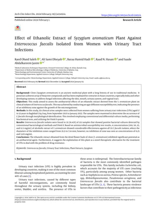
From Table 4, it is evident that the effect of the
ethanolic extract of S. aromaticum varies according to
the concentrations tested. The highest effectiveness
was observed at a concentration of 100 mg/mL against
the bacteria isolated during the current study. This
concentration resulted in an inhibition zone diameter
6 Arch Clin Infect Dis. 2024; 19(1):e134924.
7. Saleh RO et al.
Table 4. Numbers and Percentages of Enterococcus faecalis Isolates Inhibited by Different Concentrations of Clove Extract
Concentrations of Clove Extract
(mg/mL)
Isolates; No. (%)
Inhibition Zone Range (mm)
Inhibition Zone No Inhibition Zone
100 35 (100) 0 (0) 21.13 - 19.65
50 35 (100) 0 (0) 17.89 - 14.27
25 35 (100) 0 (0) 13.36 - 12.52
12.5 35 (100) 0 (0) 8.26 - 7.14
6.25 0 (0) 35 (100) -
3.125 0 (0) 35 (100) -
Figure 4. The impact of various concentrations of clove plant extract on Enterococcus faecalis isolates. The right photo displays the effects of concentrations of 100, 50, and 25
mg/mL, while the left photo shows the effects of concentrations of 12.5, 6.25, and 3.125 mg/mL. The bacterial growth on Moller Hinton agar after 24 h of incubation at 37°C is
depicted. The control group was treated with the solvent dimethyl sulfoxide.
rangingfrom21.13to19.65mm, followedbyconcentrations
of 50, 25, and 12.5 mg/mL, respectively. However,
the concentrations of 6.25 and 3.125 mg/mL had no
anti-bacterial activity, and no inhibition zone was
revealed. Moreover, the results showed that the sensitivity
of bacteria isolates to the clove extract was greater or
similar to their sensitivity to some antibiotics. This can be
explained by 2 possible mechanisms. First, it is possible
that the bacteria tested were not previously exposed to
these extracts, making them more susceptible to their
antimicrobial properties. In this case, the bacteria may
not have developed resistance mechanisms against the
specific compounds present in the extract. Second, S.
aromaticum may have a chemical affinity for interacting
with specific components of the bacterial cells. There
could be receptors or transporters on the bacterial cell
wall that facilitate the entry of the extract into the cell.
Once inside, the extract may interfere with the action of
active enzymes or other biological molecules, leading to
the inhibition of bacterial growth (25).
5. Conclusions
The ethanolic extract derived from the aromatic
dried flower buds of clove (S. aromaticum) showed great
potential as an effective antibacterial agent. Additionally,
this study suggests that the use of this plant could be
a promising therapeutic option for addressing UTIs,
especially in the context of increasing concerns about
drug resistance.
Arch Clin Infect Dis. 2024; 19(1):e134924. 7
8. Saleh RO et al.
Acknowledgments
We extend our sincere gratitude to the Medical
Laboratory Techniques Department at Al-Maarif University
College for Training in Anbar, Iraq, for their generous
support of this research.
Footnotes
Authors’ Contribution: Raed Obaid Saleh: Original draft
preparation. Ali Sami Dheyab: Reviewing and editing
and supervisor. Baraa Hamid Hadi: Conceptualization
and methodology. Raad N. Hasan: Visualization
andinvestigation. Saade Abdalkareem Jasim: Validation,
writing, and submission.
Conflict of Interests: The authors had no conflicts of
interest to declare in relation to this article.
Data Availability: The dataset presented in the study
is available on request from the corresponding author
during submission or after publication.
Ethical Approval: The Ethical approvals were obtained
from the Scientific Research Ethics Committee of the
Al-Maarif University College (No. 433 on 5/10/2021).
Funding/Support: This project was not financially
supported.
References
1. Ganesh R, Shrestha D, Bhattachan B, Rai G. Epidemiology of urinary
tract infection and antimicrobial resistance in a pediatric hospital in
Nepal. BMC Infect Dis. 2019;19(1):420. [PubMed ID: 31088380]. [PubMed
Central ID: PMC6518643]. https://doi.org/10.1186/s12879-019-3997-0.
2. Belete MA, Saravanan M. A Systematic Review on Drug Resistant
Urinary Tract Infection Among Pregnant Women in Developing
Countries in Africa and Asia; 2005-2016. Infect Drug Resist.
2020;13:1465–77. [PubMed ID: 32547115]. [PubMed Central ID:
PMC7245001]. https://doi.org/10.2147/IDR.S250654.
3. Li M, Yang F, Lu Y, Huang W. Identification of Enterococcus
faecalis in a patient with urinary-tract infection based on
metagenomic next-generation sequencing: a case report. BMC
Infect Dis. 2020;20(1):467. [PubMed ID: 32615925]. [PubMed Central ID:
PMC7330266]. https://doi.org/10.1186/s12879-020-05179-0.
4. Elamary RB, Albarakaty FM, Salem WM. Efficacy of Acacia nilotica
aqueous extract in treating biofilm-forming and multidrug resistant
uropathogens isolated from patients with UTI syndrome. Sci
Rep. 2020;10(1):11125. [PubMed ID: 32636429]. [PubMed Central ID:
PMC7341837]. https://doi.org/10.1038/s41598-020-67732-w.
5. Arias CA, Murray BE. The rise of the Enterococcus: beyond
vancomycin resistance. Nat Rev Microbiol. 2012;10(4):266–78.
[PubMed ID: 22421879]. [PubMed Central ID: PMC3621121].
https://doi.org/10.1038/nrmicro2761.
6. Adebiyi AO. Secondary Metabolites and bioactivities of two species of
Nephrolepis in Ekiti State, Nigeria. Int J Life Sci. 2019;8(3):95–9.
7. Liu C, Huang H, Zhou Q, Liu B, Wang Y, Li P, et al. Antibacterial and
antibiotic synergistic activities of the extract from Pithecellobium
clypearia against clinically important multidrug-resistant
gram-negative bacteria. Eur J Integr Med. 2019;32:100999.
https://doi.org/10.1016/j.eujim.2019.100999.
8. Jafri H, Ahmad I. In Vitro Efficacy of Clove Oil and Eugenol
against Staphylococcus spp and Streptococcus mutans on
Hydrophobicity, Hemolysin Production and Biofilms and
their Synergy with Antibiotics. Adv Microbiol. 2021;11(2):117–43.
https://doi.org/10.4236/aim.2021.112009.
9. Nandy S, Mukherjee A, Pandey DK, Ray P, Dey A. Indian
Sarsaparilla (Hemidesmus indicus): Recent progress in
research on ethnobotany, phytochemistry and pharmacology.
J Ethnopharmacol. 2020;254:112609. [PubMed ID: 32007632].
https://doi.org/10.1016/j.jep.2020.112609.
10. Wanger A, Chávez V, Huang RSP, Wahed A, Actor J, Dasgupta A.
Microbiology and Molecular Diagnosis in Pathology: A Comprehensive
Review for Board Preparation, Certification and Clinical Practice. Elsevier;
2017.
11. Ahmed O, Mohamed H, Salem W, Afifi M, Song Y. Efficacy of
Ethanolic Extract of Syzygium aromaticum in the Treatment
of Multidrug-Resistant Pseudomonas aeruginosa Clinical
Isolates Associated with Urinary Tract Infections. Evid Based
Complement Alternat Med. 2021;2021. [PubMed ID: 34221080].
https://doi.org/10.1155/2021/6612058.
12. Kumari S, Moorthi S, Kalpana S. Antimicrobial activity of different
extracts of Syzygiium aromaticum (Linn.) against food borne
pathogens. Int J Curr Microbiol App Sci. 2013;2(11):30–5.
13. Gonelimali FD, Lin J, Miao W, Xuan J, Charles F, Chen M, et al.
Antimicrobial Properties and Mechanism of Action of Some Plant
Extracts Against Food Pathogens and Spoilage Microorganisms. Front
Microbiol. 2018;9:1639. [PubMed ID: 30087662]. [PubMed Central ID:
PMC6066648]. https://doi.org/10.3389/fmicb.2018.01639.
14. Daoud A, Malika D, Bakari S, Hfaiedh N, Mnafgui K, Kadri A,
et al. Assessment of polyphenol composition, antioxidant and
antimicrobialpropertiesof variousextractsof DatePalmPollen(DPP)
from two Tunisian cultivars. Arab J Chem. 2019;12(8):3075–86. https:
//doi.org/10.1016/j.arabjc.2015.07.014.
15. Goel V, Kumar D, Kumar R, Mathur P, Singh S. Community
Acquired Enterococcal Urinary Tract Infections and Antibiotic
Resistance Profile in North India. J Lab Physicians. 2016;8(1):50–4.
[PubMed ID: 27013814]. [PubMed Central ID: PMC4785767].
https://doi.org/10.4103/0974-2727.176237.
16. Ghaly MF, Shalaby MA, Shash SMS, Shehata MN, Ayad AA. Synergistic
effect of antibiotics and plant extract to control clinical bacterial
isolates implicated in urinary tract infections. J Appl Sci Res.
2009;5:1298–306.
17. AL-Jmor SA. Detaction of some virulence factors of Vancomycin-resistant
Enterococcus faecalis and the effect of Punica granatum and Thuja
orientalis extracts on it. Baghdad University; 2012.
18. Hussain A, Sohail M, Abbas Z. Prevalence of Enterococcus faecalis
mediated UTI and its current antimicrobial susceptibility pattern in
Lahore, Pakistan. J Pak Med Assoc. 2016;66(10):1232.
19. Alebouyeh M, Mozafar NA, Forouhesh H. Evaluation of virulence
factors and plasmid-related transmissibility among different Isolates
of Enterococci. Iran Biomed J. 2005;9(2):51–5.
20. Hasson AH, Kadhem SA. Identification of Enterococcus faecalis
Isolated from Infected Human Tooth Root Canals Human by Using
Polymerase Chain Reaction. Ibn Al-Haitham J Pure Appl Sci. 2015;28(2).
21. Kadhem HS, Flayyih MT. Isolation and Identification of
Vancomycin-Resistant Enterococcus Faecalis. Iraq J Sci.
2014;55(4B):1811–6.
22. Cetinkaya Y, Falk P, Mayhall CG. Vancomycin-resistant enterococci.
Clin Microbiol Rev. 2000;13(4):686–707. [PubMed ID: 11023964].
[PubMed Central ID: PMC88957]. https://doi.org/10.1128/CMR.13.4.686.
23. Praharaj I, Sujatha S, Parija SC. Phenotypic & genotypic
characterization of vancomycin resistant Enterococcus isolates
from clinical specimens. India J Med Res. 2013;138(4):549.
8 Arch Clin Infect Dis. 2024; 19(1):e134924.
9. Saleh RO et al.
24. Nguyen M, Joshi SG. Carbapenem resistance in Acinetobacter
baumannii, and their importance in hospital-acquired
infections: a scientific review. J Appl Microbiol. 2021;131(6):2715–38.
[PubMed ID: 33971055]. https://doi.org/10.1111/jam.15130.
25. Hancock RE, Wong PG. Compounds which increase the permeability
of the Pseudomonas aeruginosa outer membrane. Antimicrob Agents
Chemother. 1984;26(1):48–52. [PubMed ID: 6433788]. [PubMed Central
ID: PMC179915]. https://doi.org/10.1128/AAC.26.1.48.
Arch Clin Infect Dis. 2024; 19(1):e134924. 9