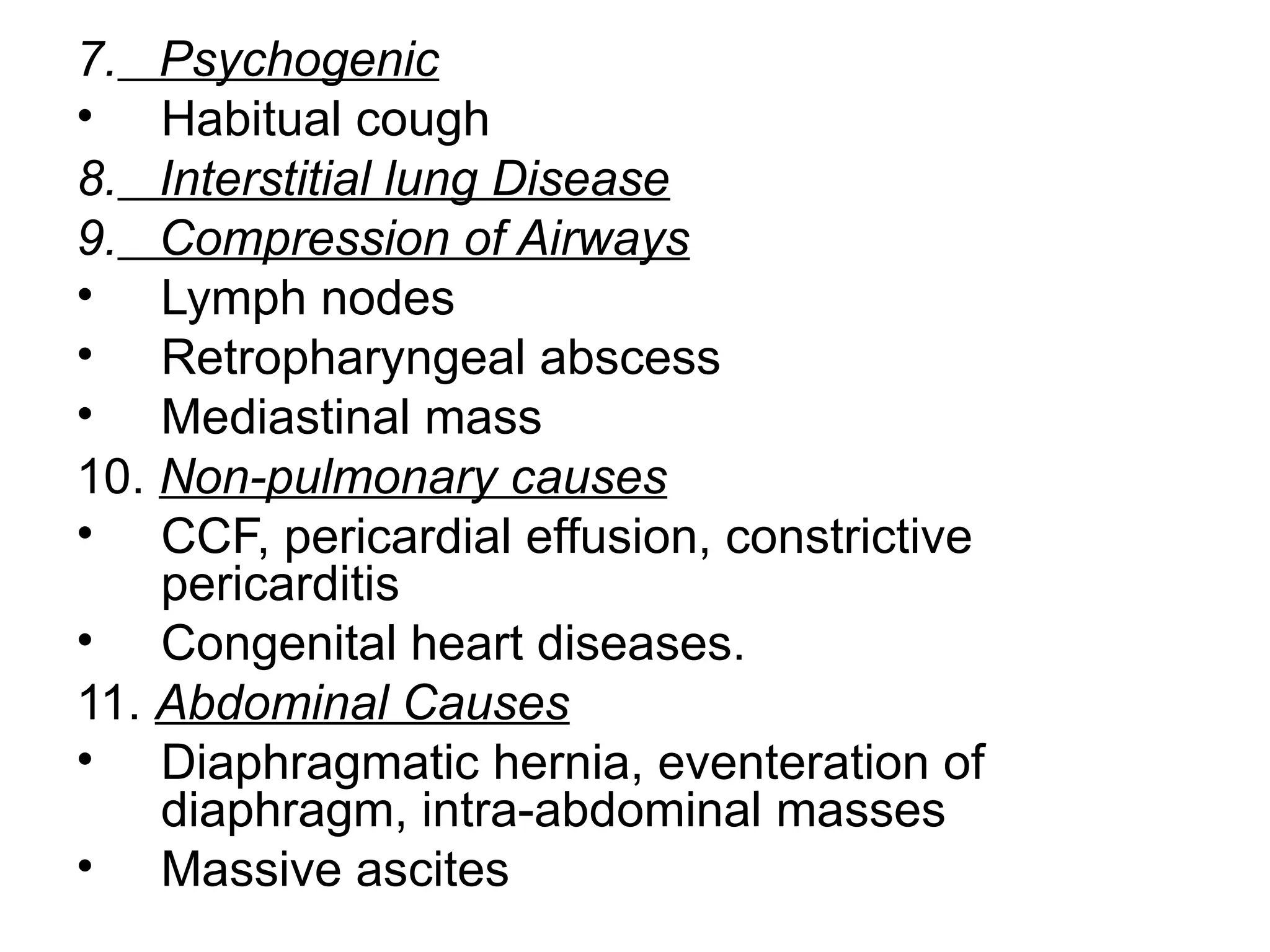
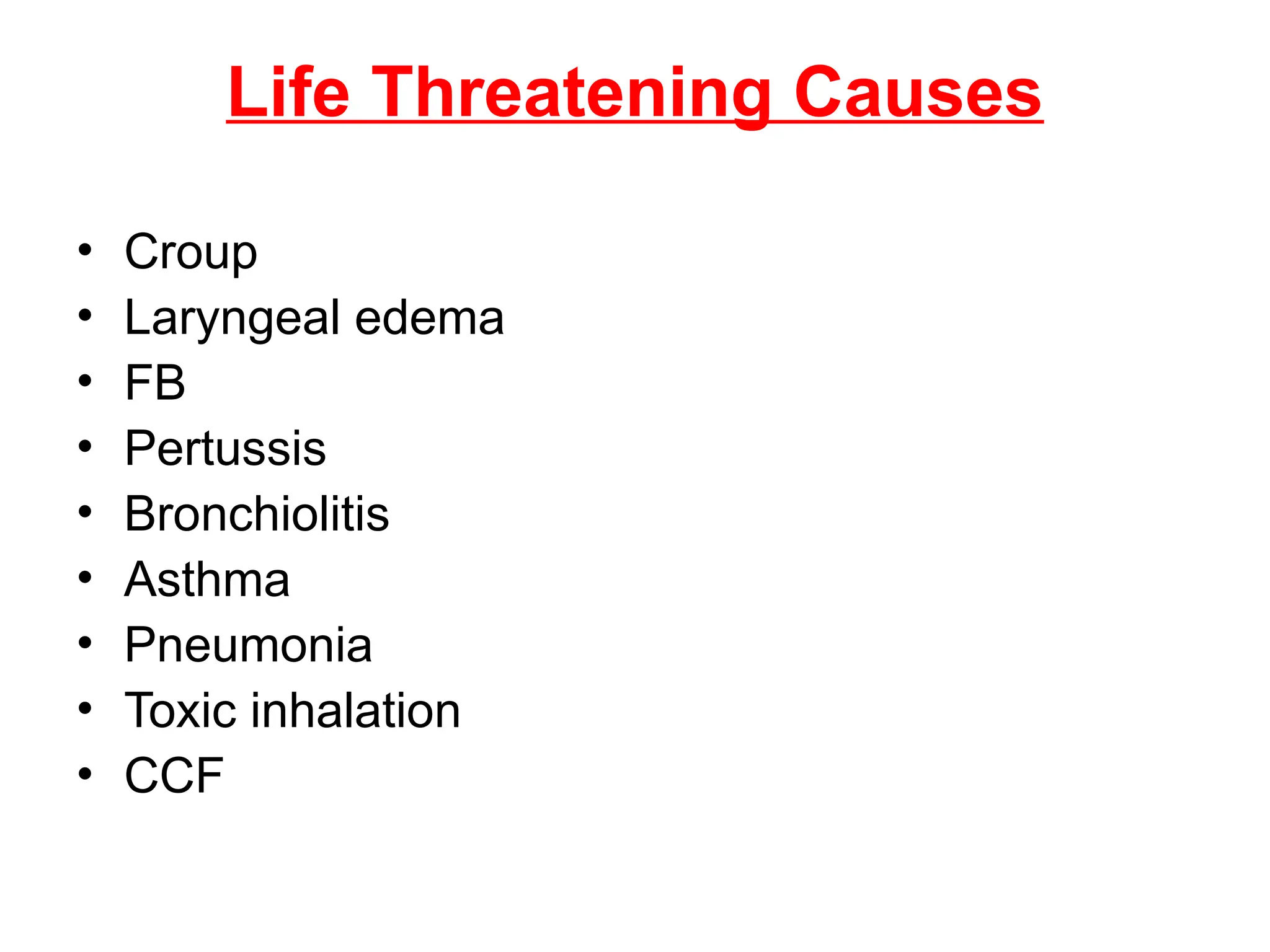
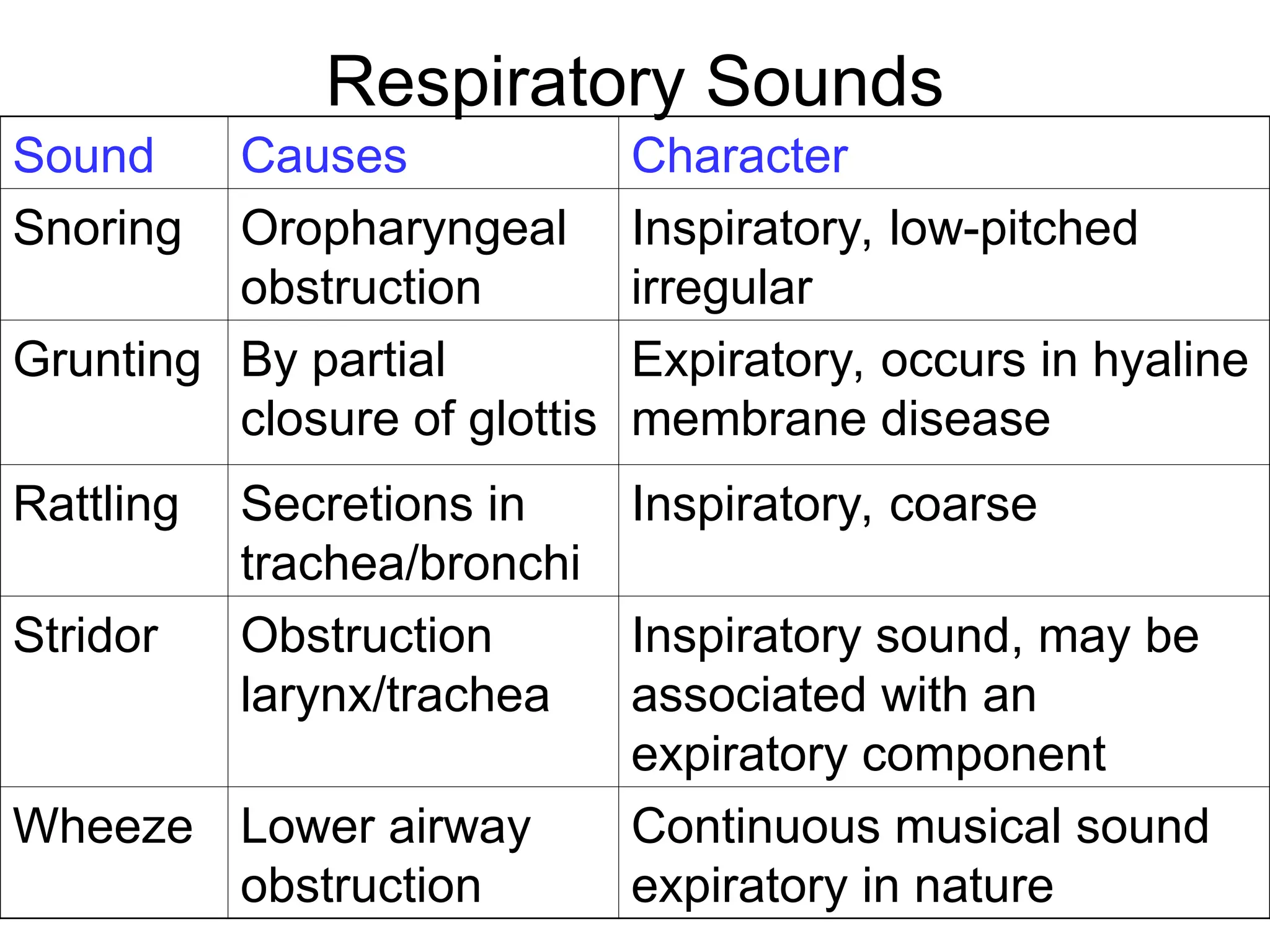
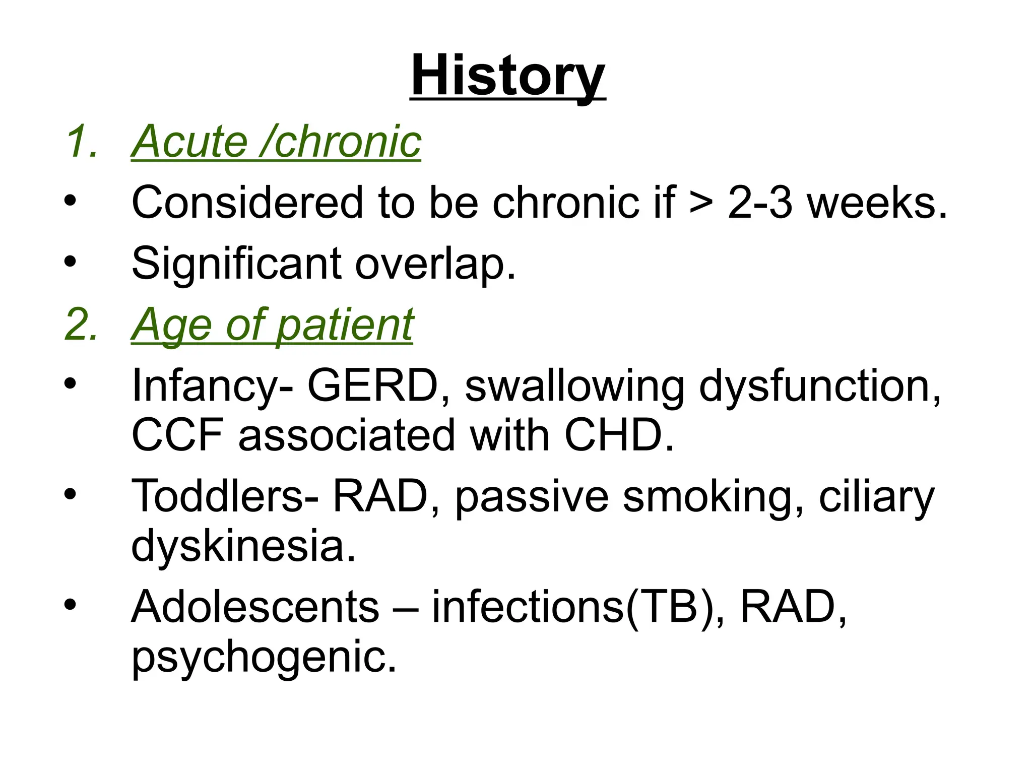
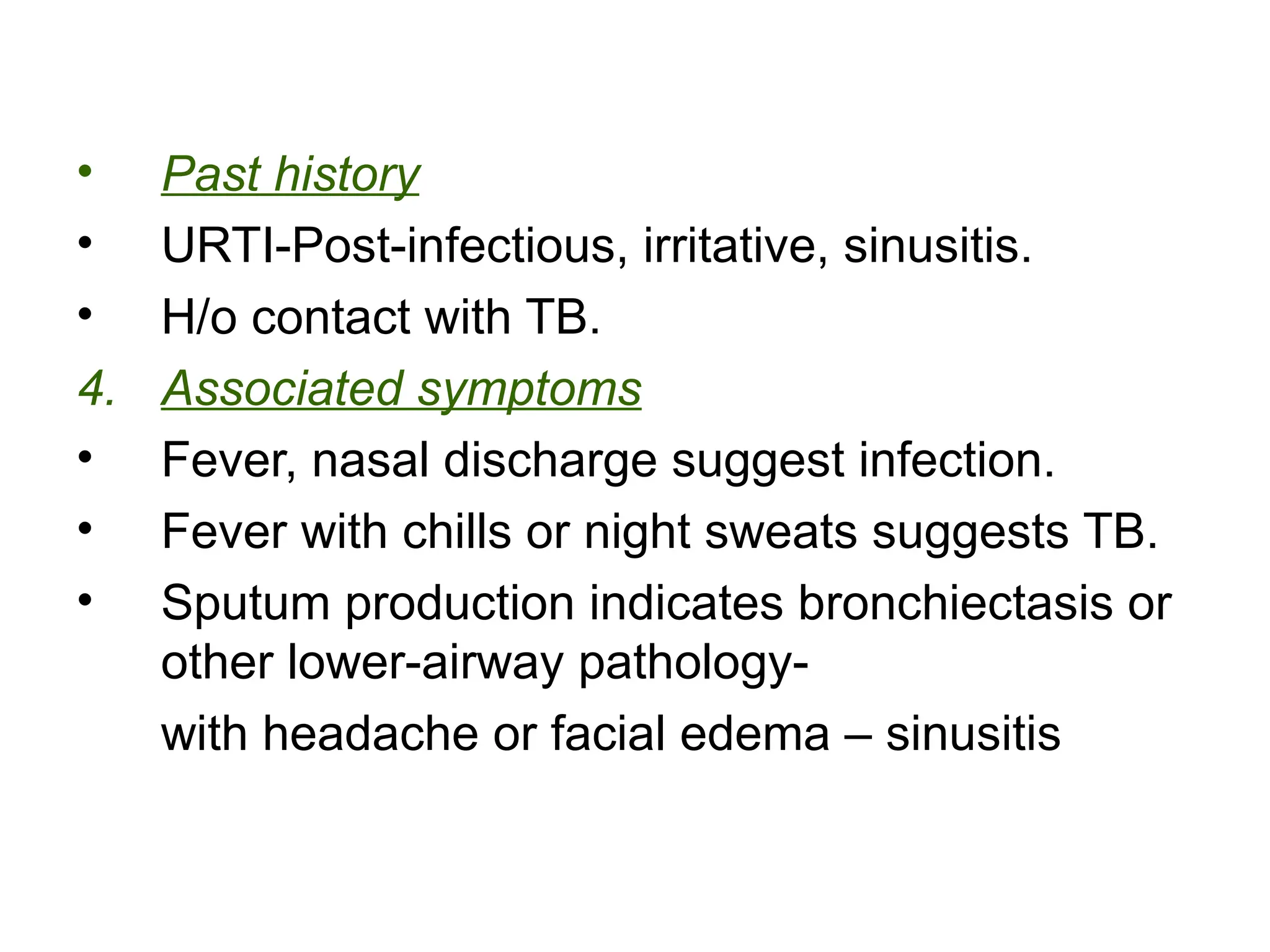
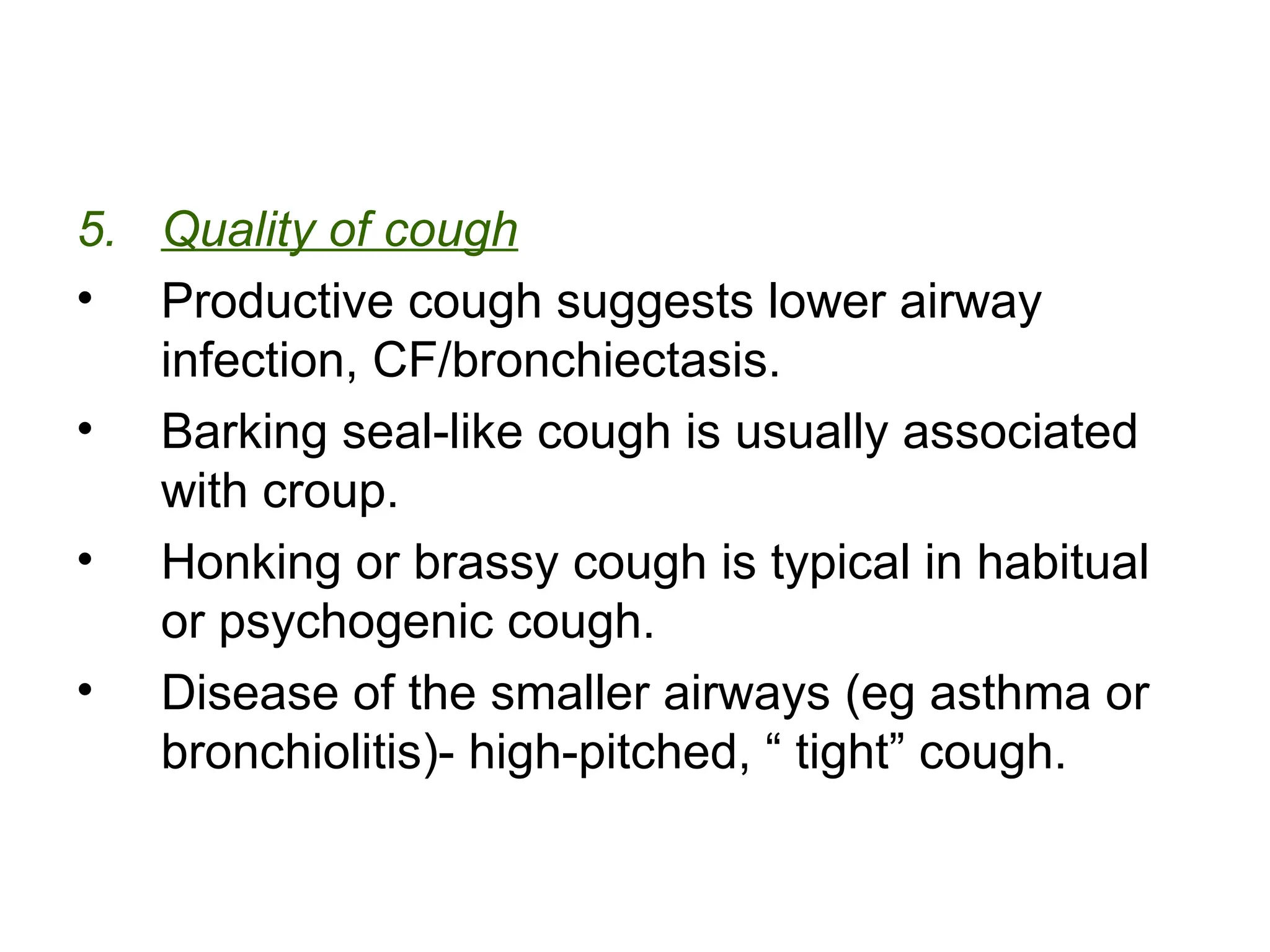
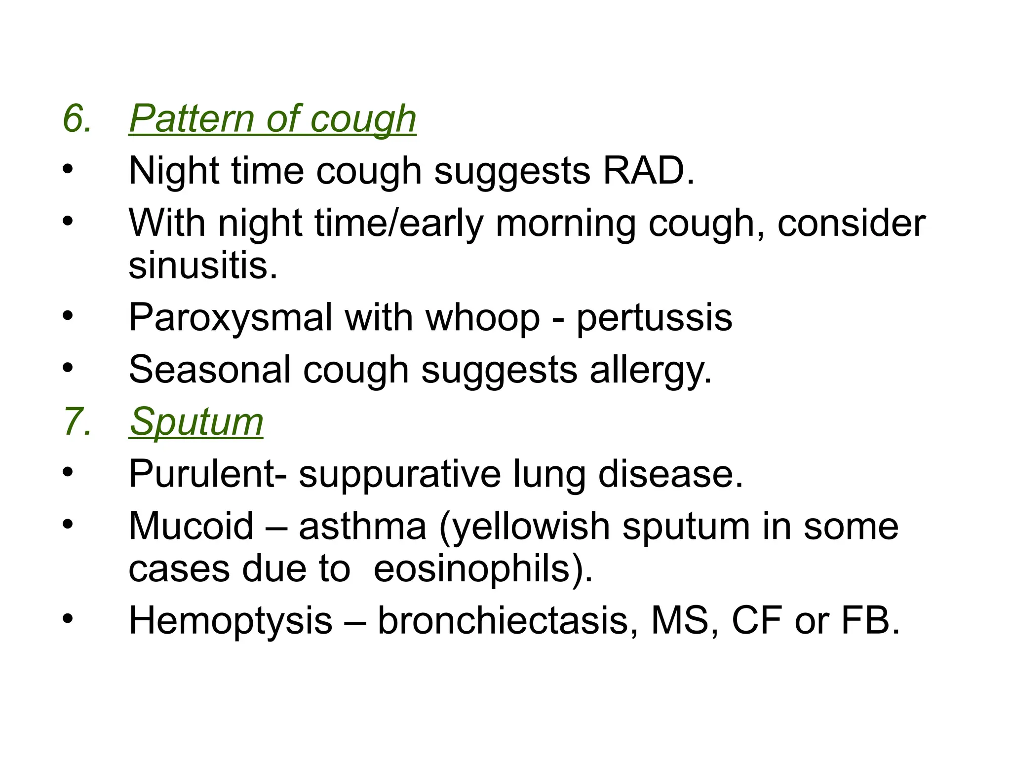
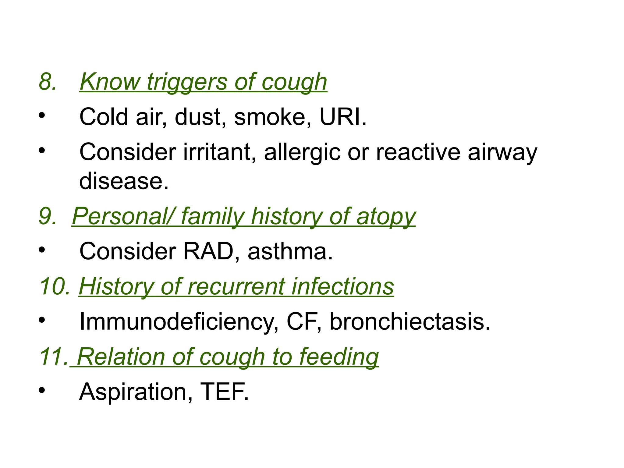
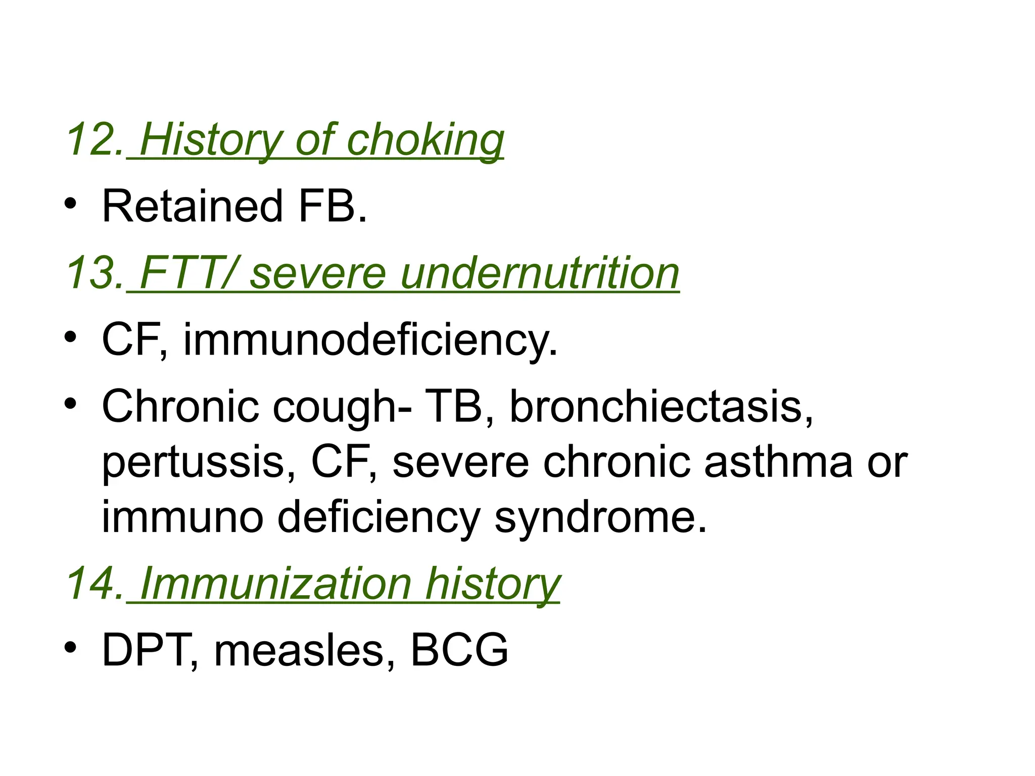
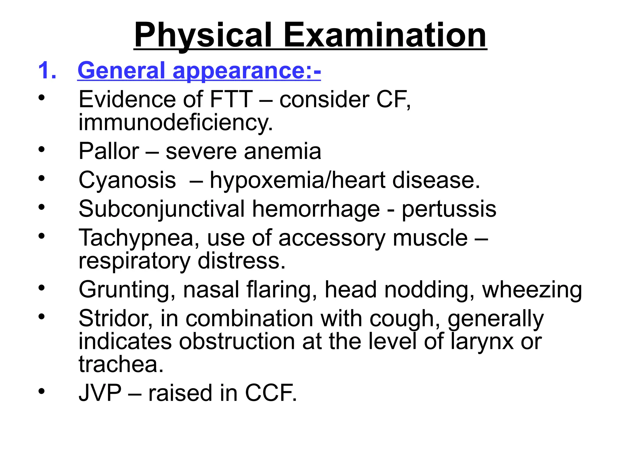
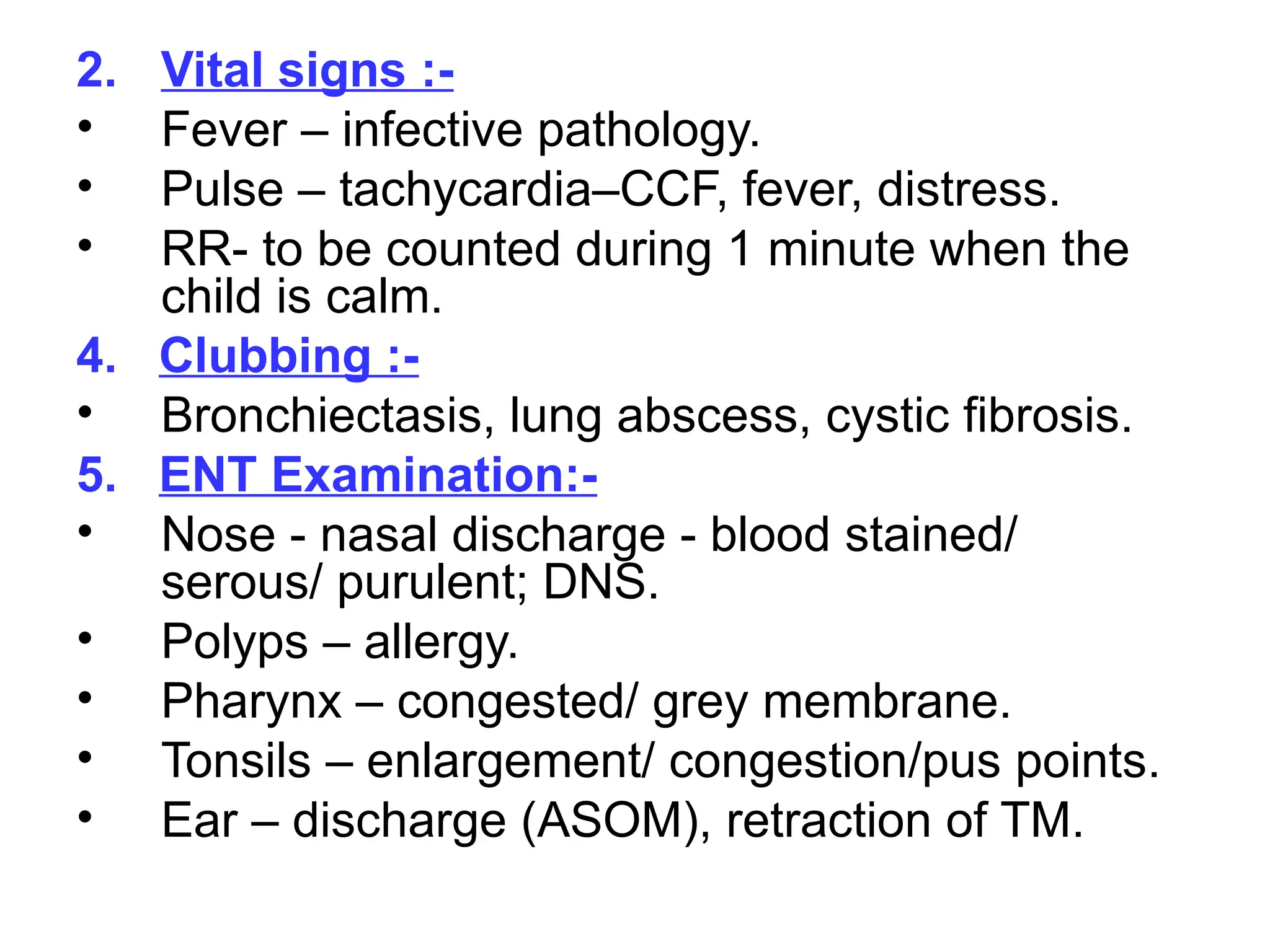
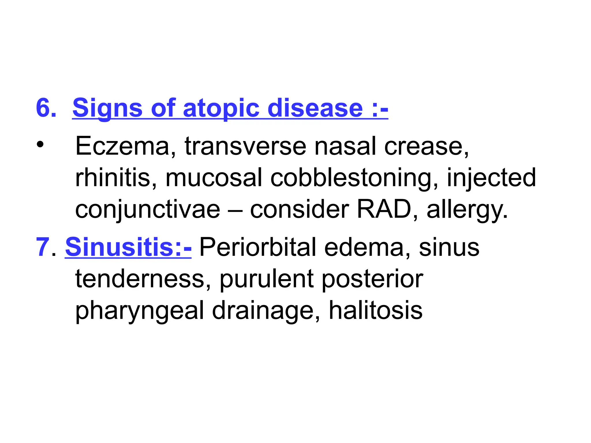
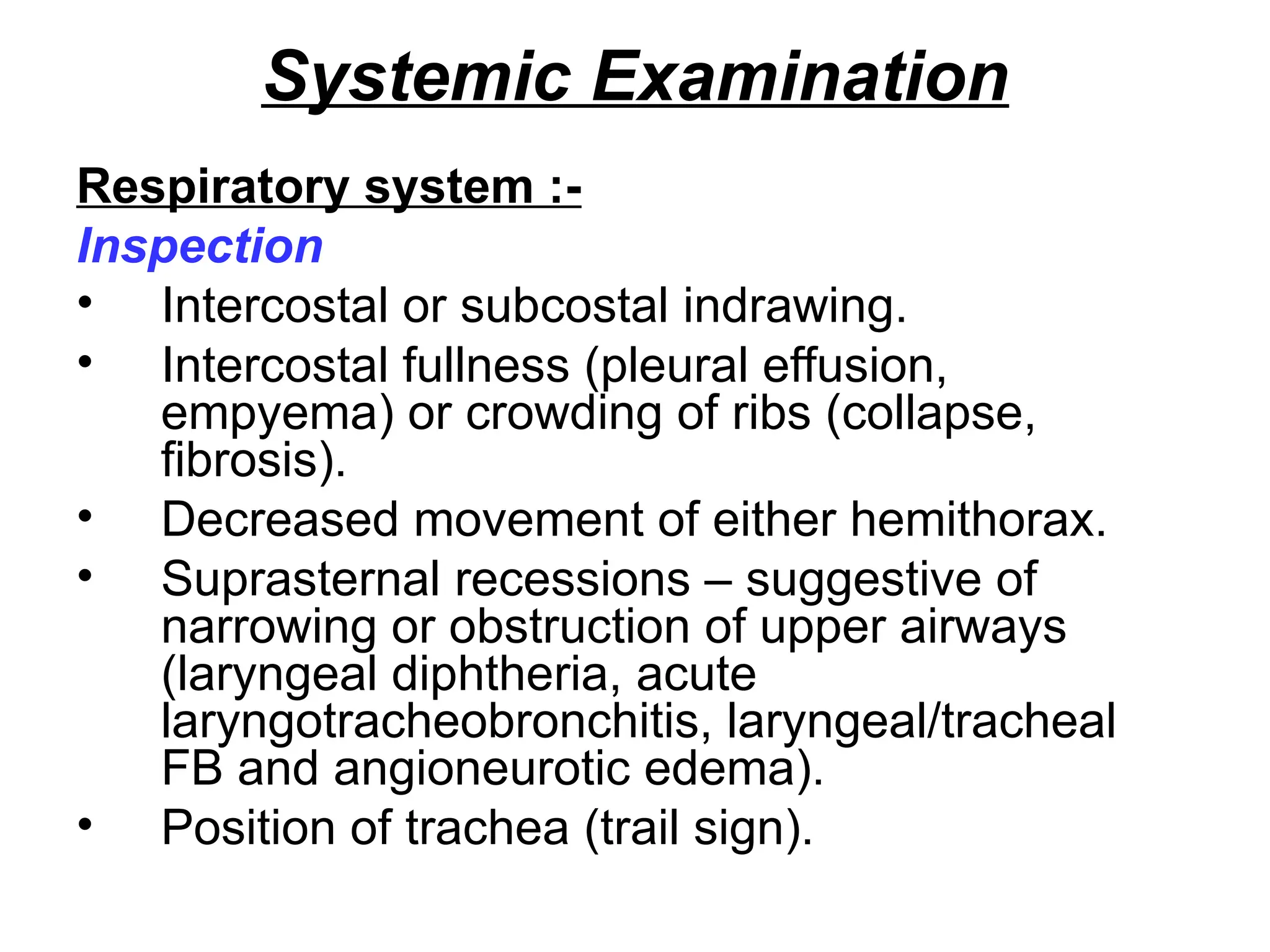
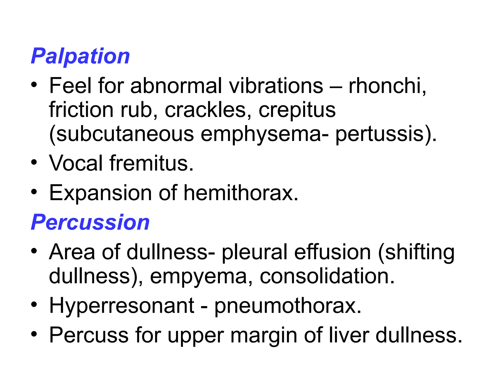
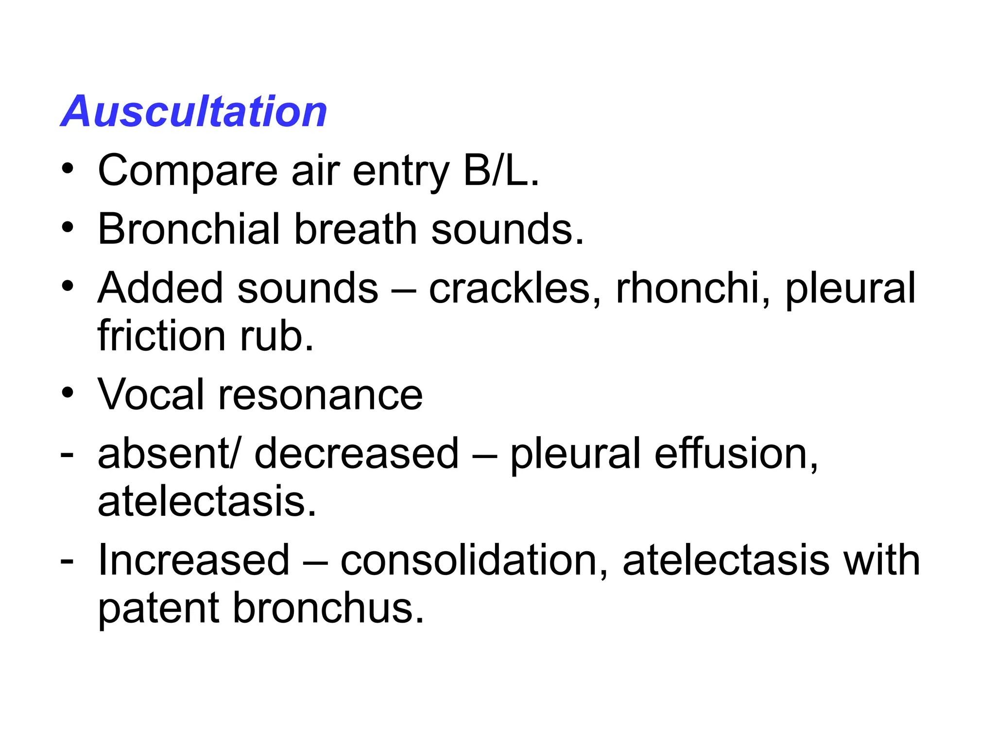
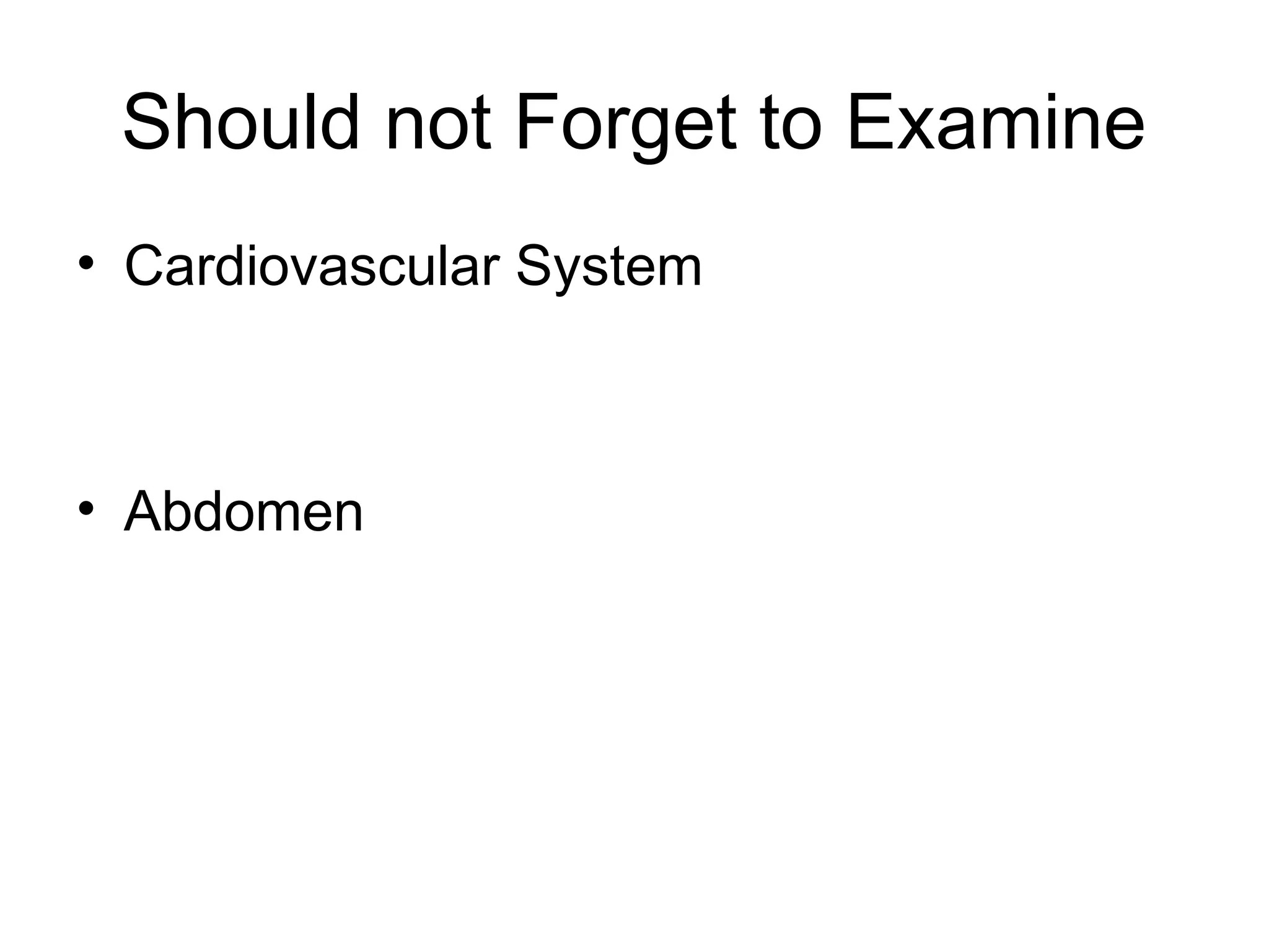
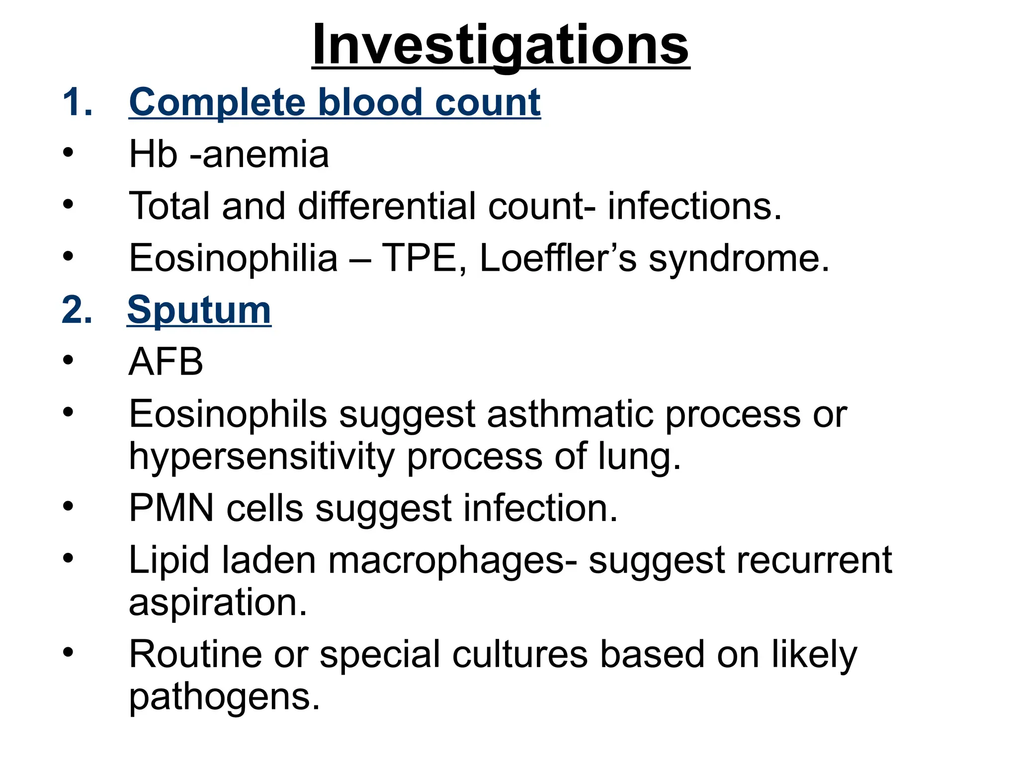
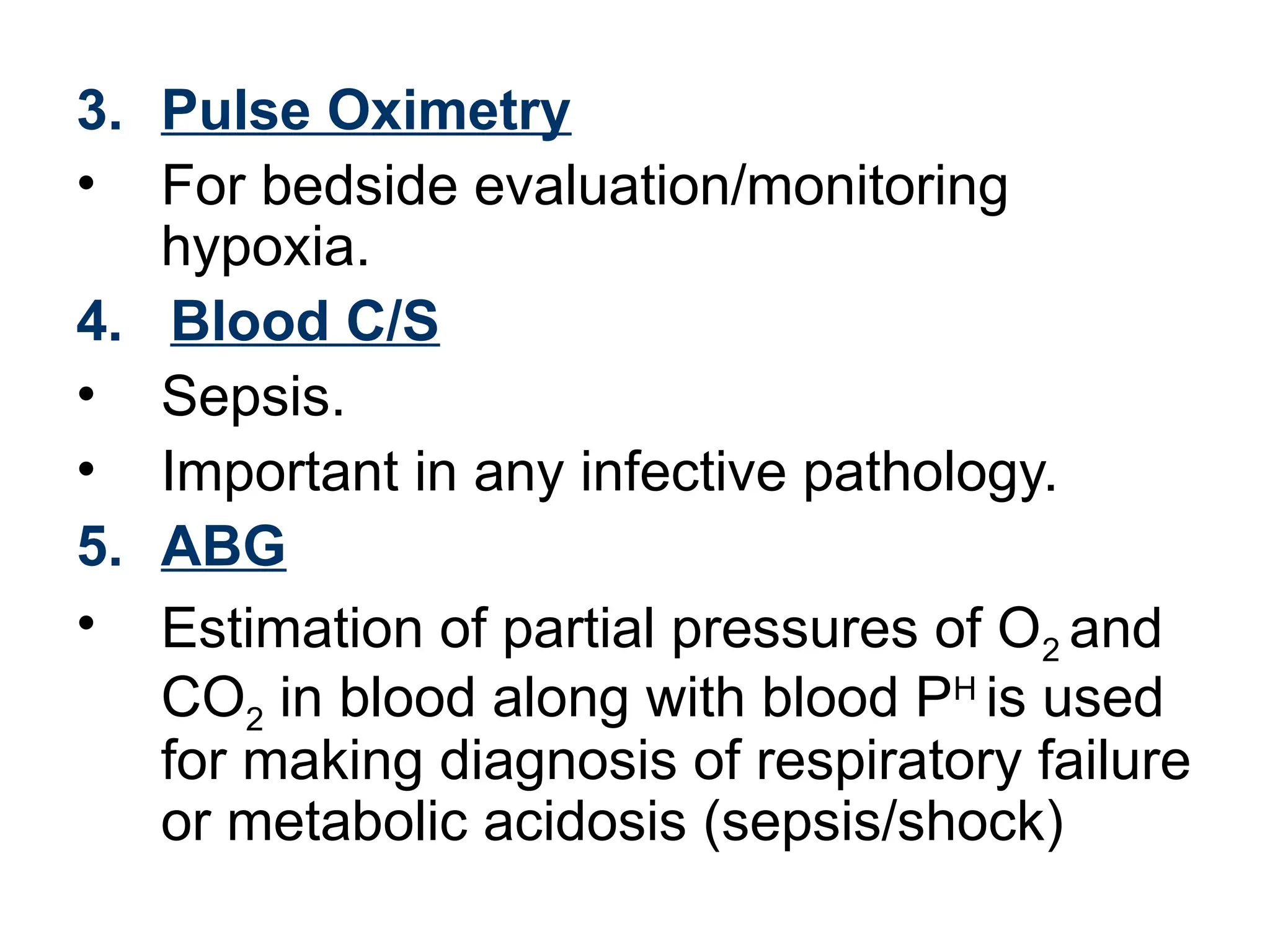
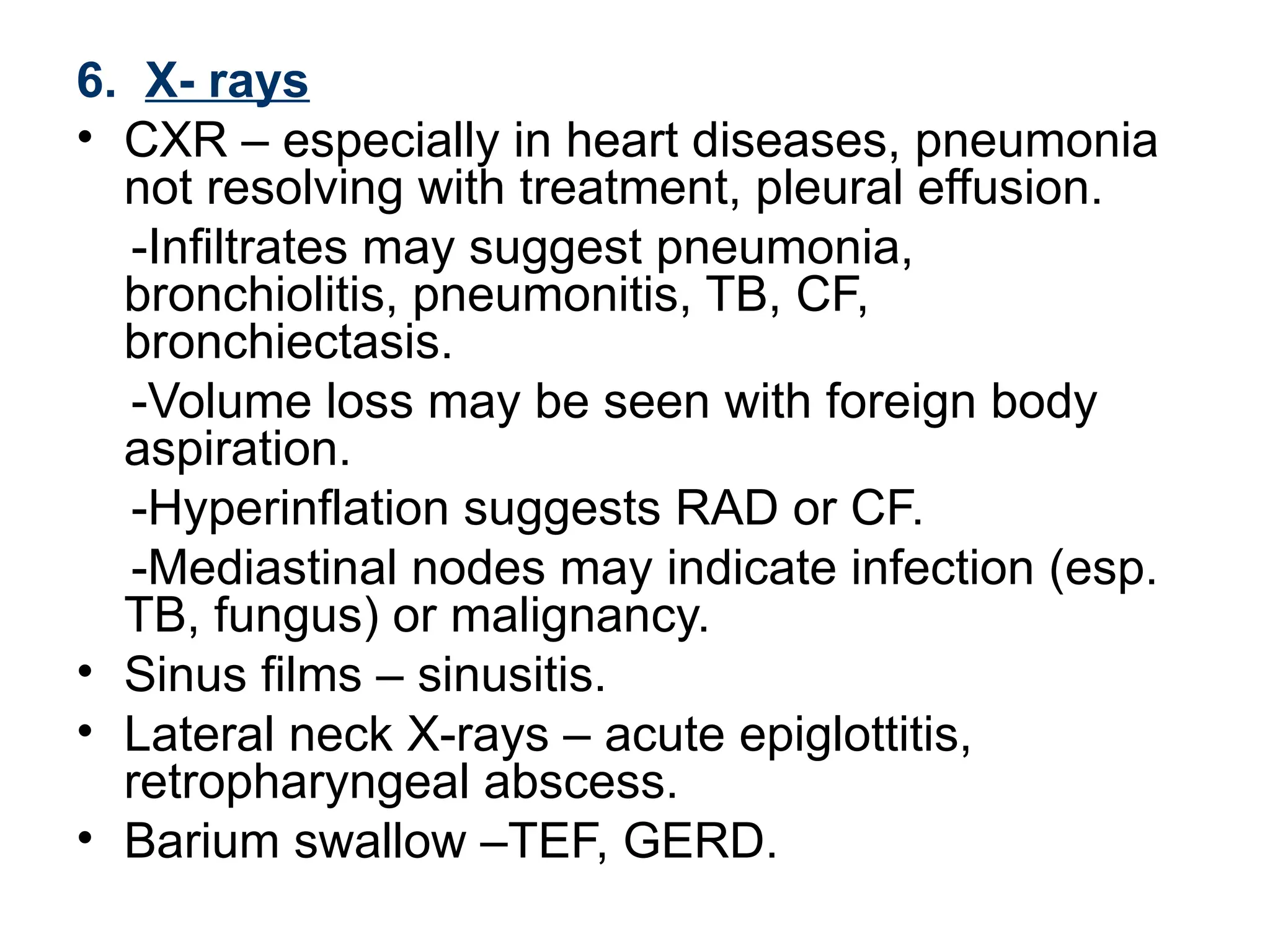
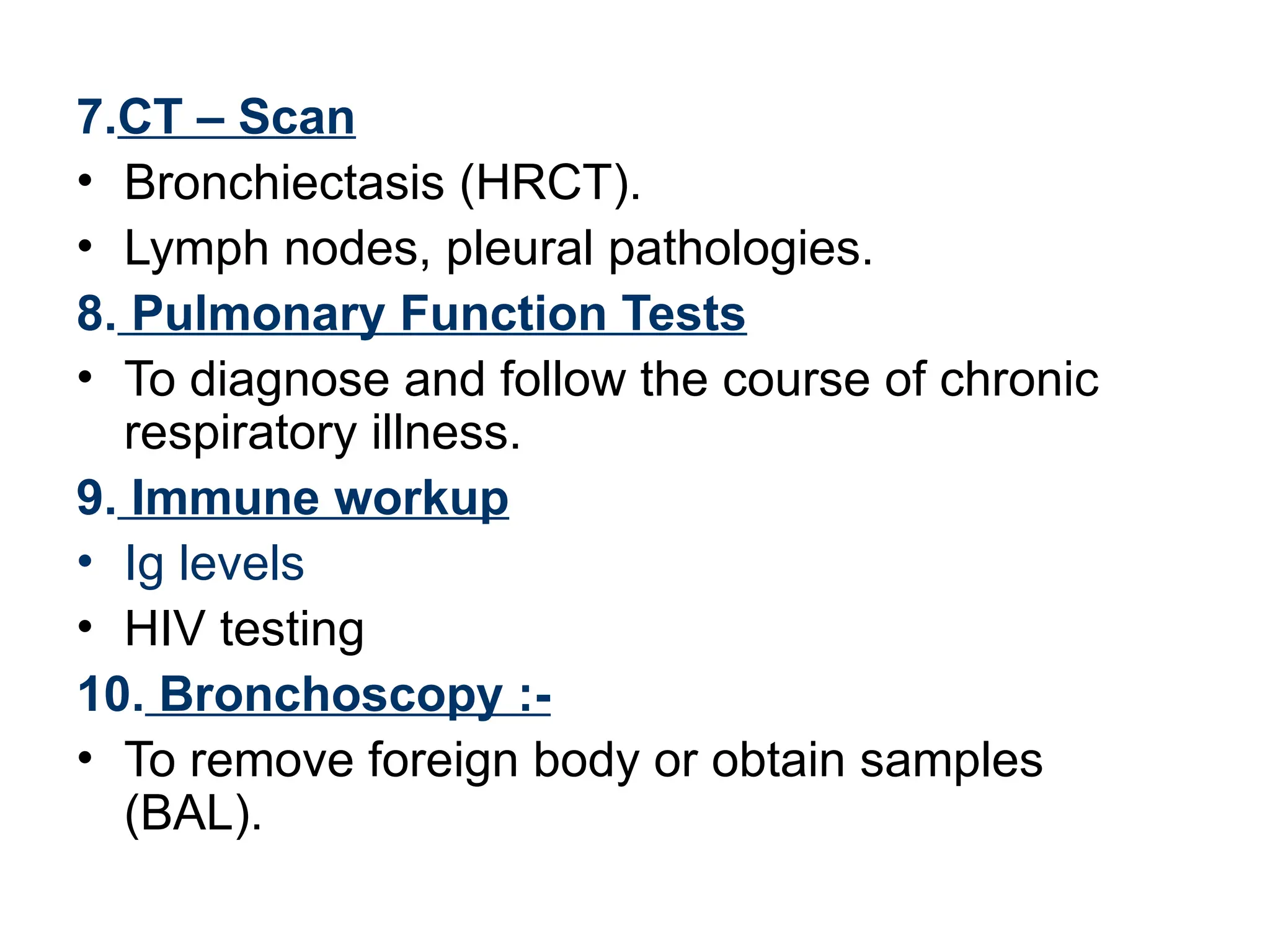
PATHOPHYSIOLOGY
• Occurs primarily as a reflex designed to clear the airway of mucus
and foreign material.
• Elicited by stimulation of receptors located throughout the
respiratory tract, from the pharynx to terminal bronchioles.
• Triggered by various inflammatory, mechanical, chemical and
thermal stimuli.
• Cough reflex (sequence of events )
Deep inspiration→closure of glottis →relaxation of diaphragm
→contraction of expiratory muscles →sudden opening of glottis
→increase in intrathoracic pressure, produce a rapid expulsion of air
through the glottis, which propels mucus and particulate matter out
of the airway
CAUSES
1. Infective Disorders of Airway
• Common cold
• Sinusitis (post-nasal discharge)
• Tonsillitis, Pharyngitis, Laryngitis
• Laryngotracheobronchitis, Bronchiolitis
• Pneumonia
• Measles, whooping cough
2. Inflammatory Disorders of Airway
• Asthma and Loeffler’s syndrome, TPE
• Inhalation of environmental irritants like tobacco, smoke dust
3. Pleural pathology
• Pleural effusion
• Empyema
4. Suppurative Lung Disease
• Bronchiectasis
• Cystic fibrosis
• Foreign body retained in bronchi
• Lung abscess
5.Anatomic Lesions
• Congenital malformations, Sequestrated lobe
• Bronchomalacia, Tumors
• Tracheal stenosis, H-type Tracheo Esophageal Fistula
• Vascular ring
• Tracheomalacia
6. Irritative
• Post-nasal discharge
• Sinusitis
• Gastro Esophageal Reflux Disease
• Irritation of External auditory meatus
7. Psychogenic
• Habitual cough
8. Interstitial lung Disease
9. Compression of Airways
• Lymph nodes
• Retropharyngeal abscess
• Mediastinal mass
10. Non-pulmonary causes
• CCF, pericardial effusion, constrictive
pericarditis
• Congenital heart diseases.
11. Abdominal Causes
• Diaphragmatic hernia, eventeration of
diaphragm, intra-abdominal masses
• Massive ascites
Life Threatening Causes
• Croup
• Laryngeal edema
• FB
• Pertussis
• Bronchiolitis
• Asthma
• Pneumonia
• Toxic inhalation
• CCF
Respiratory Sounds
Sound Causes Character
Snoring Oropharyngeal
obstruction
Inspiratory, low-pitched
irregular
Grunting By partial
closure of glottis
Expiratory, occurs in hyaline
membrane disease
Rattling Secretions in
trachea/bronchi
Inspiratory, coarse
Stridor Obstruction
larynx/trachea
Inspiratory sound, may be
associated with an
expiratory component
Wheeze Lower airway
obstruction
Continuous musical sound
expiratory in nature
History
1. Acute /chronic
• Considered to be chronic if > 2-3 weeks.
• Significant overlap.
2. Age of patient
• Infancy- GERD, swallowing dysfunction,
CCF associated with CHD.
• Toddlers- RAD, passive smoking, ciliary
dyskinesia.
• Adolescents – infections(TB), RAD,
psychogenic.
• Past history
• URTI-Post-infectious, irritative, sinusitis.
• H/o contact with TB.
4. Associated symptoms
• Fever, nasal discharge suggest infection.
• Fever with chills or night sweats suggests TB.
• Sputum production indicates bronchiectasis or
other lower-airway pathology-
with headache or facial edema – sinusitis
5. Quality of cough
• Productive cough suggests lower airway
infection, CF/bronchiectasis.
• Barking seal-like cough is usually associated
with croup.
• Honking or brassy cough is typical in habitual
or psychogenic cough.
• Disease of the smaller airways (eg asthma or
bronchiolitis)- high-pitched, “ tight” cough.
6. Pattern of cough
• Night time cough suggests RAD.
• With night time/early morning cough, consider
sinusitis.
• Paroxysmal with whoop - pertussis
• Seasonal cough suggests allergy.
7. Sputum
• Purulent- suppurative lung disease.
• Mucoid – asthma (yellowish sputum in some
cases due to eosinophils).
• Hemoptysis – bronchiectasis, MS, CF or FB.
8. Know triggers of cough
• Cold air, dust, smoke, URI.
• Consider irritant, allergic or reactive airway
disease.
9. Personal/ family history of atopy
• Consider RAD, asthma.
10. History of recurrent infections
• Immunodeficiency, CF, bronchiectasis.
11. Relation of cough to feeding
• Aspiration, TEF.
12. History of choking
• Retained FB.
13. FTT/ severe undernutrition
• CF, immunodeficiency.
• Chronic cough- TB, bronchiectasis,
pertussis, CF, severe chronic asthma or
immuno deficiency syndrome.
14. Immunization history
• DPT, measles, BCG
Physical Examination
1. General appearance:-
• Evidence of FTT – consider CF,
immunodeficiency.
• Pallor – severe anemia
• Cyanosis – hypoxemia/heart disease.
• Subconjunctival hemorrhage - pertussis
• Tachypnea, use of accessory muscle –
respiratory distress.
• Grunting, nasal flaring, head nodding, wheezing
• Stridor, in combination with cough, generally
indicates obstruction at the level of larynx or
trachea.
• JVP – raised in CCF.
2. Vital signs :-
• Fever – infective pathology.
• Pulse – tachycardia–CCF, fever, distress.
• RR- to be counted during 1 minute when the
child is calm.
4. Clubbing :-
• Bronchiectasis, lung abscess, cystic fibrosis.
5. ENT Examination:-
• Nose - nasal discharge - blood stained/
serous/ purulent; DNS.
• Polyps – allergy.
• Pharynx – congested/ grey membrane.
• Tonsils – enlargement/ congestion/pus points.
• Ear – discharge (ASOM), retraction of TM.
6. Signs of atopic disease :-
• Eczema, transverse nasal crease,
rhinitis, mucosal cobblestoning, injected
conjunctivae – consider RAD, allergy.
7. Sinusitis:- Periorbital edema, sinus
tenderness, purulent posterior
pharyngeal drainage, halitosis
Systemic Examination
Respiratory system :-
Inspection
• Intercostal or subcostal indrawing.
• Intercostal fullness (pleural effusion,
empyema) or crowding of ribs (collapse,
fibrosis).
• Decreased movement of either hemithorax.
• Suprasternal recessions – suggestive of
narrowing or obstruction of upper airways
(laryngeal diphtheria, acute
laryngotracheobronchitis, laryngeal/tracheal
FB and angioneurotic edema).
• Position of trachea (trail sign).
Palpation
• Feel for abnormal vibrations – rhonchi,
friction rub, crackles, crepitus
(subcutaneous emphysema- pertussis).
• Vocal fremitus.
• Expansion of hemithorax.
Percussion
• Area of dullness- pleural effusion (shifting
dullness), empyema, consolidation.
• Hyperresonant - pneumothorax.
• Percuss for upper margin of liver dullness.
Auscultation
• Compare air entry B/L.
• Bronchial breath sounds.
• Added sounds – crackles, rhonchi, pleural
friction rub.
• Vocal resonance
- absent/ decreased – pleural effusion,
atelectasis.
- Increased – consolidation, atelectasis with
patent bronchus.
Should not Forget to Examine
• Cardiovascular System
• Abdomen
Investigations
1. Complete blood count
• Hb -anemia
• Total and differential count- infections.
• Eosinophilia – TPE, Loeffler’s syndrome.
2. Sputum
• AFB
• Eosinophils suggest asthmatic process or
hypersensitivity process of lung.
• PMN cells suggest infection.
• Lipid laden macrophages- suggest recurrent
aspiration.
• Routine or special cultures based on likely
pathogens.
3. Pulse Oximetry
• For bedside evaluation/monitoring
hypoxia.
4. Blood C/S
• Sepsis.
• Important in any infective pathology.
5. ABG
• Estimation of partial pressures of O2 and
CO2 in blood along with blood PH
is used
for making diagnosis of respiratory failure
or metabolic acidosis (sepsis/shock)
6. X- rays
• CXR – especially in heart diseases, pneumonia
not resolving with treatment, pleural effusion.
-Infiltrates may suggest pneumonia,
bronchiolitis, pneumonitis, TB, CF,
bronchiectasis.
-Volume loss may be seen with foreign body
aspiration.
-Hyperinflation suggests RAD or CF.
-Mediastinal nodes may indicate infection (esp.
TB, fungus) or malignancy.
• Sinus films – sinusitis.
• Lateral neck X-rays – acute epiglottitis,
retropharyngeal abscess.
• Barium swallow –TEF, GERD.
7.CT – Scan
• Bronchiectasis (HRCT).
• Lymph nodes, pleural pathologies.
8. Pulmonary Function Tests
• To diagnose and follow the course of chronic
respiratory illness.
9. Immune workup
• Ig levels
• HIV testing
10. Bronchoscopy :-
• To remove foreign body or obtain samples
(BAL).
References
1. Ghai Essential Pediatrics,5th
Edition, OP
Ghai.
2. Nelson Textbook of Pediatrics, 17th
Edition.
3. Pediatric Clinical Methods, 2nd
Edition,
Meharban Singh.
4. Management of the Child with a Serious
Infection or Severe Malnutrition, WHO.
5. Pediatric Emergency Medicine, Chapter-
14: Cough, Raymond B Karasic.
approach to persistent and recurrent cough

approach to persistent and recurrent cough

  • 2.
    PATHOPHYSIOLOGY • Occurs primarilyas a reflex designed to clear the airway of mucus and foreign material. • Elicited by stimulation of receptors located throughout the respiratory tract, from the pharynx to terminal bronchioles. • Triggered by various inflammatory, mechanical, chemical and thermal stimuli. • Cough reflex (sequence of events ) Deep inspiration→closure of glottis →relaxation of diaphragm →contraction of expiratory muscles →sudden opening of glottis →increase in intrathoracic pressure, produce a rapid expulsion of air through the glottis, which propels mucus and particulate matter out of the airway
  • 3.
    CAUSES 1. Infective Disordersof Airway • Common cold • Sinusitis (post-nasal discharge) • Tonsillitis, Pharyngitis, Laryngitis • Laryngotracheobronchitis, Bronchiolitis • Pneumonia • Measles, whooping cough 2. Inflammatory Disorders of Airway • Asthma and Loeffler’s syndrome, TPE • Inhalation of environmental irritants like tobacco, smoke dust 3. Pleural pathology • Pleural effusion • Empyema
  • 4.
    4. Suppurative LungDisease • Bronchiectasis • Cystic fibrosis • Foreign body retained in bronchi • Lung abscess 5.Anatomic Lesions • Congenital malformations, Sequestrated lobe • Bronchomalacia, Tumors • Tracheal stenosis, H-type Tracheo Esophageal Fistula • Vascular ring • Tracheomalacia 6. Irritative • Post-nasal discharge • Sinusitis • Gastro Esophageal Reflux Disease • Irritation of External auditory meatus
  • 5.
    7. Psychogenic • Habitualcough 8. Interstitial lung Disease 9. Compression of Airways • Lymph nodes • Retropharyngeal abscess • Mediastinal mass 10. Non-pulmonary causes • CCF, pericardial effusion, constrictive pericarditis • Congenital heart diseases. 11. Abdominal Causes • Diaphragmatic hernia, eventeration of diaphragm, intra-abdominal masses • Massive ascites
  • 6.
    Life Threatening Causes •Croup • Laryngeal edema • FB • Pertussis • Bronchiolitis • Asthma • Pneumonia • Toxic inhalation • CCF
  • 7.
    Respiratory Sounds Sound CausesCharacter Snoring Oropharyngeal obstruction Inspiratory, low-pitched irregular Grunting By partial closure of glottis Expiratory, occurs in hyaline membrane disease Rattling Secretions in trachea/bronchi Inspiratory, coarse Stridor Obstruction larynx/trachea Inspiratory sound, may be associated with an expiratory component Wheeze Lower airway obstruction Continuous musical sound expiratory in nature
  • 8.
    History 1. Acute /chronic •Considered to be chronic if > 2-3 weeks. • Significant overlap. 2. Age of patient • Infancy- GERD, swallowing dysfunction, CCF associated with CHD. • Toddlers- RAD, passive smoking, ciliary dyskinesia. • Adolescents – infections(TB), RAD, psychogenic.
  • 9.
    • Past history •URTI-Post-infectious, irritative, sinusitis. • H/o contact with TB. 4. Associated symptoms • Fever, nasal discharge suggest infection. • Fever with chills or night sweats suggests TB. • Sputum production indicates bronchiectasis or other lower-airway pathology- with headache or facial edema – sinusitis
  • 10.
    5. Quality ofcough • Productive cough suggests lower airway infection, CF/bronchiectasis. • Barking seal-like cough is usually associated with croup. • Honking or brassy cough is typical in habitual or psychogenic cough. • Disease of the smaller airways (eg asthma or bronchiolitis)- high-pitched, “ tight” cough.
  • 11.
    6. Pattern ofcough • Night time cough suggests RAD. • With night time/early morning cough, consider sinusitis. • Paroxysmal with whoop - pertussis • Seasonal cough suggests allergy. 7. Sputum • Purulent- suppurative lung disease. • Mucoid – asthma (yellowish sputum in some cases due to eosinophils). • Hemoptysis – bronchiectasis, MS, CF or FB.
  • 12.
    8. Know triggersof cough • Cold air, dust, smoke, URI. • Consider irritant, allergic or reactive airway disease. 9. Personal/ family history of atopy • Consider RAD, asthma. 10. History of recurrent infections • Immunodeficiency, CF, bronchiectasis. 11. Relation of cough to feeding • Aspiration, TEF.
  • 13.
    12. History ofchoking • Retained FB. 13. FTT/ severe undernutrition • CF, immunodeficiency. • Chronic cough- TB, bronchiectasis, pertussis, CF, severe chronic asthma or immuno deficiency syndrome. 14. Immunization history • DPT, measles, BCG
  • 14.
    Physical Examination 1. Generalappearance:- • Evidence of FTT – consider CF, immunodeficiency. • Pallor – severe anemia • Cyanosis – hypoxemia/heart disease. • Subconjunctival hemorrhage - pertussis • Tachypnea, use of accessory muscle – respiratory distress. • Grunting, nasal flaring, head nodding, wheezing • Stridor, in combination with cough, generally indicates obstruction at the level of larynx or trachea. • JVP – raised in CCF.
  • 15.
    2. Vital signs:- • Fever – infective pathology. • Pulse – tachycardia–CCF, fever, distress. • RR- to be counted during 1 minute when the child is calm. 4. Clubbing :- • Bronchiectasis, lung abscess, cystic fibrosis. 5. ENT Examination:- • Nose - nasal discharge - blood stained/ serous/ purulent; DNS. • Polyps – allergy. • Pharynx – congested/ grey membrane. • Tonsils – enlargement/ congestion/pus points. • Ear – discharge (ASOM), retraction of TM.
  • 16.
    6. Signs ofatopic disease :- • Eczema, transverse nasal crease, rhinitis, mucosal cobblestoning, injected conjunctivae – consider RAD, allergy. 7. Sinusitis:- Periorbital edema, sinus tenderness, purulent posterior pharyngeal drainage, halitosis
  • 17.
    Systemic Examination Respiratory system:- Inspection • Intercostal or subcostal indrawing. • Intercostal fullness (pleural effusion, empyema) or crowding of ribs (collapse, fibrosis). • Decreased movement of either hemithorax. • Suprasternal recessions – suggestive of narrowing or obstruction of upper airways (laryngeal diphtheria, acute laryngotracheobronchitis, laryngeal/tracheal FB and angioneurotic edema). • Position of trachea (trail sign).
  • 18.
    Palpation • Feel forabnormal vibrations – rhonchi, friction rub, crackles, crepitus (subcutaneous emphysema- pertussis). • Vocal fremitus. • Expansion of hemithorax. Percussion • Area of dullness- pleural effusion (shifting dullness), empyema, consolidation. • Hyperresonant - pneumothorax. • Percuss for upper margin of liver dullness.
  • 19.
    Auscultation • Compare airentry B/L. • Bronchial breath sounds. • Added sounds – crackles, rhonchi, pleural friction rub. • Vocal resonance - absent/ decreased – pleural effusion, atelectasis. - Increased – consolidation, atelectasis with patent bronchus.
  • 20.
    Should not Forgetto Examine • Cardiovascular System • Abdomen
  • 21.
    Investigations 1. Complete bloodcount • Hb -anemia • Total and differential count- infections. • Eosinophilia – TPE, Loeffler’s syndrome. 2. Sputum • AFB • Eosinophils suggest asthmatic process or hypersensitivity process of lung. • PMN cells suggest infection. • Lipid laden macrophages- suggest recurrent aspiration. • Routine or special cultures based on likely pathogens.
  • 22.
    3. Pulse Oximetry •For bedside evaluation/monitoring hypoxia. 4. Blood C/S • Sepsis. • Important in any infective pathology. 5. ABG • Estimation of partial pressures of O2 and CO2 in blood along with blood PH is used for making diagnosis of respiratory failure or metabolic acidosis (sepsis/shock)
  • 23.
    6. X- rays •CXR – especially in heart diseases, pneumonia not resolving with treatment, pleural effusion. -Infiltrates may suggest pneumonia, bronchiolitis, pneumonitis, TB, CF, bronchiectasis. -Volume loss may be seen with foreign body aspiration. -Hyperinflation suggests RAD or CF. -Mediastinal nodes may indicate infection (esp. TB, fungus) or malignancy. • Sinus films – sinusitis. • Lateral neck X-rays – acute epiglottitis, retropharyngeal abscess. • Barium swallow –TEF, GERD.
  • 24.
    7.CT – Scan •Bronchiectasis (HRCT). • Lymph nodes, pleural pathologies. 8. Pulmonary Function Tests • To diagnose and follow the course of chronic respiratory illness. 9. Immune workup • Ig levels • HIV testing 10. Bronchoscopy :- • To remove foreign body or obtain samples (BAL).
  • 25.
    References 1. Ghai EssentialPediatrics,5th Edition, OP Ghai. 2. Nelson Textbook of Pediatrics, 17th Edition. 3. Pediatric Clinical Methods, 2nd Edition, Meharban Singh. 4. Management of the Child with a Serious Infection or Severe Malnutrition, WHO. 5. Pediatric Emergency Medicine, Chapter- 14: Cough, Raymond B Karasic.