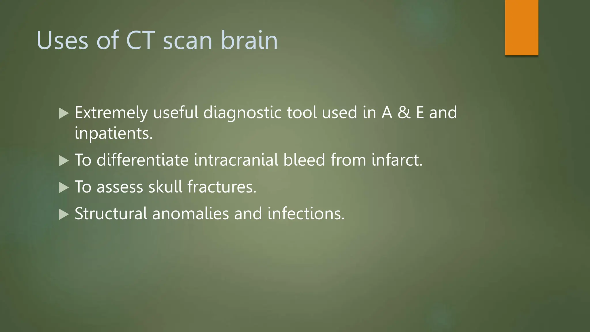
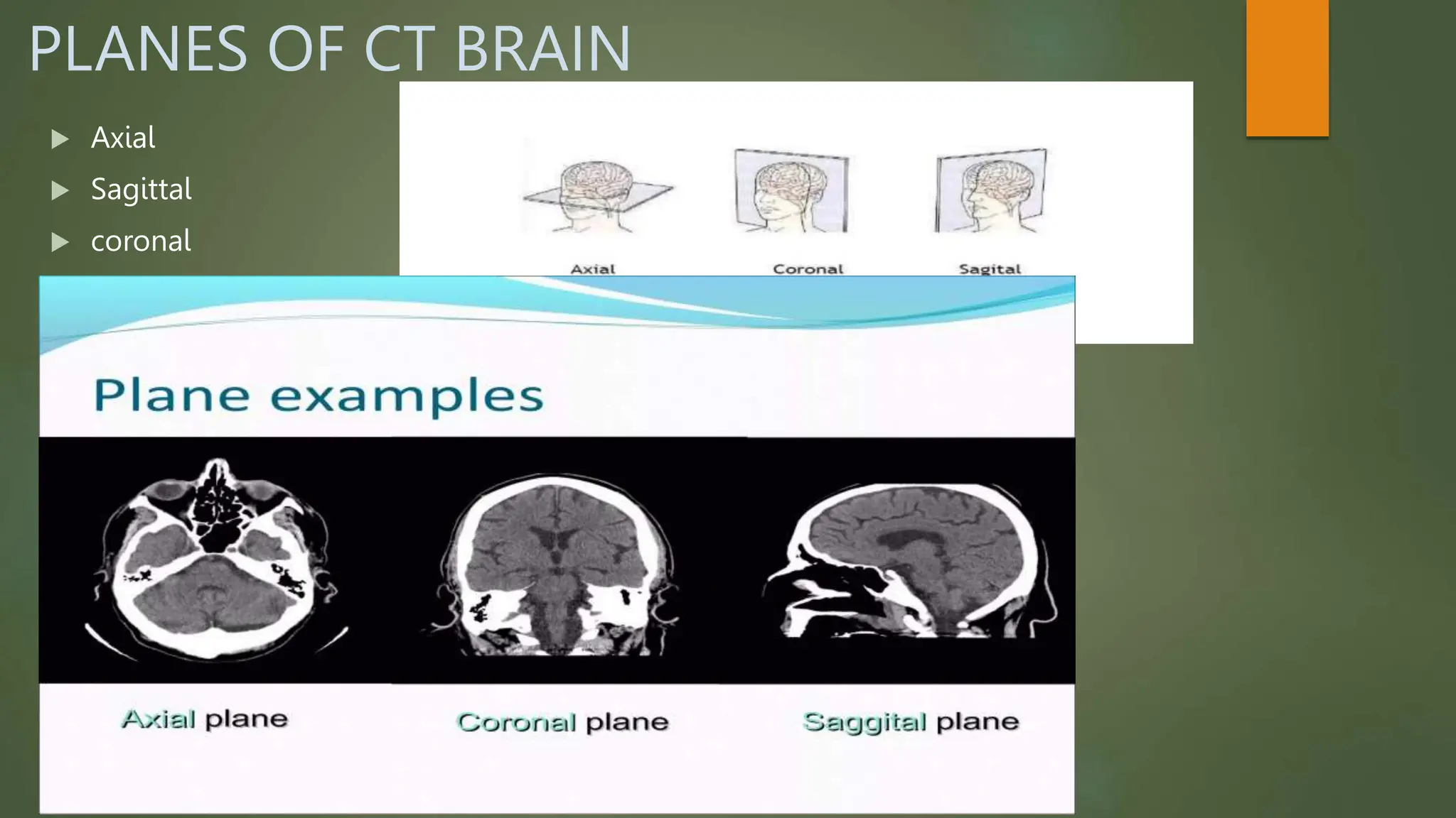
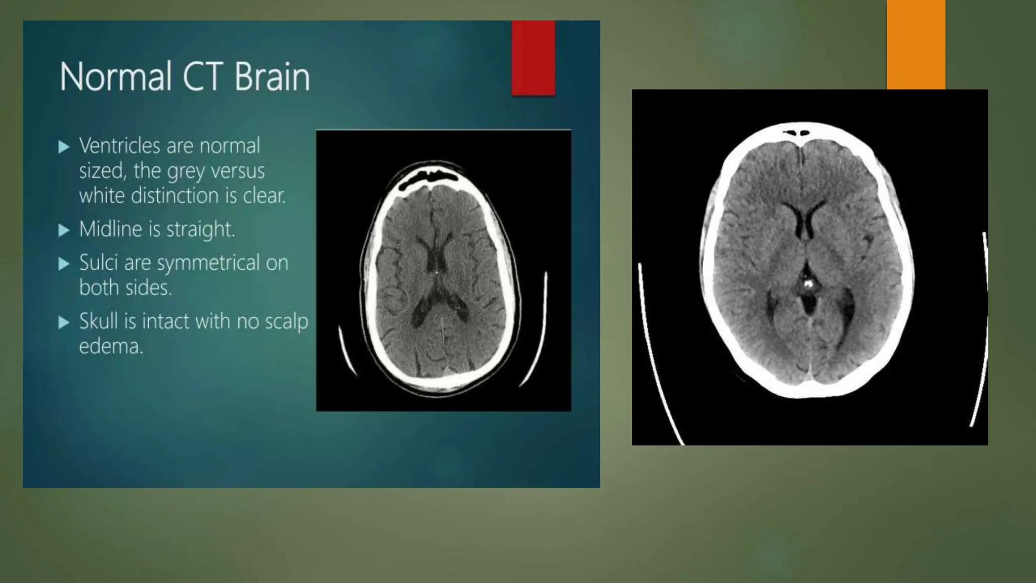
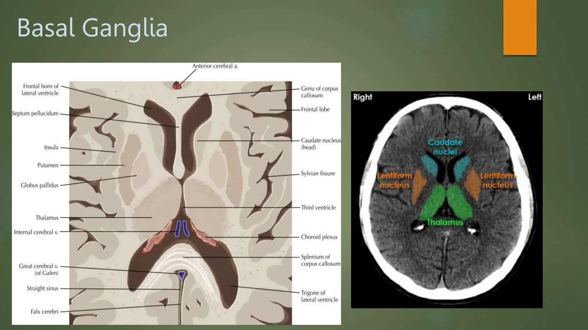
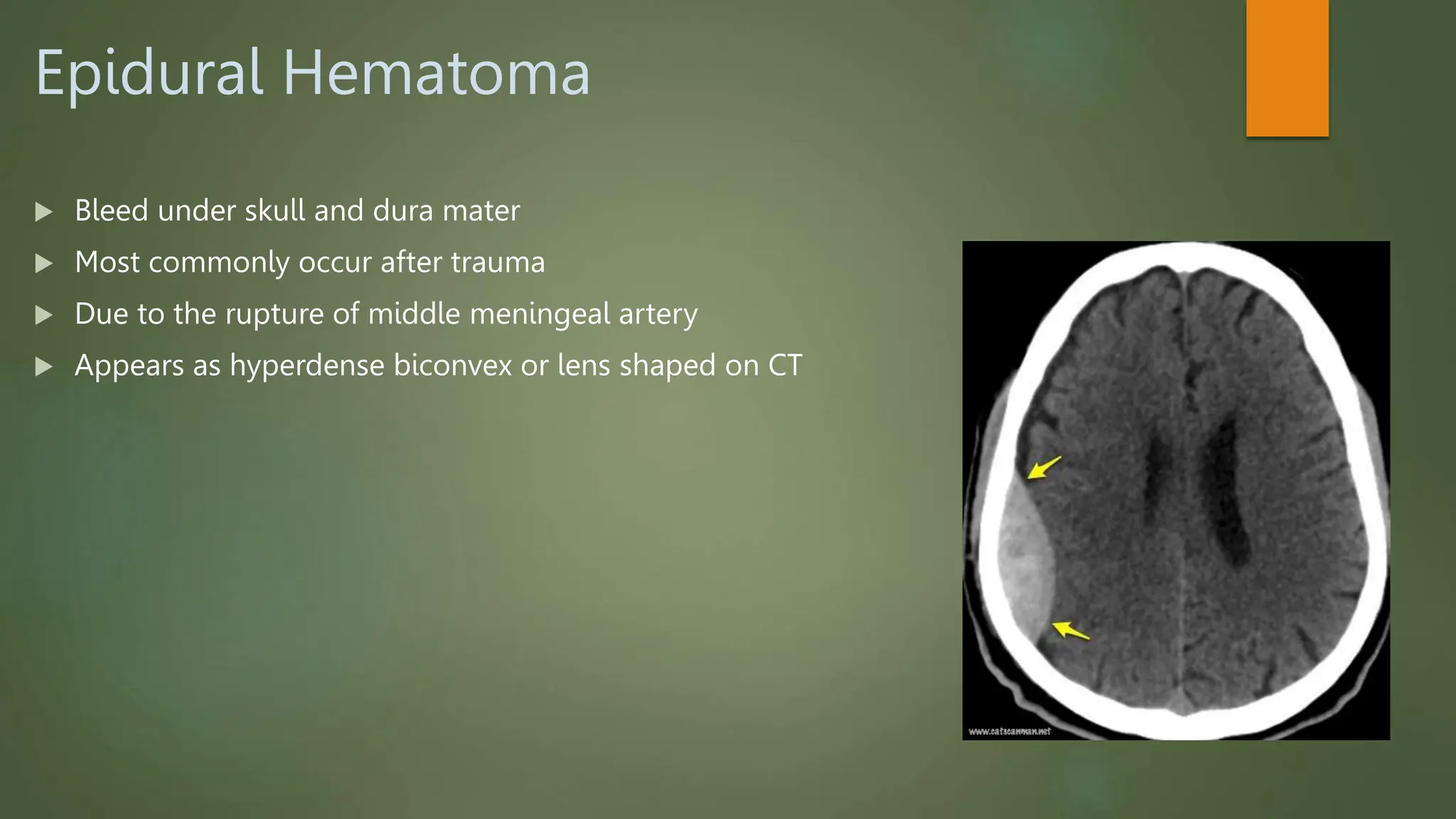
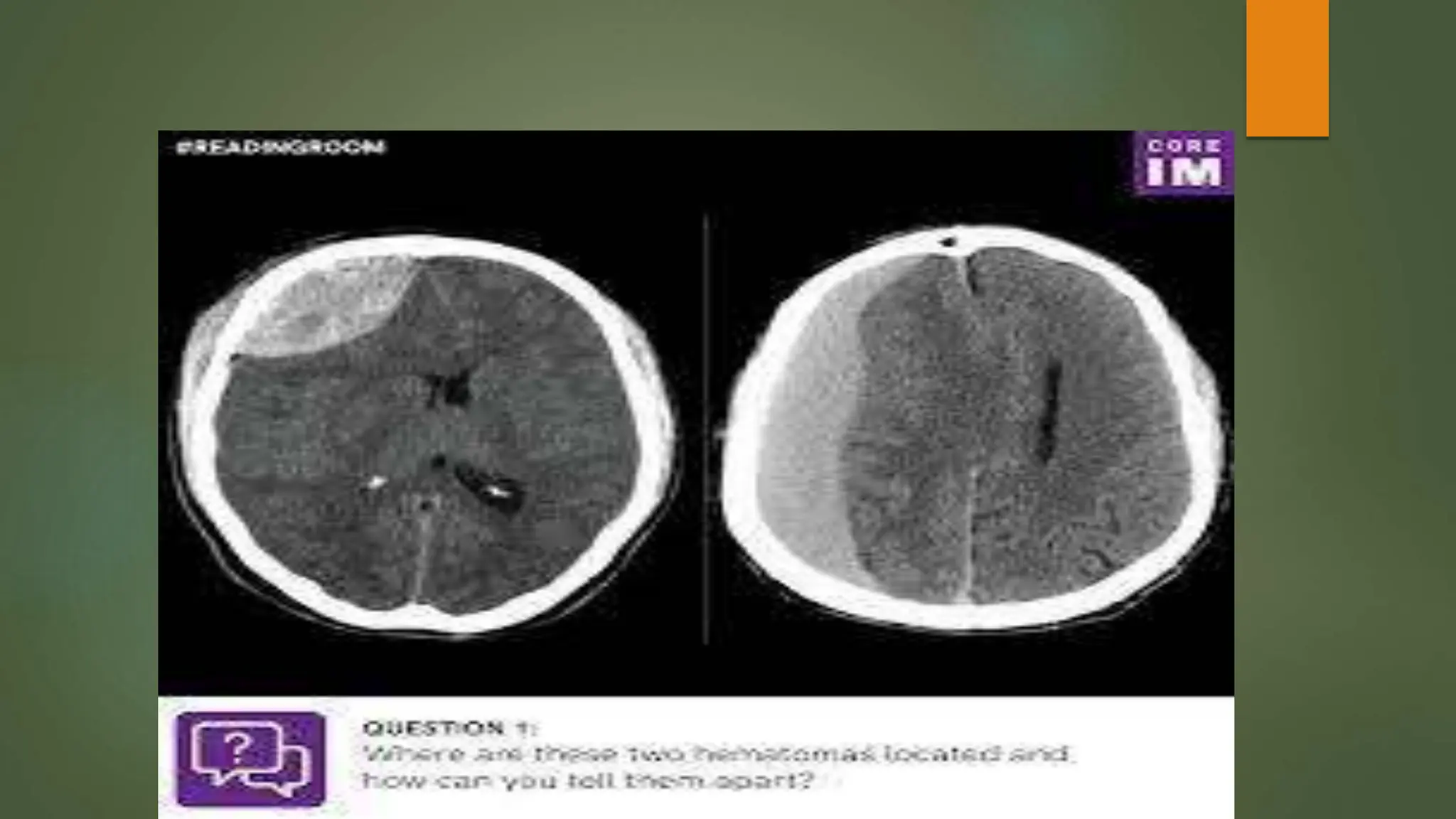
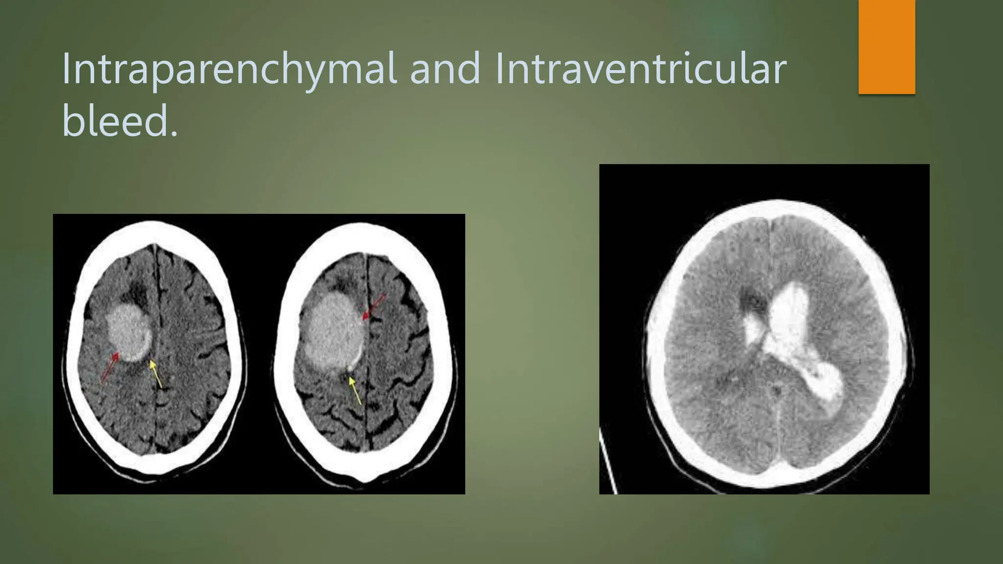
A SYSTEMATIC APPROCH TO
CT SCAN BRAIN
DR IQRA MUSTAFA
PGR RADIOLOGY
Uses of CT scan brain
 Extremely useful diagnostic tool used in A & E and
inpatients.
 To differentiate intracranial bleed from infarct.
 To assess skull fractures.
 Structural anomalies and infections.
 Unit of CT scan is Hounsfield (HU)
 It has different value in different structures.
PLANES OF CT BRAIN
 Axial
 Sagittal
 coronal
Skull sutures
Normal CT Brain
LOBES OF BRAIN
 04 LOBES
Frontal lobe
Temporal lobe
Parietal lobe
Occipital lobe
Central sulcus divides frontal lobe from parietal
lobe superiorly and frontal lobe from temporal
lobe inferiorly.

Ventricular system
 Lateral ventricle (frontal horn,body, temporal
and occiput horns)
 Third ventricle
 4th ventricle.
Fourth ventricle
Third ventricle
Basal Ganglia
Types of Hemorrhages
 Epidural hematoma
 Subdural hematoma
 Subarachnoid hematoma
 Intraparenchymal hematoma
 Intraventricular hematoma
Epidural Hematoma
 Bleed under skull and dura mater
 Most commonly occur after trauma
 Due to the rupture of middle meningeal artery
 Appears as hyperdense biconvex or lens shaped on CT
Subdural Hematoma
 Bleed under duramater and arachnoid mater
 Occurs in elderly patients or alcoholics due to rupture of bridging veins
 Appears as concave or hyperdense extra axial or crescent shaped on CT
Intraparenchymal and Intraventricular
bleed.
Subarachnoid hemorrhage
Approach to CT scan brain.pptx      Uuuuuu

Approach to CT scan brain.pptx Uuuuuu