Framing an Appropriate Research Question 6b9b26d93da94caf993c038d9efcdedb.pdf
anti tumor 1.doc
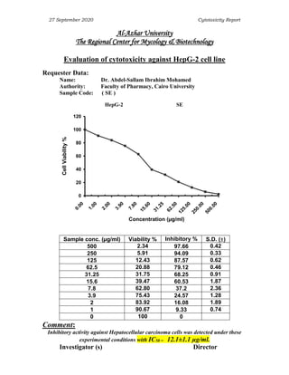
1. 27 September 2020 Cytotoxicity Report
Al-Azhar University
The Regional Center for Mycology & Biotechnology
Evaluation of cytotoxicity against HepG-2 cell line
Requester Data:
Name: Dr. Abdel-Sallam Ibrahim Mohamed
Authority: Faculty of Pharmacy, Cairo University
Sample Code: ( SE )
HepG-2 SE
0
20
40
60
80
100
120
5
0
0
.
0
0
2
5
0
.
0
0
1
2
5
.
0
0
6
2
.
5
0
3
1
.
2
5
1
5
.
6
0
7
.
8
0
3
.
9
0
2
.
0
0
1
.
0
0
0
.
0
0
Concentration (µg/ml)
Cell
Viability
%
Sample conc. (µg/ml) Viability % Inhibitory % S.D. ()
500 2.34 97.66 0.42
250 5.91 94.09 0.33
125 12.43 87.57 0.62
62.5 20.88 79.12 0.46
31.25 31.75 68.25 0.91
15.6 39.47 60.53 1.87
7.8 62.80 37.2 2.36
3.9 75.43 24.57 1.28
2 83.92 16.08 1.89
1 90.67 9.33 0.74
0 100 0
Comment:
Inhibitory activity against Hepatocellular carcinoma cells was detected under these
experimental conditions with IC50 = 12.1±1.1 µg/ml.
Investigator (s) Director
2. 27 September 2020 Cytotoxicity Report
Al-Azhar University
The Regional Center for Mycology & Biotechnology
Evaluation of cytotoxicity against HCT-116 cell line
Requester Data:
Name: Dr. Abdel-Sallam Ibrahim Mohamed
Authority: Faculty of Pharmacy, Cairo University
Sample Code: ( SE )
HCT-116 SE
0
20
40
60
80
100
120
5
0
0
.
0
0
2
5
0
.
0
0
1
2
5
.
0
0
6
2
.
5
0
3
1
.
2
5
1
5
.
6
0
7
.
8
0
3
.
9
0
2
.
0
0
1
.
0
0
0
.
0
0
Concentration (µg/ml)
Cell
Viability
%
Sample conc. (µg/ml) Viability % Inhibitory % S.D. ()
500 1.79 98.21 0.17
250 4.86 95.14 0.28
125 13.95 86.05 0.63
62.5 24.62 75.38 0.44
31.25 34.59 65.41 1.75
15.6 46.38 53.62 2.64
7.8 59.46 40.54 1.82
3.9 73.21 26.79 1.43
2 84.89 15.11 0.75
1 92.36 7.64 0.62
0 100 0
Comment:
Inhibitory activity against colon carcinoma cells was detected under these
experimental conditions with IC50 = 13.4 ± 1.8µg/ml.
Investigator (s) Director
3. 27 September 2020 Cytotoxicity Report
Al-Azhar University
The Regional Center for Mycology & Biotechnology
Evaluation of cytotoxicity against MCF-7 cell line
Requester Data:
Name: Dr. Abdel-Sallam Ibrahim Mohamed
Authority: Faculty of Pharmacy, Cairo University
Sample Code: ( SE )
MCF-7 SE
0
20
40
60
80
100
120
5
0
0
.
0
0
2
5
0
.
0
0
1
2
5
.
0
0
6
2
.
5
0
3
1
.
2
5
1
5
.
6
0
7
.
8
0
3
.
9
0
2
.
0
0
1
.
0
0
0
.
0
0
Concentration (µg/ml)
Cell
Viability
%
Sample conc. (µg/ml) Viability % Inhibitory % S.D. ()
500 4.27 95.73 0.51
250 11.82 88.18 0.64
125 26.93 73.07 0.79
62.5 41.75 58.25 1.38
31.25 60.89 39.11 2.17
15.6 78.16 21.84 0.84
7.8 91.78 8.22 0.29
3.9 98.42 1.58 0.34
2 100 0
1 100 0
0 100 0
Comment:
Inhibitory activity against Breast carcinoma cells was detected under these
experimental conditions with IC50 = 49 ± 3.9 µg/ml.
Investigator (s) Director
4. 27 September 2020 Cytotoxicity Report
Al-Azhar University
The Regional Center for Mycology & Biotechnology
Evaluation of cytotoxicity against PC-3 cell line
Requester Data:
Name: Dr. Abdel-Sallam Ibrahim Mohamed
Authority: Faculty of Pharmacy, Cairo University
Sample Code: ( SE )
PC-3 SE
0
20
40
60
80
100
120
5
0
0
.
0
0
2
5
0
.
0
0
1
2
5
.
0
0
6
2
.
5
0
3
1
.
2
5
1
5
.
6
0
7
.
8
0
3
.
9
0
2
.
0
0
1
.
0
0
0
.
0
0
Concentration (µg/ml)
Cell
Viability
%
Sample conc. (µg/ml) Viability % Inhibitory % S.D. ()
500 4.96 95.04 0.42
250 9.74 90.26 0.68
125 18.68 81.32 1.16
62.5 33.49 66.51 1.32
31.25 47.28 52.72 2.38
15.6 63.91 36.09 1.75
7.8 79.02 20.98 0.84
3.9 91.47 8.53 0.91
2 98.62 1.38 0.46
1 100 0
0 100 0
Comment:
Inhibitory activity against prostate carcinoma cells was detected under these
experimental conditions with IC50 = 28.6 ± 2.7 µg/ml.
Investigator (s) Director
5. 27 September 2020 Cytotoxicity Report
Evaluation of Cytotoxic Effects of certain Chemical compound
Mammalian cell lines: MCF-7 cells (human breast cancer cell line), HepG-2 cells (human
Hepatocellular carcinoma), HCT-116 (colon carcinoma) and PC-3 cells (human prostate carcinoma)
were obtained from VACSERA Tissue Culture Unit.
Chemicals Used: Dimethyl sulfoxide (DMSO), crystal violet and trypan blue dye were purchased from
Sigma (St. Louis, Mo., USA).
Fetal Bovine serum, DMEM, RPMI-1640, HEPES buffer solution, L-glutamine, gentamycin and 0.25%
Trypsin-EDTA were purchased from Lonza.
Crystal violet stain (1%): It composed of 0.5% (w/v) crystal violet and 50% methanol then made up to
volume with ddH2O and filtered through a Whatmann No.1 filter paper.
Cell line Propagation:
The cells were propagated in Dulbecco’s modified Eagle’s medium (DMEM) supplemented with
10% heat-inactivated fetal bovine serum, 1% L-glutamine, HEPES buffer and 50µg/ml gentamycin. All
cells were maintained at 37ºC in a humidified atmosphere with 5% CO2 and were subcultured two times
a week.
Cytotoxicity evaluation using viability assay: For cytotoxicity assay, the cells were seeded in 96-well
plate at a cell concentration of 1×10⁴
cells per well in 100µl of growth medium. Fresh medium containing
different concentrations of the test sample was added after 24 h of seeding. Serial two-fold dilutions of
the tested chemical compound were added to confluent cell monolayers dispensed into 96-well, flat-
bottomed microtiter plates (Falcon, NJ, USA) using a multichannel pipette. The microtiter plates were
incubated at 37ºC in a humidified incubator with 5% CO2 for a period of 24 h. Three wells were used for
each concentration of the test sample. Control cells were incubated without test sample and with or
without DMSO. The little percentage of DMSO present in the wells (maximal 0.1%) was found not to
affect the experiment. After incubation of the cells for at 37°C, for 24 h, the viable cells yield was
determined by a colorimetric method.
In brief, after the end of the incubation period, media were aspirated and the crystal violet solution (1%)
was added to each well for at least 30 minutes. The stain was removed and the plates were rinsed using
tap water until all excess stain is removed. Glacial acetic acid (30%) was then added to all wells and
mixed thoroughly, and then the absorbance of the plates were measured after gently shaken on
Microplate reader (TECAN, Inc.), using a test wavelength of 490 nm. All results were corrected for
background absorbance detected in wells without added stain. Treated samples were compared with the
cell control in the absence of the tested compounds. All experiments were carried out in triplicate. The
cell cytotoxic effect of each tested compound was calculated. The optical density was measured with the
microplate reader (SunRise, TECAN, Inc, USA) to determine the number of viable cells and the
percentage of viability was calculated as [(ODt/ODc)]x100% where ODt is the mean optical density of
wells treated with the tested sample and ODc is the mean optical density of untreated cells. The relation
between surviving cells and drug concentration is plotted to get the survival curve of each tumor cell line
after treatment with the specified compound. The 50% inhibitory concentration (IC50), the concentration
required to cause toxic effects in 50% of intact cells, was estimated from graphic plots of the dose
response curve for each conc. using Graphpad Prism software (San Diego, CA. USA).
References:
Mosmann, T. (1983): Rapid colorimetric assay for cellular growth and survival: application to
proliferation and cytotoxicity assays. J. Immunol. Methods; 65: 55-63.
Gomha, S.M.; Riyadh, S.M.; Mahmmoud, E.A. and Elaasser, M.M. (2015): Synthesis and
Anticancer Activities of Thiazoles, 1,3-Thiazines, and Thiazolidine Using Chitosan-Grafted-
Poly(vinylpyridine) as Basic Catalyst. Heterocycles; 91(6):1227-1243.
6. 27 September 2020 Cytotoxicity Report
Al-Azhar University
The Regional Center for Mycology & Biotechnology
Evaluation of cytotoxicity against HCT-116 cell line
Requester Data:
Sample Code: (Vinblastine Sulfate)
HCT-116 Vinblastine Sulfate
0
20
40
60
80
100
120
500.00
250.00
125.00
62.50
31.25
15.60
7.80
3.90
2.00
1.00
0.00
Concentration (µg/ml)
Cell
Viability
%
Sample conc. (µg/ml) Viability % Inhibitory %
500 4.09 95.91
250 6.76 93.24
125 12.16 87.84
62.5 15.54 84.46
31.25 18.92 81.08
15.6 28.71 71.29
7.8 39.86 60.14
3.9 47.30 52.7
2 58.11 41.89
1 66.48 33.52
0 100.00
Comment:
Inhibitory activity against colon carcinoma cells was detected under these experimental
conditions with IC50 = 3.5 µg/ml.
Investigator (s) Director
7. 27 September 2020 Cytotoxicity Report
Al-Azhar University
The Regional Center for Mycology & Biotechnology
Evaluation of cytotoxicity against HepG-2 cell line
Requester Data:
Sample Code: (Vinblastine Sulfate)
HepG-2 Vinblastine Sulfate
0
20
40
60
80
100
120
500.00
250.00
125.00
62.50
31.25
15.60
7.80
3.90
2.00
1.00
0.00
Concentration (µg/ml)
Cell
Viability
%
Sample conc. (µg/ml) Viability % Inhibitory %
500 3.27 96.73
250 5.89 94.11
125 10.92 89.08
62.5 14.36 85.64
31.25 19.24 80.76
15.6 26.85 73.15
7.8 34.19 65.81
3.9 45.06 54.94
2 54.28 45.72
1 60.94 39.06
0 100.00
Comment:
Inhibitory activity against Hepatocellular carcinoma cells was detected under these
experimental conditions with IC50 = 2.93 µg/ml.
Investigator (s) Director
8. 27 September 2020 Cytotoxicity Report
Al-Azhar University
The Regional Center for Mycology & Biotechnology
Evaluation of cytotoxicity against MCF-7 cell line
Requester Data:
Sample Code: (Vinblastine Sulfate)
MCF-7 Vinblastine Sulfate
0
20
40
60
80
100
120
500.00
250.00
125.00
62.50
31.25
15.60
7.80
3.90
2.00
1.00
0.00
Concentration (µg/ml)
Cell
Viability
%
Sample conc. (µg/ml) Viability % Inhibitory % S.D. ()
500 5.49 94.51 0.23
250 7.82 92.18 0.16
125 15.18 84.82 0.49
62.5 23.87 76.13 1.59
31.25 31.95 68.05 0.72
15.6 40.56 59.44 1.18
7.8 47.21 52.79 2.35
3.9 52.94 47.06 1.78
2 58.76 41.24 0.24
1 67.16 32.84 0.32
0 100.00
Comment:
Inhibitory activity against Breast carcinoma cells was detected under these
experimental conditions with IC50 = 5.9 µg/ml.
Investigator (s) Director