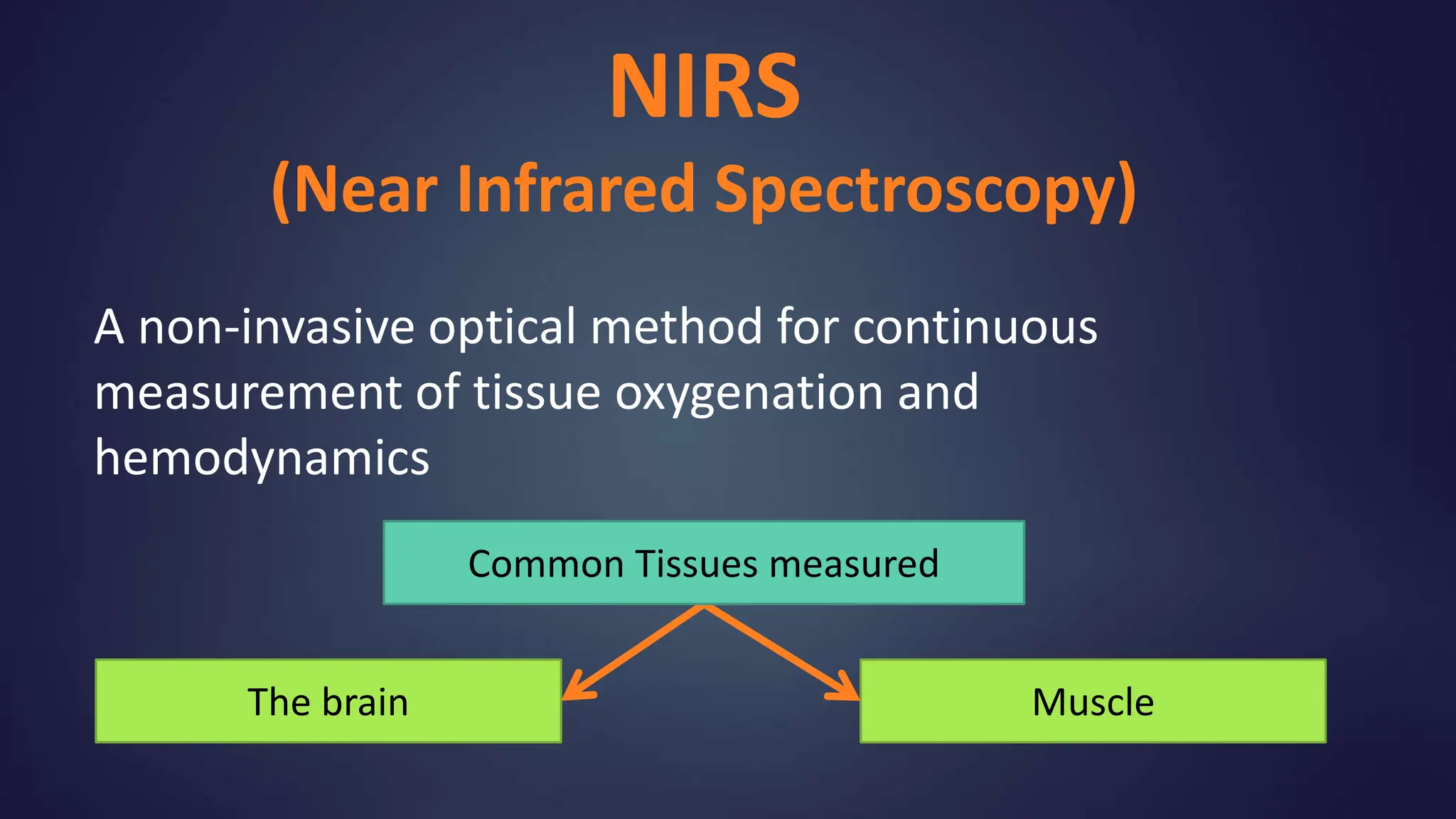
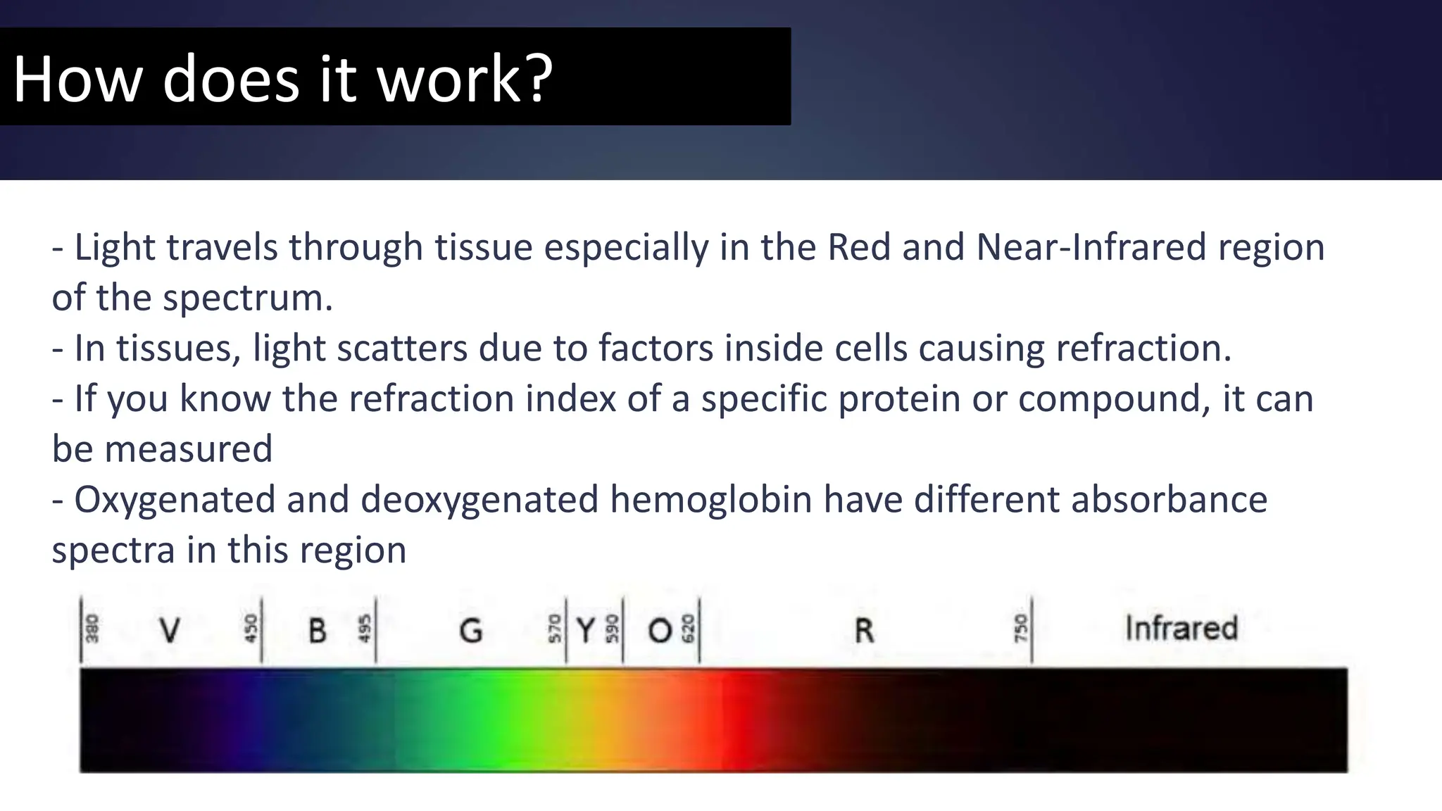
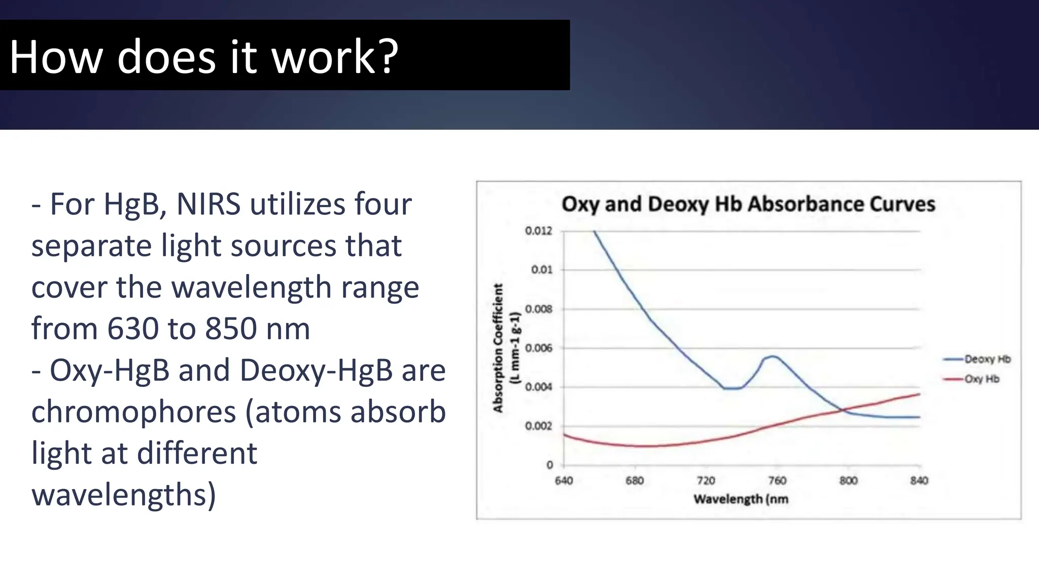
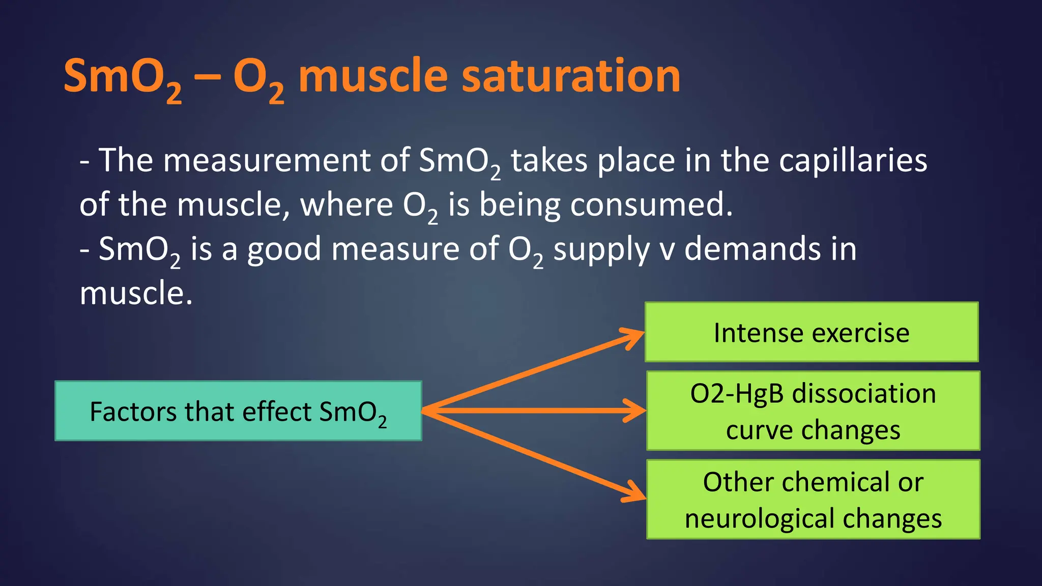
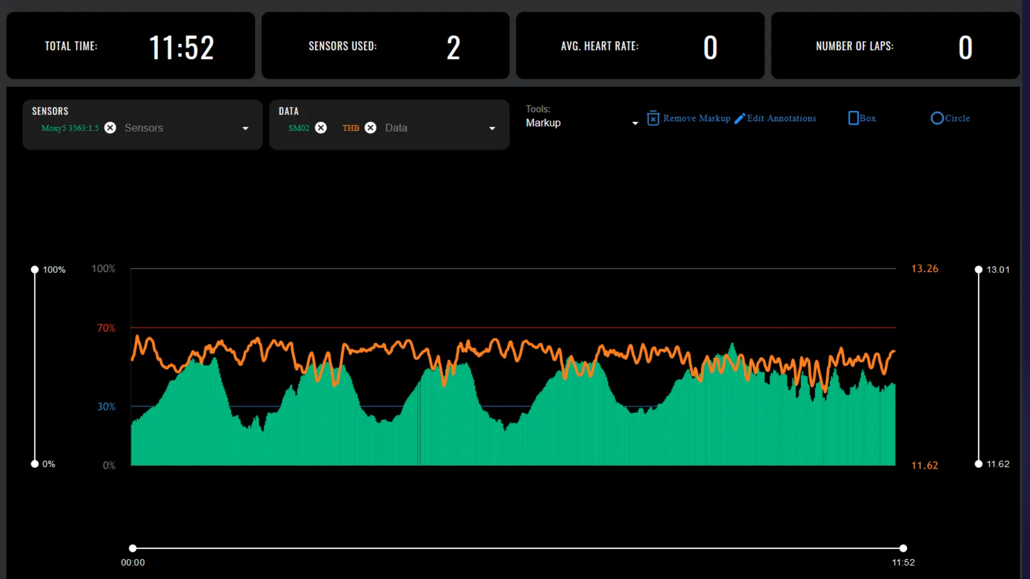
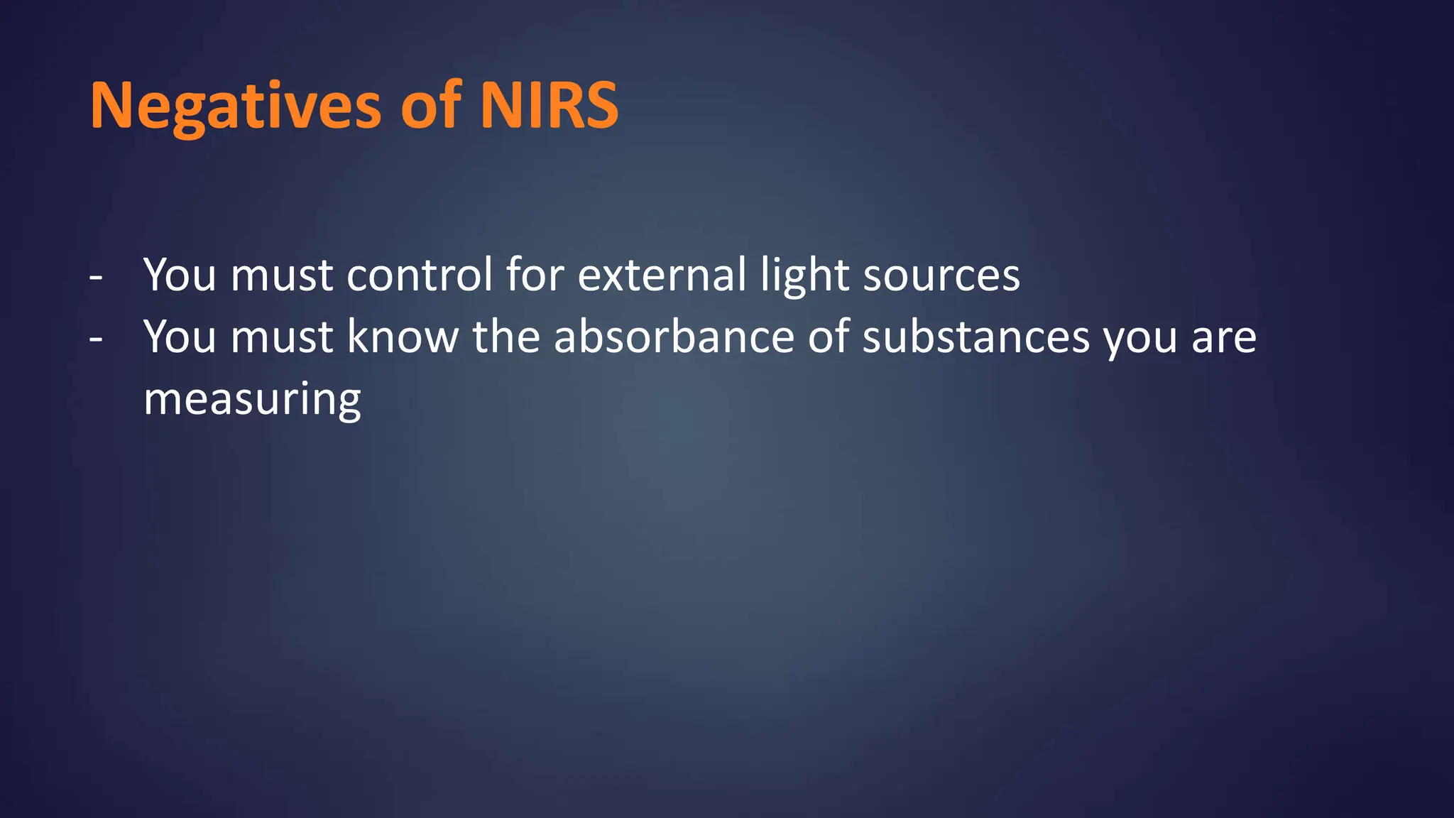
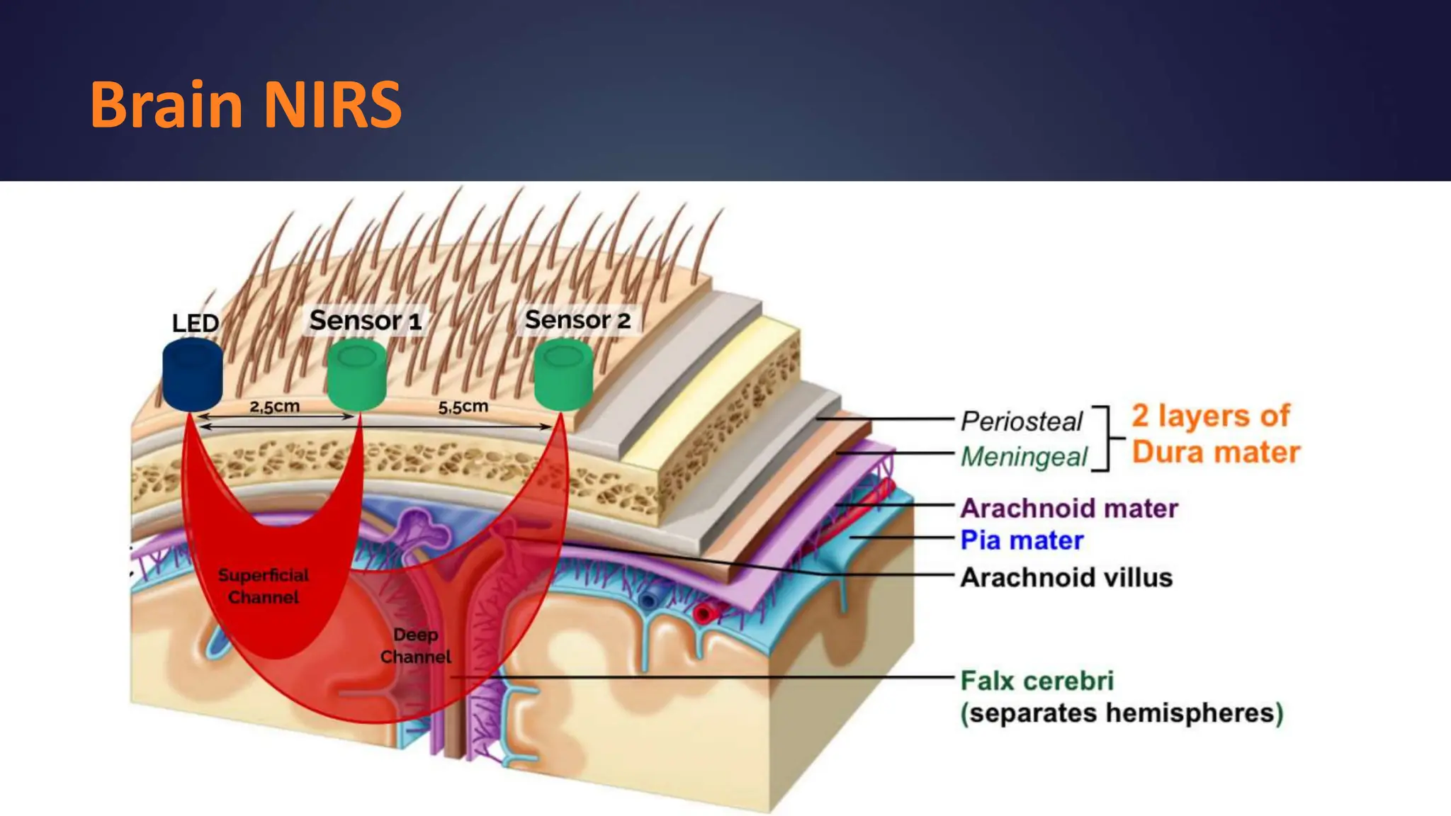
Near Infared
Spectroscopy
NIRS
(Near Infrared Spectroscopy)
A non-invasive optical method for continuous
measurement of tissue oxygenation and
hemodynamics
The brain Muscle
Common Tissues measured
How does it work?
- Light travels through tissue especially in the Red and Near-Infrared region
of the spectrum.
- In tissues, light scatters due to factors inside cells causing refraction.
- If you know the refraction index of a specific protein or compound, it can
be measured
- Oxygenated and deoxygenated hemoglobin have different absorbance
spectra in this region
How does it work?
- For HgB, NIRS utilizes four
separate light sources that
cover the wavelength range
from 630 to 850 nm
- Oxy-HgB and Deoxy-HgB are
chromophores (atoms absorb
light at different
wavelengths)
- Light is emitted into the
tissue at one location and
then is detected at two
locations, 12.5 and 25 mm
from the emitter.
- Oxy-HgB conc. to the total
HgB conc. in the muscle is
reported as a % = SmO2
How does it work?
SmO2 – O2 muscle saturation
- The measurement of SmO2 takes place in the capillaries
of the muscle, where O2 is being consumed.
- SmO2 is a good measure of O2 supply v demands in
muscle.
Intense exercise
O2-HgB dissociation
curve changes
Factors that effect SmO2
Other chemical or
neurological changes
Negatives of NIRS
- You must control for external light sources
- You must know the absorbance of substances you are
measuring
Brain NIRS
- You must control or external light sources
- You must know the absorbance of substances you are
measuring

An Introduction to Near Infared Spectroscopy (NIRS)

  • 1.
  • 2.
    NIRS (Near Infrared Spectroscopy) Anon-invasive optical method for continuous measurement of tissue oxygenation and hemodynamics The brain Muscle Common Tissues measured
  • 3.
    How does itwork? - Light travels through tissue especially in the Red and Near-Infrared region of the spectrum. - In tissues, light scatters due to factors inside cells causing refraction. - If you know the refraction index of a specific protein or compound, it can be measured - Oxygenated and deoxygenated hemoglobin have different absorbance spectra in this region
  • 4.
    How does itwork? - For HgB, NIRS utilizes four separate light sources that cover the wavelength range from 630 to 850 nm - Oxy-HgB and Deoxy-HgB are chromophores (atoms absorb light at different wavelengths)
  • 5.
    - Light isemitted into the tissue at one location and then is detected at two locations, 12.5 and 25 mm from the emitter. - Oxy-HgB conc. to the total HgB conc. in the muscle is reported as a % = SmO2 How does it work?
  • 6.
    SmO2 – O2muscle saturation - The measurement of SmO2 takes place in the capillaries of the muscle, where O2 is being consumed. - SmO2 is a good measure of O2 supply v demands in muscle. Intense exercise O2-HgB dissociation curve changes Factors that effect SmO2 Other chemical or neurological changes
  • 8.
    Negatives of NIRS -You must control for external light sources - You must know the absorbance of substances you are measuring
  • 9.
    Brain NIRS - Youmust control or external light sources - You must know the absorbance of substances you are measuring