Adolescent idiopathic scoliosis (AIS) is a type of scoliosis that typically develops during adolescence, between the ages of 10 and 18, and is characterized by an abnormal sideways curvature of the spine. "Idiopathic" means that the cause of the condition is unknown.
Here are some key points about adolescent idiopathic scoliosis:
Onset: AIS usually becomes noticeable during the growth spurt that occurs just before puberty. It can progress during the adolescent growth phase, but it typically stabilizes once the child reaches skeletal maturity.
Curvature: The degree of curvature can vary widely among individuals, ranging from mild to severe. Curves may occur in different regions of the spine and can be classified as thoracic (affecting the upper back), lumbar (affecting the lower back), or both.
Symptoms: In mild cases, AIS may not cause any symptoms or may only cause mild back pain or discomfort. In more severe cases, it can lead to noticeable spinal deformity, uneven shoulders or hips, and difficulty breathing if the curvature compresses the chest cavity.
Diagnosis: AIS is usually diagnosed through a physical examination, including a forward bend test
2. ADOLESCENT IDIOPATHIC SCOLIOSIS
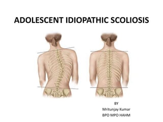
• AIS is defined as a scoliosis that starts after the age of 10 year up to
18 years and has no clear underlying disease as reason for its
development[1].
• It is usually located at the thoracic level and at this site almost
without exception involves a right-convex curve.
• Adolescent idiopathic scoliosis (AIS) is the most common type of
scoliosis, accounting for more than 80 % of scoliosis cases [2].
• It occurs less commonly at the thoracolumbar and lumbar levels,
and such cases show a marked tendency to go out of balance.
• It always involves rotation, whereby the posterior parts of the
vertebral bodies are always rotated towards the concave side of the
curve.
• For a given degree of curvature, the rotation is always more
pronounced at the lumbar level than the thoracic level [1].
1. Pathogenesis and biomechanics of adolescent idiopathic scoliosis (AIS). Fritz Hefti . J Child Orthop (2013) 7:17–24 DOI 10.1007/s11832-
012-0460-9
2. Wick JM, Konze J, Alexander K, Sweeney C. Infantile and juvenile scoliosis: the crooked path to diagnosis and treatment. AORN J.
2009;90:347–80.
3. PREVALENCE OF AIS:
Overall prevalence:
• There are not many studies that provide data of high
relevance regarding prevalence of AIS. Data from available
studies indicate prevalence of 0.47–5.2 % for AIS [1].
• The prevalence of AIS is related to geography. AIS is more
prevalent in areas located at high northern latitudes than in
regions of lower latitude.
• One meta-analysis from more than 17 countries evaluated
scoliosis screening in that the prevalence rate in Asia is the
range was 0.4–2.5% [2]
1. Epidemiology of adolescent idiopathic scoliosis Markus Rafael Konieczny • Hu ̈sseyin Senyurt • Ru ̈diger Krauspe, J Child Orthop (2013) 7:3–
9 DOI 10.1007/s11832-012-0457-4
2. Adolescent idiopathic scoliosis Jack C. Cheng, René M. Castelein, Article number:15030 doi:10.1038/nrdp.2015.30 Published online 24
September 2015;
4. Prevalence according to race or genetic factors:
• Genetic factors do influence the incidence and progression of
scoliosis. 97 % of AIS patients are related to other family members
with AIS.
• Children of families of a high or middle social status had a higher
prevalence (6.2 % high status, 5.6 % middle status) than children of
a lower social status (3.5 %).
Prevalence according to age:
• Daruwalla found a higher prevalence for adolescents than for
young children: 0.12 % in the 6–7 years-group, 1.0 % in the 11–12
years group and 3.12 % in the 16–17 years group (only girls
investigated in the latter group).
• In Germany Kamtsiuris found a prevalence of 6.5% in the age of
11–13 years and a prevalence of 11.1 % in the age of 14–17 years.
• These data indicate a higher prevalence of scoliosis in patients
older than 15 years (after puberty).
5. Prevalence according to gender:
• Kamtsiuris1 found a prevalence ratio between female and male of 1.5:1, with slight
increase with age.
• Daruwalla2 found a prevalence ratio female to male of 2:1, rising up to 3:1 in the age of
11–12 years.
• Nery3 found a prevalence ratio of 2:1 without differentiation of different age groups.
Gender and severity:
• Several studies2,3 report about higher Cobb angels in girls than in boys, indicating that
scoliosis in girls progresses to a higher grade of severity.
• For patients with a Cobb angle of more than 30° the prevalence ratio gets as high as
10:1
Cobb angle
(degree)
Prevalence
(%)
Female: Male
11-20 1.5-3 1.4:1
21-40 0.2-0.5 2.8-5.4:1
>40 0.04-0.3 7.2:1
1. Kamtsiuris P, Atzpodien K, Ellert U, Schlack R, Schlaud M (2007) Prevalence of somatic diseases in German children and adolescents. Results of the
German Health Interview and Examination Survey for Children and Adolescents (KiGGS). Bundesgesundheitsblatt Gesundheitsforschung Gesundheitss-
chutz 50(5–6):686–700
2. Daruwalla JS, Balasubramaniam P, Chay SO, Rajan U, Lee HP (1985) Idiopathic scoliosis. Prevalence and ethnic distribution in Singapore schoolchildren. J
Bone Joint Surg Br 67(2):182–184
3. Nery LS, Halpern R, Nery PC, Nehme KP, Stein AT (2010) Prevalence of scoliosis among school students in a town in southern Brazil. Sao Paulo Med J
128(2):69–73
6. • Overall prevalence of different curve types:
• Thoracic curves are the most common (48 %), followed by
thoracolumbar/lumbar curves (40 %).
• Double curves (9 %) and double thoracic curves (3 %) are less
common.
• 80 % of all children have thoracic or thoracolumbar/ lumbar curves.
• Curve type according to gender:
7. MECHANISMS & PATHOPHYSIOLOGY:
• Multiple abnormalities have been found, yet none has been
conclusively linked to all cases.
• The pathophysiology of AIS can be loosely grouped into six
main categories on the basis of the type of abnormality they
describe: Genetics, Central Nervous System (CNS), Skeletal
spinal growth and Bone metabolism, Metabolic pathways,
Biomechanics, and other [1].
1. Rinsky RA, Gamble JG. Adolescent idiopathic scoliosis. West J Med 1988;148:182-91.
8. Genetic basis
• Individuals affected with IS having an AA genotype had lower
mean maximum Cobb angle as compared to patients with AG
and GG genotypes [1].
• There is also increased expression of NTF3 messenger RNA in
paravertebral muscle in IS [2].
• Over transmission of the CHD7 associated polymorphism &
rs4738824 are associated with IS patients [2].
1. Y. Qiu, S.-H. Mao, B.-P. Qian et al., “A promoter polymorphism of neurotrophin 3 gene is associated with curve severity and bracing
effectiveness in adolescent idiopathic scoliosis,” Spine, vol. 37, no. 2, pp. 127–133, 2012.
2. R. Wang, Y. Qiu, and B. Rui, “Neurotrophin-3 mRNA expres- sion in paravertebral muscles of patients with idiopathic sco- liosis,” Chinese
Journal of Spine and Spinal Cord, vol. 15, pp. 532–534, 2007.
9. CNS AND NEUROPHYSIOLOGICAL DYSFUNCTION
• Abnormal neurophysiological functions have been reported in
patients with AIS, also include postural instability under both
static and dynamic conditions, abnormal proprioceptive
function, which is the ability to sense the relative positions of
different body parts.
• Patients with AIS have reduced spinal cord to vertebral length
ratios. In these individuals, the position of the spinal cord
tends to be shifted to the concave side, which is accompanied
by a distorted spinal cord shape at the apex of the curve.
10. The cascade concept of AIS
• The leptin is the linked to the development of the CNS and to the
asynchronous neuro-osseous growth mechanism.
• According to the cascade concept of AIS, during the late childhood fat
mass has low circulating leptin level.
• This effects on the CNS including impaired growth of the CNS axis or
neuro-axis.
• During the early adolescent growth spurt, these issues translate into
changes in spinal conformation, which initially occurs in the sagittal
plane by the tethering of anterior vertebral growth; this, together
with tension in the tether, induces a more backward vertebral tilt.
• Increasing backward vertebral tilt with thoracic axial vertebral
rotation leads to increasing spinal instability and torsion.
Burwell, R. G., Clark, E., Dangerfield, P. H. & Moulton, A. Adolescent idiopathic scoliosis (AIS): cascade concept of pathogenesis. Clin. Anat.
11. Skeletal growth and bone quality
• The pathogenesis of AIS interrelated with specific genetic and
multiple environmental factor that could lead to abnormal
regulation and modulation of systemic bone growth, bone
metabolism and bone modeling and remodeling.
• These abnormalities might function through different
biological and biomechanical pathways and might be
phenotypically expressed as systemic osteopenia, abnormal
bone mineralization and abnormal bone micro-architecture.
12. Abnormal skeletal growth
• AIS occurs in children during their pubertal growth spurt.
• Patients with AIS tend to be taller than those without AIS and have longer
arm spans and leg lengths, which can be transitory or persistent1.
• Longitudinal growth studies have revealed a significant increase in the peak
height velocity in patients with AIS2.
• Disproportionate growth and asymmetric morphology of skeletal features
including periapical ribs, upper arm length, left-right symmetry and iliac
height, with apical vertebral rotation and curve severity1.
• Disproportionate and asynchronous neuro-osseous growth of the spinal
column and spinal cord, leads to relative anterior spinal overgrowth.3
• These vertebral changes during adolescence, combined with various other
contributing factors, lead to the relative anterior spinal overgrowth that
results in the formation of a three-dimensional (3D) scoliosis deformity.
1. Burwell, R. G. et al. Patterns of extra-spinal left-right skeletal asymmetries in adolescent girls with lower spine scoliosis: relative
lengthening of the ilium on the curve concavity and of right lower limb segments. Stud. Health Technol. Inform. 123, 57–65 (2006).
2. Normelli, H., Sevastik, J. & Akrivos, J. The length and ash weight of the ribs of normal and scoliotic persons. Spine 10, 590–592 (1985).
3. Chu, W. C. et al. Morphological and functional electrophysiological evidence of relative spinal cord tethering in adolescent idiopathic
scoliosis. Spine 33, 673–680 (2008).
13. Abnormal body composition
• Studies shown that AIS correlates with abnormal body
composition with lower body weight and lower body mass
index1.
• Lower body weight is caused decreased body fat and fat free
mass.
• Decreases in leptin, lean mass and fat mass were associated
with an increased risk of scoliosis2.
1. Shohat, M. et al. Growth and ethnicity in scoliosis. Acta Orthop. Scand. 59, 310–313 (1988).
2. Clark, E. M. et al. Association between components of body composition and scoliosis: a prospective cohort study reporting differences
identifiable before the onset of scoliosis. J. Bone Miner. Res. 29, 1729–1736 (2014).
14. Osteopenia
• Low bone mineral density (osteopenia) is defined using the z-
score.
• Individuals with a z-score of <1 are considered to have
osteopenia, and in one study, this threshold was met by 36–
38% of girls with AIS1.
• Osteopenia can persist into adulthood if not treated and
constitutes an important prognostic factor for curve
progression in AIS2.
• A recent study proposed that the low bone mass in patient
with AIS affect both cortical and trabecular bone
compartment3.
• AIS was also associated with abnormal bone mineralization,
bone morphology, trabecular micro-architecture, volumetric
bone density, overall bone quality and mechanical strength3.
1. Cheng, J. C. et al. Generalized low areal and volumetric bone mineral density in adolescent idiopathic scoliosis. J. Bone Miner. Res. 15, 1587–
1595 (2000).
2. Hung, V. W. et al. Osteopenia: a new prognostic factor of curve progression in adolescent idiopathic scoliosis. J. Bone Joint Surg. Am. 87,
2709–2716 (2005).
3. Yu, W. S. et al. Bone structural and mechanical indices in adolescent idiopathic scoliosis evaluated by high- resolution peripheral quantitative
computed tomography (HR-pQCT). Bone 61, 109–115 (2014).
15. • Although spinal deformities have been
observed in animals, but true idiopathic
scoliosis has only been diagnosed in humans.
• With a lordotic curvature; the human center of
gravity directly above the pelvis.
• All other vertebrates lack these lordotic
curvatures putting the trunk’s center of gravity
in front of the hips.
• The spine able to withstand axial and anterior
load but certain areas in the human spine are
posteriorly inclined and subject to posteriorly
directed loads.
• The posterior shear loads decreased the
rotational stiffness of the spine.
• Rotational stability apparently depends on
whether vertebra are loaded in an anterior or
posterior direction or anteriorly or posteriorly
inclined in the sagittal plane.
Bipedalism and 3D spinal–pelvic deformity
Sarwark, J. F., Castelein, R. M., Maqsood, A., & Aubin, C.-E. (2019). The Biomechanics of Induction in Adolescent Idiopathic Scoliosis. The Journal
of Bone and Joint Surgery, 101(6), e22. doi:10.2106/jbjs.18.00846
16. • This profile differs considerably between girls and boys
especially during their pubertal growth spurt, with girls having
a larger posteriorly inclined area of the spine, and thus less
rotational stability, than boys 1.
• Once the spinal curve develops, its rotational direction follows
an in-built pattern that is already present in the normal, non-
scoliotic spine. The magnitude of rotation depends on the
position of the spine relative to gravity line2.
1. Schlösser, T. P., Vincken, K. L., Rogers, K.,Castelein, R. M. & Shah, S. A. Natural sagittal spino-pelvic alignment in boys and girls before,at and
after the adolescent growth spurt. Eur. Spine J. 24,1158–1167 (2014).
2. Janssen, M. M. et al. Pre-existent vertebral rotation in the human spine is influenced by body position. Eur. Spine J. 19, 1728–1734 (2010).
17. • Mechanical factors that could predispose individuals to the initiation
of scoliosis during growth include an abnormal sagittal curvature such
as hypokyphosis and a ‘slender’ spine.
• Spines with these features are more susceptible to ‘buckling’ in the
frontal plane and rotation in the axial plane, which is greatest at the
apex of the spinal curve1.
• It is also possible that uncoupled neuro-osseous growth causes the
spine to buckle into a scoliosis deformity2.
• Ligamentous and muscular structures have also been implicated in
‘tethering’ of the spine, which could produce scoliosis and rotation of
the spine3.
• In addition, it is possible that anomalies of neuromuscular control
produce asymmetrical spinal loading and hence initiation and
progression of scoliosis3.
1. Millner, P. A. & Dickson, R. A. Idiopathic scoliosis: biomechanics and biology. Eur. Spine J. 5, 362–373 (1996).
2. Veldhuizen, A. G., Wever, D. J. & Webb, P. J. The aetiology of idiopathic scoliosis: biomechanical and neuromuscular factors. Eur. Spine J. 9,
178–184 (2000).
3. Jarvis, J. G., Ashman, R. B., Johnston, C. E.& Herring, J. A. The posterior tether in scoliosis. Clin. Orthop. Relat. Res. 227, 126–134 (1988).
Dorsal shear concept for AIS pathogenesis
18. • A ‘vicious cycle’ of scoliosis progression has
been proposed, in which asymmetrical
stresses that act on a laterally curved spine
and the vertebral growth plates produce
asymmetrical spinal growth1.
• This asymmetry, in turn, leads to progression
of the lateral curvature through the Hueter–
Volkmann principle, in which increased
compressive loading retards growth and
decreased loading results in accelerated
growth2.
• Other study also described the sensitivity of
growth plate to compression stress and
geometric changes that result from
asymmetrical growth.3
1. Roaf, R. Vertebral growth and its mechanical control. J. Bone Joint Surg. Br. 42-B, 40–59 (1960).
2. Mehlman, C. T., Araghi, A. & Roy, D. R. Hyphenated history: the Hueter–Volkmann law. Am. J. Orthop. (Belle Mead NJ) 26, 798–800 (1997).
3. Stokes, I. A., Aronsson, D. D., Dimock, A. N., Cortright, V. & Beck, S. Endochondral growth in growth plates of three species at two anatomical locations modulated by
mechanical compression and tension. J. Orthop. Res. 24, 1327–1334 (2006).
19. Abnormal metabolic pathways in AIS:
• Significantly higher serum OST(osteocalcin) levels were
observed.
• Significantly higher serum RANKL(nuclear factor-κB ligand)
levels & decrease in circulating leptin levels were observed.
• Plasma MATRILIN-1 levels were reduced. Plasma MATRILIN-1
levels were significantly lower in patients whose AIS
progressed than for those whose AIS was stable
• Plasma COMP(cartilage oligomeric Matrix protein) levels were
lower in children with idiopathic scoliosis.
20. PATHOGENESIS AND BIOMECHANICS
GROWTH
• There is a disparity between the growth of the vertebral bodies
anteriorly and that of the posterior elements.
• The vertebral bodies grow faster than the posterior elements,
resulting primarily in a lordosis.
• The diminished dorsal growth impedes the ventrally located
vertebral bodies from increasing in height, forcing them to
rotate. This produces a rotational lordosis.
• Porter1 in 2000 was found The relative shortening of vertebral
canal with respect to anterior vertebral body height in the
scoliosis spines was correlated with the Cobb angle and the
degree of rotation.
• The kyphotic spines had canals significantly longer than the
vertebral length.
1. Porter R (2000) Idiopathic scoliosis: the relation between the vertebral canal and the vertebral bodies. Spine 25:1360–1366
21. • Guo et al 2003 also reported the scoliotic spines had longer
vertebral bodies between T1 to T12 in the anterior column
and shorter pedicles with a larger interpedicular distance in
the posterior column.
• There was also a significant positive correlation between the
scoliosis severity score and the ratio of differential growth
between the anterior and posterior columns for each thoracic
vertebra
Guo X, Chau W-W, Chan Y-L, Cheng JC-Y (2003) Relative anterior spinal overgrowth in adolescent
idiopathic scoliosis. Results of disproportionate endochondral–membranous bone growth. J
Bone Joint Surg [Br] 85:1026–1031
22. • FORCE
• The vertebral column in humans is still
predominantly loaded in an axial direction.
• Certain segments of the human spine
(especially the backward inclined segments)
are subject to dorsally directed shear loads
as well.
• The facet joints play an important role in
providing rotational stability to the spine
and counteract ventrally directed shear
loads(A).
• The vertebrae are not well designed to
resist dorsally directed shear loads taking
the anatomy of the facet joints, and the
posterior location of the spinal muscles and
ligaments into consideration(B).
Castelein RM, van Dieen JH, Smit TH (2005) The role of dorsal shear forces in the pathogenesis of adolescent idiopathic scolio- sis—a hypothesis.
Med Hypotheses 65:501–508
23. • In a in vitro biomechanical study showed that at the mid and
lower thoracic spine more axial vertebral rotation occurred
under dorsally directed shear loads than under ventrally
directed shear loads.
• The dorsally shear load can act as a enhancer of slight
preexistent vertebral rotation, where as ventrally directed
shear load counteract the rotation.
• This force could result in a progressive deformation of
individual vertebrae because of Hueter–Volkmann’s law, and
ultimately lead to progressive scoliosis.
Kouwenhoven JW, Castelein RM (2008) The pathogenesis of adolescent idiopathic scoliosis:
review of the literature. Spine 33(26):2898–2908
24. • Wever et al. suggested that the
vertebral deformation is caused by
bone remodeling due to an
imbalance between forces in an
anterior and posterior spinal
column.
• In the anterior column the
compressive forces result in a force
driving the apical vertebra body out
of the midline,
• The tension forces of the posterior
column result in a force attempting
to keep the posterior complex in
normal position.
25. Natural History & Prognosis
• The three main determinants of progression are patient gender,
future growth potential and curve magnitude at the time of
diagnosis.
• Females have a risk of curve progression 10 times higher than
males.
• The pubertal growth spurt begins with accelerated longitudinal
growth of limbs, which causes a temporary disproportion of the
body (long limbs and short trunk).
• Then, longitudinal growth is seen in the axial skeleton. It is the
period of the most marked progression of IS.
• The greater the growth potential and the larger the curve, the
greater the likelihood of curve progression.
• Evaluation of growth potential is done by assessing the Tanner
stage and the Risser grade.
26. Risk of Curve Progression
CURVE
(DEGREE)
GROWTH POTENTIAL
(RISSER SIGN)
RISK
10-19 LIMITED (2-4) LOW RISK
10-19 HIGH (0-1) MODERATE
20-29 LIMITED (2-4) LOW/MODERATE
20-29 HIGH (0-1) HIGH
>29 LIMITED (2-4) HIGH
>29 HIGH (0-1) VERY HIGH
Low risk = 5 to 15 percent;
Moderate risk = 15 to 40 percent;
High risk = 40 to 70 percent;
Very high risk = 70 to 90 percent.
27. Diagnosis and evaluation of patient
with AIS
• SCREENING:
• Screening for scoliosis was common in schools and
communities in past years.
• The forward bending test with a scoliometer is used
in the early detection of scoliosis with school
screening.
• The school nurse and school physical education
teachers at the regional workshops organized by the
state health or education department aided by an
orthopedic surgeon.
• The American Academy of Orthopedic Surgeons
recommends screening girls at ages 11 and 13 and
screening boys at age 13 or 14 year of age.
• An angle of trunk rotation more than 5 to 7 degree in
scoliometer is often the threshold for referral for
radiography.
28. Patient History
• The information should include a history of the problem,
patient general condition and health, family history.
• The child is being examined, the examiner must also obtain
prenatal and postnatal histories.
• Injuries experienced by the mother during pregnancy and any
complication during pregnancy or delivery, drug taken by the
mother during that period, especially during first trimester.
• How old is the patients is, In the child has there been a
growth spurt. For female when did menarche begin.
• His/her total treatment history including therapeutic, bracing,
medication, history of surgery etc.
29. • Observation :Body built, Assess the posture in anterior,
posterior and lateral view.
• Physical examination:
– TRACE examination, POTSI (Posterior Trunk Symmetry Index),
ATSI (Anterior Trunk Symmetry Index)
– Plumb line test
– Range of motion of trunk
– Joint laxity/subluxation
– Palpation of pain
– Forward bending test & scoliometer
– Spinal flexibility test by side bending and traction.
– Skin lesion, motor examination, reflex (Neuro condition)
examination
30. SPINAL FLEXIBILITY TEST ADAM’S FORWARD
BENDING TEST
PLUMB LINE
Shoulder asymmetry Scapula asymmetry
Hemi thorax Waist asymmetry
TRACE EXAMINATION
31. • It is most valuable, reliable and
most used diagnostic tool for
evaluation of scoliosis.
• Mostly PA or AP and Lateral view
is used in standing position is
used.
• To evaluate flexibility PA or AP
view in side bending on the
convex side is used.
Radiographic Examination:
32. • The parameter we check in X ray are:
– Apex or apical vertebra
– Side of curve
– Number of curve present and its location
– Angle of curve by cobb method
– Curve progression- RVAD angle or Mehta’s angle
– Rotation of vertebrae- Nash-Moe grade
– Chronological or bony age- Risser sign
– Trunk compensation (plumb line & CSVL line)
35. King-Moe classification
• Published in 1983
• Five curve types as a guide to surgical
treatment
• Type 1: S-shaped curve in which both
the thoracic and lumbar curves cross
the midline. Both curves are structural,
and the lumbar curve may be larger or
less flexible than the thoracic curve
• Type 2: S-shaped curve in which the
thoracic curve is larger or less flexible
than the lumbar curve (also called a
“false” double major curve)
36. • Type 3: Single
thoracic curve
without a structural
lumbar curve
• Type 4: Long thoracic
curve in which L5 is
centered over the
sacrum and L4 is
tilted into the
thoracic curve
• Type 5: Double
thoracic curve with
T1 tilted into the
convexity of the
upper curve
37. LENKE CLASSIFICATION
Structural criteria
Proximal thoracic: cobb angle >25, T2-T5 kyphosis>+20
Main thoracic: cobb angle>25 degree, T10-L2 kyphosis>+20
Thoracolumbar/Lumber: cobb angle >25, T10-L2 kyphosis>+20
Location of Apex
Thoracic:T2-T11/12 disk
Thoracolumbar/Lumber:L1/2 disk – L4
38. Augmented Lehnert-Schroth (ALS)
Classification
This classification is used for the application of Schroth Best
Practice (SBP) exercises and for the application of the
Gensingen brace (GBW) worldwide.
1. 3CH (functional 3-curve, with hip prominence)
2. 3CTL (functional 3-curve, thoracolumbar)
3. 3CN (functional 3-curve, compensated)
4. 3CL (functional 3-curve with long lumbar counter curve)
5. 4C (functional 4-curve, double curvature)
6. 4CL (functional 4-curve with major lumbar curvature)
7. 4CTL (functional 4-curve with major thoracolumbar
curvature)
39. 3CH (functional 3-curve, with hip
prominence)
Radiological features:
• Patients with this pattern typically have a major
curvature in the thoracic region with one cranial
and one caudal equalization curvature
respectively.
• Thoracic cobb angle> lumbar cobb angle
• Lumbar apex not crossing the CSV line.
Clinical signs:
• Decompensation to thoracic convex side
• Long thoracic curve no clear lumber counter
curve
• Significant hip prominences on thoracic concave
side
40. 3CTL (functional 3-curve,
thoracolumbar)
Radiological features:
• Thoracolumbar curve longer than
lumbar
• TL Cobb angle> Lumbar cobb angle
• No Lumbar curvature
• Apex at TH 12
Clinical signs:
• Decompensation to TL convex side.
• Long TL curve no lumbar counter
curve
• Significant hip prominence on TL
concave side.
41. 3CN (functional 3-curve, compensated)
Radiological features:
• Thoracic curve longer than lumbar
• Thoracic cobb angle>lumber cobb
angle
• Lumbar apex is crossing the CSV line.
Clinical signs:
• Decompensation to thoracic convex
side.
• Thoracic longer than lumbar counter
curve.
• Hip prominence on thoracic concave
side or centered.
42. 3CL (functional 3-curve with long
lumbar counter curve)
Radiological features:
• Thoracic and lumber curve about same
length
• Thoracic cobb angle> lumbar cobb angle
• Lumbar apex is crossing the CSV line
• No wedging of the disc spaceL4/L5/S1
Clinical signs:
• Decompensation to thoracic convex side
• Thoracic and lumbar curve about same
length
• Hip prominences on thoracic convex
side.
43. 4C (functional 4-curve, double
curvature)
Radiological features:
• Thoracic and lumbar curve about same
length
• Thoracic cobb angle=lumber cobb angle
• Lumbar apex is crossing the CSV line
• Wedging of the disc space L4/L5/S1.
Clinical signs:
• Decompensation to the thoracic
concavity or balanced.
• Thoracic and lumbar curve about same
length.
• Hip prominences on the thoracic convex
side.
44. 4CL (functional 4-curve with major
lumbar curvature)
Radiological features:
• Lumbar curve with short thoracic
curvature.
• Lumbar cobb angle > Thoracic cobb angle
• Main curve apex L2 or below.
• Wedging of the disc space L5/S1.
Clinical signs:
• Decompensation to thoracic concave side
• Lumbar curve bigger than thoracic curve.
• Hip prominence on thoracic convex side
• Ventral rib hump on the side of lumbar
convexity.
45. 4CTL (functional 4-curve with major
thoracolumbar curvature)
Radiological features:
• TL curve & short thoracic curve
• TL cobb angle > Thoracic cobb angle
• TL curve apex at L1
• Wedging of the disc space L4/L5/S1
Clinical signs:
• Decompensation to TL concave side
• TL curve bigger and longer than thoracic
curve
• Hip prominences on thorax convex side
• Ventral rib hump on the side of the TL
concavity.
46. RIGO CLASSIFICATION SYSTEM:
• This classification was developed by Dr. Manuel d Rigo to
correlate with scoliosis specific treatment and brace design.
• Classification is based on –
1. Clinical observation.
2. Radiological correlation.
• On the basis of clinical observation the curve classified into 4
basic types:
1. Three curve (3C)===A1, A2, A3
2. Four curve (4C)===B1, B2
3. Non three –Non four (N3N4)===C1, C1
4. Single lumbar or thoracolumbar.===E1, E2
47. Three curve (3C)
TYPE A1:
Clinical signs:
• Pelvis lateral to thoracic concave and outward
projected.
• Trunk imbalance, COG in thoracic convex side.
• If the pelvis does not move laterally, the sternum
tends to move laterally to the thoracic concave.
• The rib protrusion on the back of the chest is longer,
and the protrusion on the same side of the waist is
also seen.
Radiological features:
• Single long thoracic curve (apical vertebra in T19-T11)
involved the upper segment of lumbar curve.
• L3 in the thoracic convex side was inclined, L4 in the
horizontal or inclined to the thoracic convex side, L5
in the horizontal position.
• TP, T1 in the thoracic convex side.
• The entire spine in the CSL side; may be accompanied
by proximal thoracic curve (top vertebra in T2-T4).
48. Three curve (3C)
TYPE A2:
Clinical signs:
• Pelvis translated to the concave thoracic
side.
• Trunk imbalance to the convex thoracic side
• Noticeable rib hump, no or minimal lumbar
prominence.
Radiological features:
• Main thoracic curvature (apical vertebra at
T5-T9), no or only upper lumbar curvature,
L4-5 in horizontal position.
• Single thoracic/ no or minimal functional
lumbar curve.
• Transitional position imbalance to the
convex thoracic side.
• T1 imbalance to the convex thoracic side.
• L4 L5 horizontal
49. Three curve (3C)
TYPE A3:
Clinical signs:
• Pelvis translated to the concave thoracic
side.
• Trunk imbalance to the convex thoracic
side.
• Noticeable rib hump & minor lumbar
prominence
Radiological features:
• Single major thoracic & lumbar minor.
• Transitional position imbalance to the
convex thoracic side.
• T1 imbalance to the convex thoracic
side.
• L4 tilted to the concave thoracic side
with negative L5-L4 counter tilting.
50. Four curve (4C)
TYPE B1
Clinical signs:
• Pelvis translated to the convex thoracic
side.
• Trunk imbalance to the concave
thoracic side.
• Noticeable rib hump and lumbar or TL
prominence.
Radiological features:
• Double thoracic and lumbar or thoracic
and thoracolumbar.
• Transitional position imbalance to the
concave thoracic side
• T1 imbalance to the concave thoracic
side.
• Positive L5-L4 counter-tilting.
51. Four curve (4C)
TYPE B2
Clinical signs:
• Pelvis translated to the convex thoracic side.
• Trunk imbalance to the concave thoracic
side.
• Noticeable TL prominence associated to a
major thoracic hump.
Radiological features:
• Major TL combined with a minor thoracic
curve.
• TP imbalance to the concave thoracic side.
• T1 imbalance to the concave thoracic side
• Positive L4-L5 counter-tilting often positive
L4-L3 counter tilting.
52. Non three –Non four (N3N4)
Subtype-C1
Clinical signs:
• Pelvis centered
• Trunk centered
• Noticeable rib hump with
lumbar spine rectilinear.
Radiological features:
• Single thoracic with no
lumbar curve
• TP on the CSL
• T1 on the CSL
53. Non three –Non four (N3N4)
Subtype-C2
Clinical signs:
• Pelvis centered
• Trunk balanced
• Noticeable rib hump combined
with a noticeable lumbar
prominence.
Radiological features:
• Thoracic major and lumbar
minor or double thoracic and
lumbar.
• TP on the CSL
• T1 on the CSL
• Negative L4-L5 counter tilting
54. Single lumbar or thoracolumbar
Subtype-E1
Clinical signs:
• Pelvis translated to the concave
lumbar side.
• Trunk imbalanced to the convex
lumbar side.
• Noticeable lumbar prominence
with no thoracic hump
Radiological features:
• Single lumbar with no thoracic
curve
• TP imbalance to the convex
lumbar side according the CSL
• T1 imbalance to the convex
lumbar side
55. Single lumbar or thoracolumbar
Subtype-E2
Clinical signs:
• Pelvis translated to the concave
thoracolumbar side.
• Trunk imbalanced to the convex
TL side
• Noticeable TL prominence with
no thoracic hump.
Radiological features:
• Single TL with no thoracic curve
• TP imbalanced to the convex TL,
lumbar side according to the CSL
• T1 imbalanced to the convex TL
lumbar side
56. Treatment
• The treatment modalities used for AIS are:
–Non operative treatment
• Therapeutic treatment
• Traction and casting
• Spinal bracing/ orthotic treatment
–Operative/Surgical treatment
57. Non-operative Treatment
• Therapeutic & Occupational treatment:
• It include postural strategies, such as posture training in sitting,
standing, and sleeping positions, and prescribing scoliosis specific
exercises as strengthening and breathing exercise .
• Scoliosis specific exercises include methods such as “Schroth” aim to
correct aesthetic differences and strengthened muscles and
connective tissue that may have atrophied as a result of scoliosis and
asymmetric posture.
• Occupational therapists are often involved in the process of selection
and fabrication of customized cushions.
• Several self care strategies can be used as a part of occupational
therapy treatment. Environmental adaptations for bathing could
include a bath bench, grab bars installed in the shower area, or a
handheld shower nozzle.
58. Traction and casting
• Casting is mainly use for
early onset scoliosis or
juvenile scoliosis.
• Halo traction frame has been
use for AIS.
• Noninvasive HALO traction
therapy also use for
postsurgical AIS to reduce
the back pain.
59. Spinal Bracing
• BRACE TREATMENT: WHEN TO BEGIN:
• According to SRS (THE SCOLIOSIS RESEARCH SOCIETY) the indication of
bracing are as follows: Since adolescent idiopathic scoliosis progresses
most often in patients who are growing and have curves which are above
20 degrees, this is the time to use a brace modality.
• Indications for brace treatment are patients with a curve of 25° to 40° still
growing child or with curves less than 25° and a documented progression
of 5° to 10° in six months (progression of more than 1° per month).
• Patients with scoliosis of 20° to 25° with pronounced skeletal immaturity
(Risser 0, Tanner 1 or 2) should also be treated immediately.
RISSER SIGN CURVE ACTION
0-1 0-20 DEGREE OBSERVED
0-1 20-40 DEGREE BRACE
2-3 0-30 DEGREE OBSERVED
2-3 30-40 DEGREE BRACE
0-3 40-50 DEGREE GRAY
0-4 50 DEGREE AND HIGHER SURGERY
60. • CONTRAINDICATION OF BRACING:
• contraindications for brace treatment are a child who is skeletally
mature, or a growing child with a curve of over 45°, or under 25°
without documented progression.
• Real thoracic lordosis is also a contraindication for orthotic
treatment.
• Patients unable to cope emotionally with treatment
• USING PROTOCAL:
• The braces or corrective orthosis is wear for a specified period of
time each day.
• Usually it is worn until maturity. According to SOSORT, the use of a
rigid brace implies the use of exercises when out of the brace.
– Night Time Rigid Bracing (8–12 h per day) (NTRB): wearing a brace
mainly in bed.
– Soft Bracing (SB): it includes mainly the Spine-Cor brace but also
other similar designs.
– Part Time Rigid Bracing (12–20 h per day) (PTRB): wearing a rigid
brace mainly outside school and in bed.
– Full Time Rigid Bracing (20–24 h per day) or cast (FTRB): wearing a
rigid brace all the time.
61. Evaluation of Braces for scoliosis
EUROPEAN BRACE
BRACE 0RIGIN CONSTRUCTION RIGIDITY CONSTRUCTION OF
THE ENVELOP/opening
Plane of
action
MAIN
INDICATION
INITIAL HOUR OF
PRESCRIPTION
LYON BRACE 1958,
P. Stagnara
Rigid Asymmetric/Anterior 3D Single and
double curves
Full time
More than 20 hour/ Part
time 10 to 18 hour
CHENEAU 1960,
J. Cheneau
CAD/CAM or
hand made
Rigid Asymmetric/Anterior 3D Single and
double curves
Full time
More than 20 hour
Progressive
Action Short
Brace(PASB)
1976
L Aulisa
Hand made Rigid Asymmetric/Anterior 3D Single lumbar
and
thoracolumbar
Full time
More than 20 hour
DYNAMIC
DEROTATING
BRACE (DDB)
1982,
N Vastatzidis
CAD/CAM or
hand made
Rigid Asymmetric/
Posterior
Frontal
horizont
al
Single and
double curves
Full time
More than 20 hour
RIGO-CHENEAU 1990,
J. Cheneau
CAD/CAM or
hand made
Rigid Asymmetric/Anterior 3D Single and
double curves
Full time
More than 20 hour
TriaC 2000
Dr. Albert Gerrit
Prefabricated Low Rigid Asymmetric
NA
3D Single and
double curves
Full time
More than 20 hour/ Part
time 10 to 18 hour
SFORZESCO
BRACE
2004
STEFANO
NEGRINI
CAD/CAM or
hand made
VERY
RIGID
Symmetric
Anterior
3D Single and
double curves
Full time
More than 20 hour
SCOLIOLOGIC
CHENEAU LIGHT
2005,
Dr. Hans-Rudolf
Weiss
CAD/CAM or
hand made
Rigid Asymmetric/Anterior 3D Single and
double curves
Full time
More than 20 hour
TLI Brace 2015
Piet Van Loon
Prefabricated
model
Rigid Symmetric
posterior
Sagittal Single and
double curve
Full time
More than 20 hour
62. AMERICAN BRACES
BRACE 0RIGIN CONSTRUCTIO
N
RIGIDIT
Y
CONSTRUCTION
OF THE
ENVELOP/openi
ng
Plane of
action
MAIN
INDICATION
INITIAL HOUR OF
PRESCRIPTION
Milwaukee
brace
1946
Walter Blount
Hand made Rigid Asymmetric
Posterior
3D Single and
double curves
Full time
More than 20 hour/
Part time 10 to 18
hour
Wilmington
brace
1969
D MacEwen
Custom made Rigid Asymmetric/
Anterior
Frontal
Hrizontal
Single and
double curves
Full time/ Part time
BOSTON
BRACE
1972
J Hall, W Miller
Prefabricated
models
hand made
Rigid Symmetric
Posterior
3D Single and
double curves
Full time
More than 20 hour/
Part time 10 to 18
hour
Charleston
bending brace
1979
F Reed, R
Hooper
Rigid Asymmetric/
Anterior
Frontal Single curves
T,TL,L
Night time
less than 10, only
during sleeping
Providence
brace
1990
D’Amato,
Griggs and
McCoy
CAD/CAM or
hand made
Rigid Asymmetric/
Anterior
Frontal Single and
double curves
Night time
Rosemberger
brace
1983
Richard
Rosenberger
Rigid Asymmetric/
Anterior
Frontal Single and
double curves
Full time
>20 hour/ Part time
10 to 18 hour
SpineCor 1993
C Coillard, C
Rivard
Prefabricated Elastic Asymmetric 3D Single and
double curves
Full time
63. 1. Milwaukee brace
• Curve apex: above T8
• Curve pattern: double thoracic, , double thoracic and
lumbar, or double thoracic and thoracolumbar
patterns.
• Success rate:
• Curve between 20-29 degree & Risser sign 0&1
progressed 28% & Risser sign 2 or more progressed
10% less than untreated curve
• Curve between 30-39 degree Risser sign 0&1
progressed 14% less & with Risser sign 2 or more
progressed 21% less than untreated curve.
2.Lyonnaise brace
• It is composed of adjustable pelvic section with
axillary, thoracic and lumbar plates connected in units
by two vertical aluminum rods, one anterior and one
posterior.
• It is usually prescribed for progressive scoliosis with
lumbar or low thoracolumbar curves between 30° to
50° in between 11 to 15 year old patient.
• The overall efficacy of the Lyon brace is 95%.
However, it drops to 87% for thoracic curves and to
80% in patients with Risser sign 0-1.1
64. Cheneau Brace
• Used in patients with scoliosis &
hypokyphosis.
• It utilizes large sweeping pads to
push the body against its curve into
blown out space.
• The brace principally aims to allow
lateral and longitudinal rotation
and movement.
Wilmington Brace
• Curves between 25° and 39° with apices at or
inferior to T7.
• It provide total contact with the trunk.
• Its low profile, light-weight construction, and under-
shoulder design
• A long-term retrospective study of 55 patients by
Gabos et al demonstrated the effectiveness of the
brace in preventing curve progression in 83% of
patients.
65. Boston brace
• Curve apex: below T7 (20-49 degree)
• Curve pattern: Thoracic & thoracolumbar &
lumbar curve.
• Success rate: By the use of Boston Brace
System treatment, 49% of the curves
remain unchanged, 39% of the scoliosis
have a permanent correction of 5° to 15°,
4% are stabilized with a correction of at
least 15°, 4% lose between 5° to 15°, and
3% progress more than 15°.
PASB brace
• The PASB being a low brace is indicated only for the
treatment of thoraco-lumbar and lumbar curves of 25
to 39 degree.
• In casting phase the maximal correction was achieved
by head-halter of casting frame and derotation and
lateral pressure pad.
• For thoracolumbar curve correction was accomplished
in 94% of patients, whereas a curve stabilization was
obtained in 6% of patients.
• For lumbar curve correction was accomplished in
82.5% of patients, whereas a curve stabilization was
obtained in 17.5% of patients.
66. CHARLESTON BRACE
• It is a night time brace for single correctible thoraco-
lumbar and lumbar curves.
• This brace is prescribed before structural maturity, who
have reducible scoliosis from 25-35 degree.
• Katz et al. retrospectively recommended the use of the
Boston Brace System in curves between 36 to 45 degrees
because it prevented curve progression of 6 degrees or
more in 57% of patients, as compared with only 17%
success in using the Charleston Orthosis.
MIAMI BRACE
• Provide corrective force by pads
and velcro.
• Prescribed in both single and
double curve with apex on or below
T7.
• Success rate is highest for lumber
curve of 63% correction at initial
visit and 19 % correction at last
visit.
67. Dynamic Derotation Brace
• Featuring aluminum blades set to produce
derotating and anti-rotating effects on the
thorax and trunk of patients with scoliosis.
• These function as a force couple, which is
added to the side forces exerted by the brace
itself. Corrective forces are also directed
through pads.
• DDB is prescribing for thoracic, thoracolumbar,
lumbar and double major curve.
• Making a bivalve cast of the supine patient
while reducing curves with translatory and
derotational loads, which were
subsequently incorporated into the TLSO.
• Selective contact and expansion areas and
utilizes adjustable slings for optimal 3-
point pressure system application.
• In a group of 71 patients treated with the
Rosenberger brace, 61% failed (were fused
or progressed) at the end of treatment1.
Rosenberger brace
68. PROVIDENCE BRACE
• The orthosis applied a direct, lateral
and rotational forces on the apex of the
curve to move the spine toward the
midline even beyond the midline.
• Providence brace is effective for the
control of curves progression for
deformities less than 35° and apex
lower T9.
• Studies showed that the Providence
night brace achieves about 90%
correction of the primary curve and at
long term the scoliosis progression of
the curve of more than 5° degrees is
expected in about 25% of cases.
Spine-Cor brace
• This system has a pelvic girdle made of soft plastic,
and strong elastic bands are wrapped around the
trunk, the thighs and the shoulders, pulling against
the spine curvatures, rotations, and imbalances.
• The correction is more in young patient with small,
flexible and single curvature up to 20 degree.
• The system is usually applied 20 h a day and the
patient cannot remove it for more than 2 h at
time.
• A 2003 study reported that after 2 years, the
Spine-Cor system is able to correct adolescents
and children scoliosis by 5° degrees in 55% of
patients. 45% are stabilized or worsened by more
than 5° (7%).
69. TriacTM brace
• Developed by Dr. Albert Gerrit.
• “TriaCTM” refers to the three Cs: comfort, control, and cosmesis.
• The TriaCTM orthosis, which has a flexible coupling module connecting
thoracic and lumbar parts, exerts a transverse force system consisting
of an anterior progression force counteracted by a posterior force
and torque, and acts on the vertebrae of a scoliotic spine.
• In the frontal plane, the force system in the TriaCTM brace is in
accordance with the force system of conventional braces.
• Prescribed for IS cobb angle 20-40 degree, Risser sign0-1 with
primary thoracic apex between 7th – 11th thoracic vertebra and
primary lumbar apex between 2nd -5th lumbar vertebra, in flexible
spinal column as evidenced by at least 40% correction on bending
films.
• An initial 22% correction is reported for the primary curves within the
brace and 35% for the secondary curves. The improvement remained
after bracing and in a mean follow-up of 1.6 years, as long as it was
above a threshold of 20%.
70. Sforzesco brace
• Developed by Stefano Negrini and Gianfranco
Marchini in 2004.
• It is based on the SPoRT concept (Symmetric, Patient-
Oriented, Rigid, Three-Dimensional, Active).
• Its main action is to push scoliosis from the pelvis up,
essentially to deflexed, derotated, and restore the
sagittal plane (3-dimensional action).
• This brace is reported to be more effective than the
Lyon brace after six months of treatment (38° Cobb
curves on average): 80% improved and none
worsened.
TLI (Thoracolumbar Lordotic
Intervention) Brace
• It is a 3D printed flexible brace.
• It use elastic band and flexible frame.
• This brace is use most effectively with lower
cobb angle of 15 degree and less chance of
progression.
• The brace is use with Scoliosis specific exercise.
• Flexpine and its new development was to make
the brace simpler, finer, easier to wear, and by
this to lead a quality of life for the patients
with scoliosis under brace treatment.
71. RIGO SYSTEM CHENEAU BRACE (RSCB)
• Developed during the early 1990s, Rigo-
Cheneau is basically a Cheneau type brace
designed according to redefined
biomechanical principles and a new curve-
pattern specific classification.
• Biomechanical principles are: 1) three-point
systems; 2) pair of forces for derotation and
counter-rotation; 3) breathing mechanics to
pre- vent the morphological thoracic at back;
4) sagittal physiological pro le.
SCOLIOLOGIC “CHÊNEAU LIGHT”
• Aim to make the brace as light as
possible
• Weiss et al., 2007, reported 51%
correction of Cobb angle (Cobb angle
average 16.4°), 62% correction for
lumbar and thoracolumbar curve
pattern, 36% correction for thoracic
scoliosis, and 50% correction for
double-major curve pattern.
72. TLI (Thoracolumbar Lordotic Intervention) Brace
It is a modified Boston brace that ensure only
forced lordosis at the thoracolumbar spine.
The TLI brace is applied when cobb angle of at
least one curve is equal or more than 25 degree
and kyphosis with or with out a curve is less than
25 degree.
The initial in-brace radiographs show a strong
reduction of the Cobb angles in different curves in
kyphosis and scoliosis groups After one year of
brace treatment in the scoliosis and kyphosis
group, the measurements on radiographs made
without brace revealed an improvement in all
sagittal and coronal measurements.
Scoli-Smart
• Developed in 2011, used with
scoliSMART exercises.
• All IS can be treated with this
brace mainly single primary
curve.
• Made out of elastic cotton fabric
and velcro attachment.
73. Biomechanics of brace treatment
• There is mainly two type of deformation must be take into
consideration:
• Functional curve: it disappear by active muscle strain as the
patient bending toward the convex side of the curve. This
curve maintain by less rigid ligaments, muscle and
gravitational force.
• Structural curve: it is more rigid and cannot be corrected by
active muscle action. As it is associated with deformation of
vertebra as wedging, distortion or rotation of the osseous
structure and the ligamentous components of the curve are
stiff.
• The corrective force are vary in frequency, amplitude,
duration, and mode of application.
74. Creep and relaxation
• The phenomenon is due to the
viscoelastic properties of the muscles,
ligaments, and bones.
• When a force is applied to correct a
spinal deformity, and the force
continues to work after the initial
correction, the subsequent correction
that occurs over a period of time as a
result of the same load is due to
creep.
• When a load is applied to a
viscoelastic material and the
deformation remains constant, the
observed subsequent decrease in
load with time is relaxation.
75. Comparison of Axial, Transverse, and
Combined Loads
• Scoliosis spine is modeled by
three components: two rigid
links AC and BC, connected by
way of a torsional spring C.
• The links are oriented to
simulate spine deformity in e
degrees as measured by
Cobb's method.
76. AXIAL LOADING
• Fig-A shows the spine being stretched by the axial force.
• Fig-B shows a three-component model for this loading.
• Fig-C shows a free body diagram of the model.
77. TRANSVERSE LOADING
• Use of lateral pads to provide transverse load.
• The lateral force is applied at C, and reactive forces half its size
are taken up at points A and B.
• The angular correction is again obtained by creating corrective
bending moments at the disc spaces.
78. COMBINATION OF AXIAL AND
TRANSVERSE LOAD
• The axial component provides most of the corrective bending
moment when deformity is severe, and the trans verse
component takes over the correcting function when
deformity is mild.
• Schultz and Hirsch found that, with the mild curves, axial
loading with the distraction rods provided relatively little
incremental correction and required relatively large forces.
• The axial loads will tilted toward the center force for the
equilibrium. A tilting of 30 degree of this two force will create
maximum bending moment to correct the curve.
Schultz. A. B.. and Hirsch. c.: Mechanical analysis techniques for improved correction of
idiopathic scoliosis. Clin. Drthop.. 100:66. 1974.
81. MECHANICAL PRINCIPLE:
1. BALANCING THE HORIZONTAL FORCE:
• Here 3 point pressure system apply to
correct a single curve.
• Two are in one direction. and one is in
the opposite direction.
F1
F3
F4
82. • system is in equilibrium. the
sum of the forces and the sum
of the bending moments they
create must be equal to zero.
• F1=F2+F3
• DB=2DC
• So, the magnitudes of forces at
points A, B. and C must be in
the ratio of 3:2:1.
A F1
C F2
B F3
DB
DC
83. 2. FLUID COMPRESSION:
• It is possible to use soft tissues to support a
compressive load. Naturally has used the
diaphragm and abdominal muscles to
compress the contents of the trunk cavity.
• Thus, the turgor of fluid under pressure is
employed to support the spine.
• The orthotist makes valuable use of this
concept by applying compression externally,
through the use of a corset or an abdominal
support, to apply distraction force between
the vertebra. In that way it also unloading
the spine.
84. 3. DISTRACTION:
• By providing a traction at both end
of the spine, it is possible to achieve
a certain amount of immobilization
and stability of the spine.
• It is possible in Milwaukee brace by
pelvic girdle inferiorly and chin
support superiorly.
85. 4. SLEEVE PRICIPLE:
• This essentially involves the
construction of a cage around the
patient.
• There are basically two
semicircular fixation points. one
above the other.
86. 5. skeletal fixation:
• The prime and perhaps only
examples are the halo fixation
and the halo pelvic fixation
devices.
• These appliances provide the
most effective methods of
applying reliable controls of
the spine.
87. Weaning brace treatment
• When the patient reaches full skeletal maturity. Risser sign 3 in girls
and 4 in boys.
• The time of brace wear is decreased progressively over a period of
2 to 6 months.
• If the curve remain stable, brace weaning continues to night brace
for some months then to stop.
• If at the end of the weaning process there is progression of the
residual curve is continue, this can be an indication for surgical
correction.
• For MIAMI Brace the schedule of weaning over a 12 months period
with visits every 4 months is 4 hr out, 8 hr out, 12 hr out, and night
time only. The brace is discontinued after 1 year of night time wear.
88. Risk factor of bracing
• Patient education.
• Poor brace compliances may stimulate the
deformity.
• Osteopenia.
• Respiratory problem as initial decrease of VC.
• Skin irritation, increase heat and sweat.
89. SURGICAL TREATMENT
• About 10% of adolescents with idiopathic scoliosis will
progress to a level requiring consideration of surgery.
• The decision to proceed with surgical correction therefore
needs to take into consideration the clinical assessment,
comorbid conditions, the wishes of the patient, and the
effects the scoliosis has on the patient’s quality of life.
• The aims of surgery may be to arrest curve progression by
achieving a solid fusion, to correct the deformity, and to
improve cosmetic appearance.
• It is not clear that surgery is an effective treatment for back
pain associated with scoliosis.
90. • Indications for surgery :
<30 failed conservative treatment
30 - 45, with progression
>30 with cosmetically unaccepted in skeletal maturity
Increasing curve in growing child
asymmetry of trunk in adolescent
Pain uncontrolled by nonoperative treatment
Thoracic lordosis
Significant cosmetic deformity
91. Posterior Spinal Fusion
• First spine fusion by Hibbs in 1911,
posterior spinal instrumentation is
started by Harrington in 1962
• Currently use pedicle screw fixation
with remarkable improvement of of
apical vertebra correction
• Pedicle screw instrumentation has
become the standard of care in the
management of AIS.
•
92. Distraction-Based Techniques
Growing Rod –
• Child younger than 10 year by the use of single subperiosteal
distraction rod.
• Akbarnia et al, described the dual rod technique with pedicle screw
or hook which is the preferred method today.
• Rods are contoured for the sagittal plane and placed submuscularly
without periosteum violation over the noninstrumented segments.
Magnetically Controlled Growing Rods-
• The MCGR device consists of an implantable rod containing a
magnetic actuator that couples with external magnets on a remote
controller converting rotational motion to longitudinal growth of
the rod.
• Noninvasive lengthening is possible.
• Akbarnia et al comparing MCGR (n = 12) to TGR (n = 12)
demonstrated similar curve correction (32% in MCGR vs 31% in
TGR)
93.
94. Compression-Based Techniques
Vertebral Body Stapling :
• Vertebral staples are made of Nitinol, a nickel-
titanium alloy that changes its configuration
when in contact with body temperatures.
• Restrict less spinal motion in sagittal plane thus
cause harmful effect on the operated spine.
• Thoracic curves <35◦ had a 77% success rate
while those ≥35◦ only a 25%. Lumbar curves
demonstrated a success rate of 86.7%.
Vertebral Body Tethering -
• Tethers consist of bony anchors attached to
flexible cords which provide compressive forces
around the convexity of a curve, potentially
correcting the scoliosis.
• Till now it is tested over animal model with
success.
![ADOLESCENT IDIOPATHIC SCOLIOSIS
• AIS is defined as a scoliosis that starts after the age of 10 year up to
18 years and has no clear underlying disease as reason for its
development[1].
• It is usually located at the thoracic level and at this site almost
without exception involves a right-convex curve.
• Adolescent idiopathic scoliosis (AIS) is the most common type of
scoliosis, accounting for more than 80 % of scoliosis cases [2].
• It occurs less commonly at the thoracolumbar and lumbar levels,
and such cases show a marked tendency to go out of balance.
• It always involves rotation, whereby the posterior parts of the
vertebral bodies are always rotated towards the concave side of the
curve.
• For a given degree of curvature, the rotation is always more
pronounced at the lumbar level than the thoracic level [1].
1. Pathogenesis and biomechanics of adolescent idiopathic scoliosis (AIS). Fritz Hefti . J Child Orthop (2013) 7:17–24 DOI 10.1007/s11832-
012-0460-9
2. Wick JM, Konze J, Alexander K, Sweeney C. Infantile and juvenile scoliosis: the crooked path to diagnosis and treatment. AORN J.
2009;90:347–80.](data:image/gif;base64,R0lGODlhAQABAIAAAAAAAP///yH5BAEAAAAALAAAAAABAAEAAAIBRAA7)