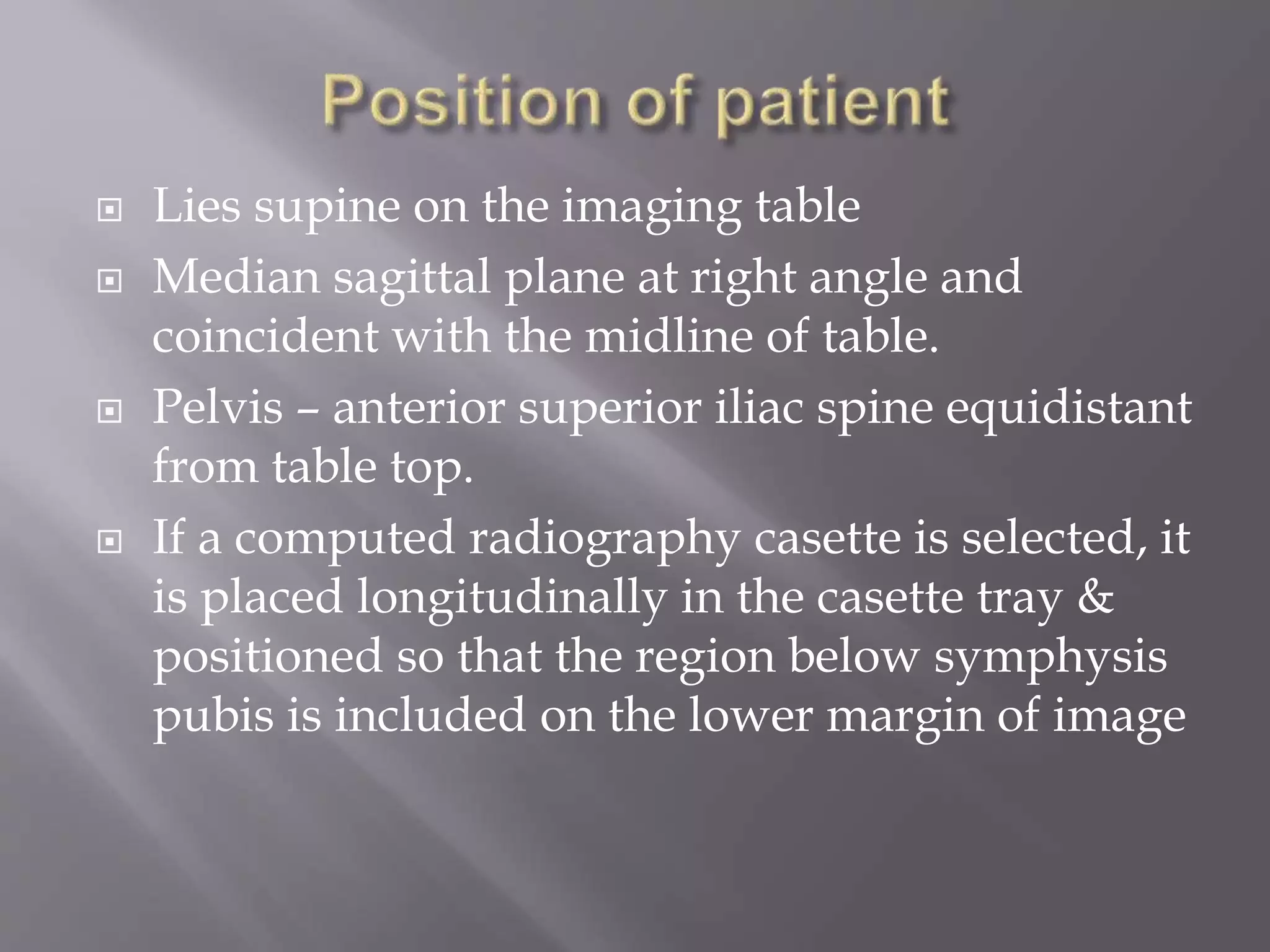
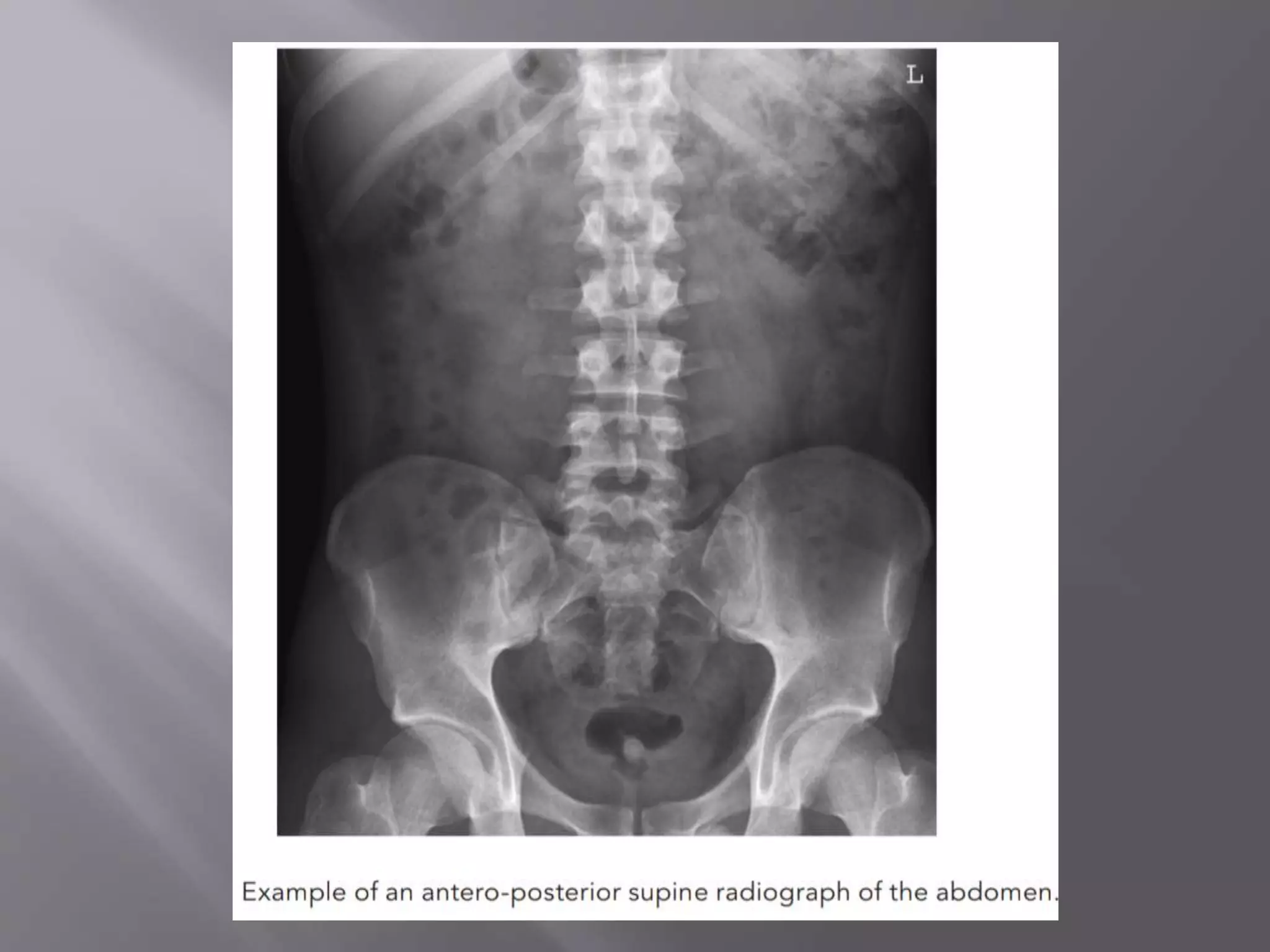
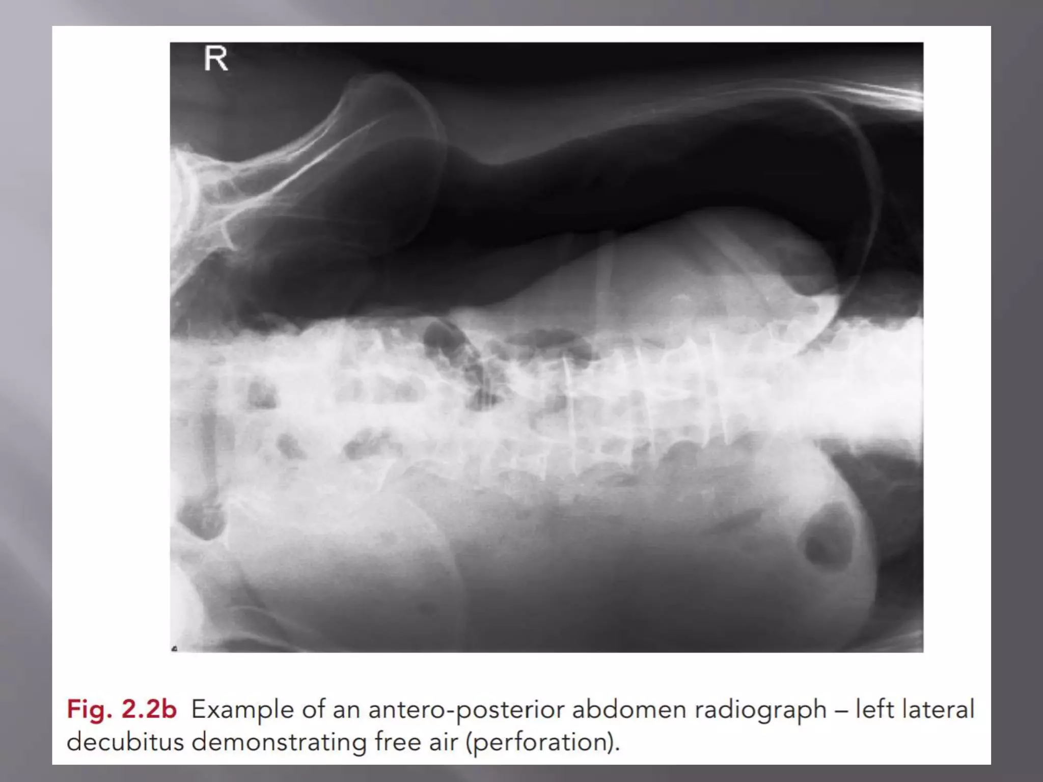
Manyata Tech Park ( Call Girls ) Bangalore ✔ 6297143586 ✔ Hot Model With Sexy...
Abdominal x ray- views.pptx
1.
2.
3. Lies supine on the imaging table
Median sagittal plane at right angle and
coincident with the midline of table.
Pelvis – anterior superior iliac spine equidistant
from table top.
If a computed radiography casette is selected, it
is placed longitudinally in the casette tray &
positioned so that the region below symphysis
pubis is included on the lower margin of image
4.
5. The centre of the image receptor will be
approximately at the level of a point located
1cm below the line joining the iliac crests.
This will ensure that the region inferior to
pubic symphysis is included on the image.
6. The collimated vertical beam is directed to the
centre of the image receptor to include the lateral
margins of abdomen.
Using a short exposure time the exposure is made
on arrested respiration.
Ideally, respiration should be arrested on full
expiration to allow the abdominal contents to lie in
their natural position.
Depending on patient’s height, respiration may
need to be arrested on full inspiration to include the
whole abdomen.
7. The bowel pattern should be demonstrated
with minimal lack of sharpness.
8. In case of a large abdomen an immobilisation
or compression band maybe applied to reduce
the dose to the patient as well as the negative
effect of scatter on image quality.
If required, two images can be taken in
landscape mode to ensure the coverge.
9.
10.
11. This projection is used if the patient cannot be
positioned erect for a chest x ray & is required
to confirm the presence of sub diaphragmatic
air.
Only when other modalities like USG or CT
cannot be used.
May also used for bowel obstruction.
12. The patient lies on their left side on a trolly or a
bed with their elbows and arms flexed so that
the hands can rest near the patient’s head.
The patient is positioned with the posterior
aspect of the trunk against a vertical Bucky
Direct Digital Radiography system with the
upper border of the image receptor high
enough to project above the right lateral
abdominal and thorasic walls.
Alternatively, a 35*43cm CR grid casette is
supported vertically against the patient’s back.
13.
14. When the patient lying on the left side, free gas
will rise to be located between the lateral
margin of the liver & right lateral abdominal
wall.
Before the exposure is made ,the patient should
remain lying on left side for a short period -
10min to allow the gas to collect .
15. The image receptor is positioned at right angles
to the median sagittal plane of the patient.
16. The collimated horizontal central beam is
directed to the anterior aspect of the patient
and centred on the centre of image receptor.
17. The lateral abdominal wall and costophrenic
angle of right lung must be included with the
patient resting on their left side for minimum
10 min. prior to exposure.