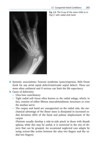
The document is a comprehensive guide titled 'Orthopedic Principles ± A Resident's Guide' by Dr. David Ip, focusing on essential knowledge and practices in orthopaedics. It offers organized chapters covering various specialties including pediatric orthopaedics, hand surgery, and total joint surgery, with illustrations to aid understanding. The guide is tailored for a wide range of readers, including trainees and experienced practitioners, and emphasizes the importance of foundational knowledge for effective patient care and examination preparation.


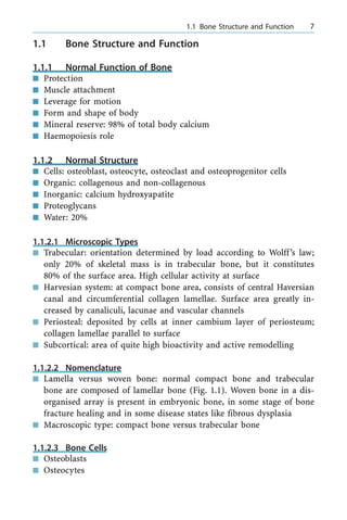
![n Osteoclasts
n Marrow with pluripotential cells
1.1.2.3.1 Osteoblasts
n Functions
± Synthesise matrix (collagen and non-collagenous proteins)
± Work in unison with osteoclasts in bone modelling and remodel-
ling
± Mineralise bone (matrix vesicles production)
± Control electrolyte flux between extracellular fluid (ECF) and the
bone fluid
[act via the receptor activator of NFjB (RANK)/RANK-L system to
exert its action on the osteoclasts]
n Origin likely from undifferentiated mesenchymal cells in connective
tissue and cambium layer of periosteum
n Locations in the bone
e.g. endosteum, under periosteum, at ends of long bones with growth
plate and on surface of newly formed trabecular bone
n Contain alkaline phosphatase that may be used as an indicator of
bone formation
n Transient cells only
1.1.2.3.2 Osteocytes
n Functions
± Possibly act as mechanoreceptor, via its multiple long processes
± Cyclic loading releases chemical mediators, such as insulin growth
factors (IGFs), causing osteoblast formation
± Coordinate formation and resorption of bone
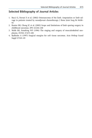
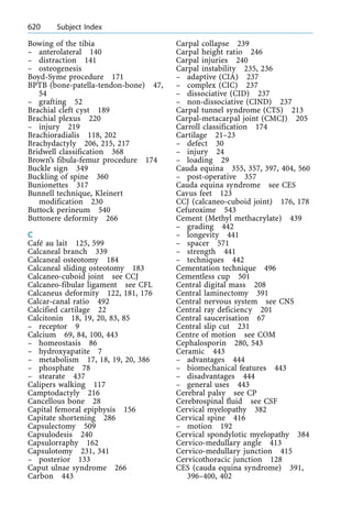
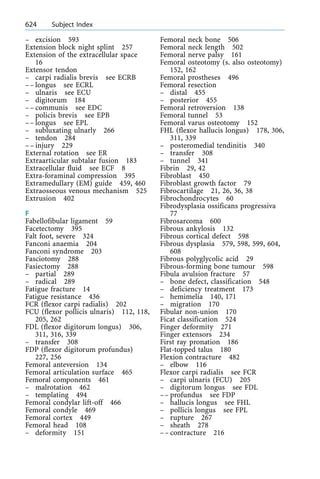
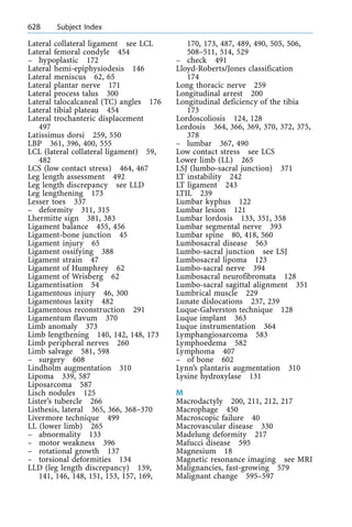
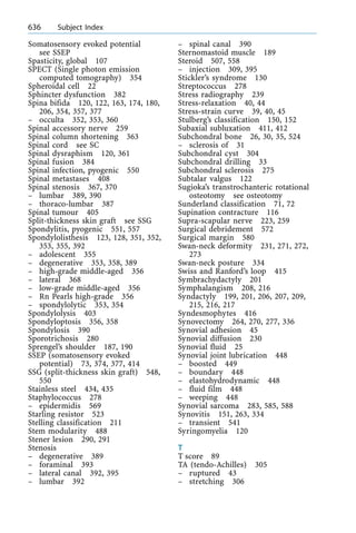
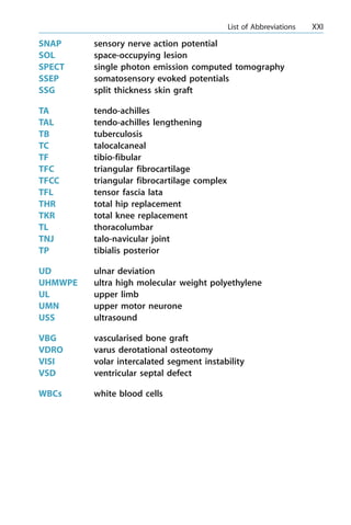
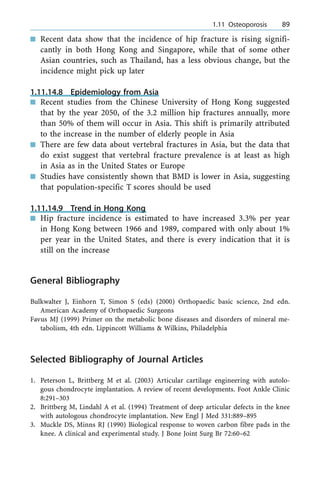
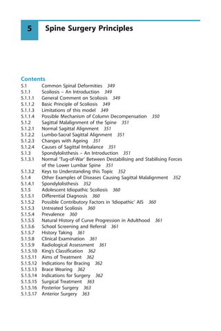
8 1 Orthopaedic Basic Science and Commmon Injuries
Fig. 1.1. Cross-section of lamellar bone](https://image.slidesharecdn.com/aresidentsguideorthopaedics-250108225802-331d4fe3/85/A-Residents-Guide-orthopaedics-surgery-pdf-26-320.jpg)
![± Help in minute-to-minute control of bone mineral homeostasis via
exchange between cell and bone fluid compartment
n Origin
± Incarcerated in bone matrix/lacunae during osteoid formation
± Linkage with one another by fine cellular processes, hence in-
creased surface area of bone matrix. Osteocytes are surrounded by
ECF
1.1.2.3.3 Osteoclasts
n Functions
± Bone remodelling and modelling (remodelling involves sequential
action of osteoclasts and osteoblasts at same site. Modelling in-
volves simultaneous action of osteoclasts and osteoblasts at adja-
cent locations)
± Releasing calcium and digesting collagen
n Origin: From fusion of monocytes of macrophagic lineage in haemo-
poietic marrow
n Transient and very short lived
Mechanism of Action of Osteoclasts
n First decalcify, then digest collagen via:
± First, adhere to bone surface to seal off a space
± Ruffled border formation with nearby clear zone with no organelle
immediately adjacent to the ruffled border
± Endosomes move to ruffled border, transport protons into the dead
space; decalcify bone by making pH 4
± Then, organic matrix degraded by acidic protease and pro-colla-
genase
± Note: osteoclasts have calcitonin receptors which will shrink the
cell and not be able to form ruffled border
Regulation of Osteoclast Activity
n General factors: parathyroid hormone (PTH), 1,25-dihydroxy vitamin
D3, calcitonin and others [oestrogen, androgen, growth hormone (GH)]
n Local chemical factors: cytokines and growth factors
n Local mechanical stimuli
(Note: PTH and vitamin D are unable to stimulate osteoclastic bone
resorption in vitro in the absence of osteoblastic cells ± there is no vi-
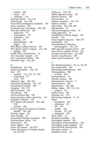
a 1.1 Bone Structure and Function 9](https://image.slidesharecdn.com/aresidentsguideorthopaedics-250108225802-331d4fe3/85/A-Residents-Guide-orthopaedics-surgery-pdf-27-320.jpg)
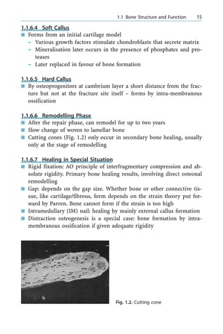
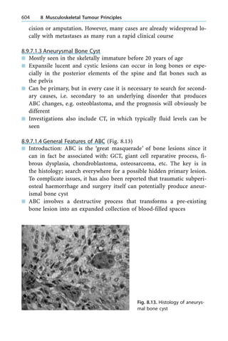
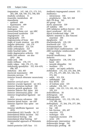
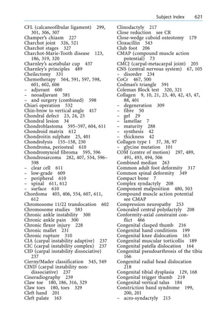
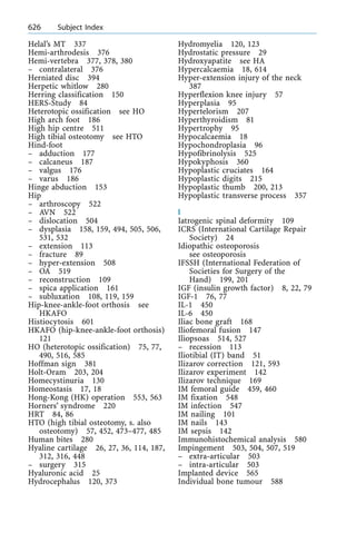
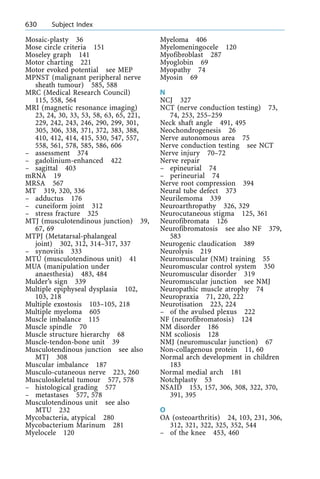
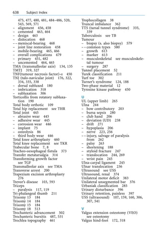
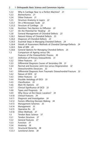
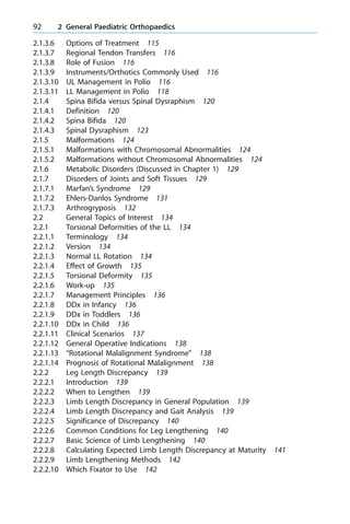
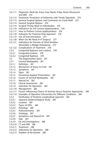
![[By contrast, natural healing by callus can be strong, although often
malaligned. (Fig. 1.3)]
1.1.6.8 `Primary Bone Healing'
n Primary bone healing ± direct osteonal remodelling ± cutting cones at
cortical bone and vascularity and osteoblasts at its trailing end to re-
create the Harversian model
1.1.7 Metabolic Bone Disease
1.1.7.1 Introduction
n To really understand the role of bone in calcium and phosphorus me-
tabolism and in metabolic bone disease, one must note that bone is
really an extension of the extracellular space
n The surface area of exchange is much increased by the hydroxyapatite
crystals ± dimension 200´100 angstroms
16 1 Orthopaedic Basic Science and Commmon Injuries
Fig. 1.3. Natural healing by external calluses](https://image.slidesharecdn.com/aresidentsguideorthopaedics-250108225802-331d4fe3/85/A-Residents-Guide-orthopaedics-surgery-pdf-34-320.jpg)
![1.1.7.2 Basic Rules of Calcium Metabolism
n Bone formation and destruction is coupled
n Unlike PO4, calcium difficult to get across cells and need transport
system ± affected by PTH that creates `holes' in cells and 1,25-dihy-
droxy vitamin D to provide calcium-binding proteins inside cells
1.1.7.3 Normal and Abnormal Calcium and Phosphate Metabolism
1.1.7.3.1 Calcium
n Function: neuromuscular conduction/activation, coagulation cascade,
enzyme co-factor and formation of the rigid mineral content of bone
n Daily requirement: total body calcium 1100 g, 98% of which is in
bone and teeth. Requirement of adult 1000 mg, adolescent 1300 mg
n Absorption from gastrointestinal (GI) tract include: active transport
in duodenum and facilitated diffusion in jejunum. Active transport re-
quires both vitamin D and PTH. PTH is needed for transport across
the cellular membrane and to stimulate production of 1,25-dihydroxy
vitamin D3, while vitamin D helps form proteins to bind calcium in-
side cells
n Of dietary calcium, 35% is absorbed. Excretory routes include both
the GI tract and kidneys. In the kidney, two-thirds is reabsorbed in
the proximal tubule, one-third adjustable absorption in the distal tu-
bule
1.1.7.3.2 Calcium Regulation (Homeostasis)
n Minute-to-minute control (rapid process)
± Involves transfer of calcium from bone surface to ECF in response
to PTH. Involves osteocytes and osteoblasts, especially the former
n Draining from bone reserve (slow process)
± Really a balance between osteoblastic bone formation and osteo-
clastic resorption
n Hormonal control
1.1.7.3.3 Details of Hormonal Control
n PTH: stimulated by low [Ca]. Acts by decreasing renal excretion and
stimulating 1,25-dihydroxy vitamin D, hence increasing GI absorption
of calcium. Acts on osteoclasts to release calcium from bone mainly
indirectly via osteoblast cells
a 1.1 Bone Structure and Function 17](https://image.slidesharecdn.com/aresidentsguideorthopaedics-250108225802-331d4fe3/85/A-Residents-Guide-orthopaedics-surgery-pdf-35-320.jpg)
![n Calcitonin: stimulated by high [Ca]. Acts by decreasing the number
and activity of osteoclasts. Osteoclasts have calcitonin receptors and
calcitonin will shrink the osteoclast in in vitro experiments
n The role of vitamin D was mentioned. Other hormones include GH,
cortisol etc., but are not the main players
1.1.7.3.4 Effect of Phosphates and Magnesium on Calcium Metabolism
n Phosphates: stimulate PTH production, bind to calcium rendering it
inert. Insoluble calcium phosphate crystals deposit to bone and are
removed in stool
n Magnesium: if [Mg] low, suppresses PTH secretion even if [Ca] is low
n Drugs: substances like thiazide diuretics can increase calcium excre-
tion
1.1.7.3.5 Other Issues Concerning Calcium
n Concentration of ionised calcium in blood depends on:
± pH: e.g. acidosis decreases protein binding and causes increased
ionised calcium, etc.
± Albumin concentration: decrease of [albumin] by 1 g/dl changes
protein-bound calcium by 0.8 mg/dl. Thus, hypercalcaemia may be
missed in cases of hypoalbuminaemia
n The reader is referred to physiology texts as regards causes and man-
agement of hypercalcaemia and hypocalcaemia
1.1.7.3.6 Phosphorus
n Functions: component of cell membrane, messenger for hormone
adenosine monophosphate (AMP), energy transfer (adenosine tri-
phosphate/adenosine diphosphate), components of fats and proteins
n Daily requirement: 800 mg
n Distribution: total body contains 600 g; 90% in bone and teeth as re-
servoir, 10% intracellular
n Absorption: in duodenum and small bowel as inorganic phosphate,
depends on amount taken
1.1.7.3.7 Homeostasis
n PTH stimulated by high [P]: acts by increasing urinary excretion and
decreasing absorption. Also increases mobilisation from bone and
stimulates increased 1,25-dihydroxy vitamin D
18 1 Orthopaedic Basic Science and Commmon Injuries](https://image.slidesharecdn.com/aresidentsguideorthopaedics-250108225802-331d4fe3/85/A-Residents-Guide-orthopaedics-surgery-pdf-36-320.jpg)
![n Increased 1,25-dihydroxy vitamin D in turn increases phosphate ab-
sorption in the gut
n Other regulators: e.g. GH increases renal absorption, calcitonin de-
creases its excretion in urine
1.1.7.3.8 Appendix 1
Role of Vitamin D in Calcium and Phosphate Metabolism
n UV light converts dietary pro-vitamin D2 and D3 to cholecalciferol
n Then undergoes 25- and 1-hydroxylation at the liver and kidney re-
spectively (in cases with high calcium, mainly 24,25-dihydroxy vita-
min D will be made in the kidney)
n Acts on targets by diffusion through cytoplasmic membrane; bound
to cellular protein and transported to the nucleus
n In bone, stimulates both osteoblasts and osteoclasts, increases miner-
alisation and resorption and increases collagen production
n Kidney: like PTH, vitamin D increases calcium absorption. However,
unlike PTH, increases phosphate absorption
n GI tract: synthesis of calcium-binding proteins helps calcium absorp-
tion and increases phosphate absorption
Role of PTH in Calcium and Phosphate Metabolism
n Produced in parathyroid gland, prepro-PTH made from mRNA,
changed to pre-PTH in rough endoplasmic reticulum, then PTH in
Golgi apparatus and packed for secretion
n Acts as a sensitive control of [Ca]
n Mechanism: most actions via membrane receptor attachment, in turn
activating cAMP and protein kinase and increasing intracellular cal-
cium
n Bone: (short-term) induces increased permeability of osteocyte cell
membrane to calcium, promotes exchange between calcium and ECF.
(Longer term) induces osteoclasts (via osteoblasts) to mobilise cal-
cium from bone
n Kidney: increased calcium but decreased phosphate absorption, in-
creased 1,25-dihydroxy vitamin D
n GI tract: increased calcium and phosphate absorption via increased
vitamin D
a 1.1 Bone Structure and Function 19](https://image.slidesharecdn.com/aresidentsguideorthopaedics-250108225802-331d4fe3/85/A-Residents-Guide-orthopaedics-surgery-pdf-37-320.jpg)
![Role of Calcitonin in Calcium and Phosphate Metabolism
n Produced from the C cells of thyroid as a large precursor, then
cleaved into 32 amino acids
n Stimulated by increased [Ca]
n Mechanism: binds to cell membrane receptor and activates adenyl cy-
clase
n Bone: shrinks osteoclasts, prevents ruffled border formation and in-
hibits formation of osteoclasts
n Kidney: decreases absorption of calcium and phosphate
n GI tract: uncertain, storage of calcium in peri-lacunar space after
meal
1.1.7.3.9 Appendix 2
Types of Rickets
n Common types include:
± Nutritional: supplements needed
± Vitamin D-resistant rickets: PO4 lost in urine ± PO4 and vitamin D
needed in treatment (Rn)
± RTA: Needs alkaline in Rn
± End organ non-response: needs activated 1,25-dihydroxy vitamin
D3
± Renal rickets: loss of the normal natural adjustments of calcium
absorption and PO4 excretion ± Rn not easy, consult renal physi-
cian
Adult Osteomalacia
n Massachusetts General Hospital study: 27% of patients coming in for
hip fracture with osteomalacia
n Sometimes co-exists with osteoporosis
n Looser's zones and milkman's lines at the concave side of the long
bone if present are characteristic
20 1 Orthopaedic Basic Science and Commmon Injuries](https://image.slidesharecdn.com/aresidentsguideorthopaedics-250108225802-331d4fe3/85/A-Residents-Guide-orthopaedics-surgery-pdf-38-320.jpg)
![n (Collagen fibres of cartilage not anchored into bone; but the cartilage
tissue is `keyed' into the irregular surface of bone like a jigsaw puz-
zle)
1.2.6 Nutrition: Two Barriers to Diffusion
n Synovial linings
n Different layers of cartilage
1.2.7 On the Potential for `Healing'
n Unlike adult cartilage, foetal cartilage can heal spontaneously (seen in
foetal lamb animal studies)
n PT defects ± transient cell reaction in the edge of lesion, usually does
not heal (except in the rare case when mesenchymal cells can be in-
duced to migrate from the synovial membrane across the articular
surface into the defect)
n Osteochondral lesions heal with fibrocartilage: combined type 2 and 1
collagen, not tough enough to withstand high compression stresses
n Salter had shown a positive effect of continuous passive motion
(CPM) on cartilage healing (e.g. in studies with periosteal grafts) ±
immobilization is detrimental to cartilage repair and development
1.2.8 General Management of Chondral Defects
1.2.8.1 Natural History of Chondral Defects
n (Arthroscopy 1997) ± In over 30,000 knee arthroscopies for knee inju-
ries, a chondral lesion was found in 60%
n Other studies report 23% when associated with anterior cruciate liga-
ment (ACL) injuries, increasing to 54% with chronic ACL injuries
n But, exact natural history of isolated lesions not known for certainty
1.2.8.2 Diagnosis of Chondral Defects
n With an arthroscope
n Magnetic resonance imaging (MRI)
n Others [physical examination (P/E) not too reliable]
n New method: Finland originated the use of articular cartilage stiffness
tester/probe (trade name ArtScan) ± potentially useful since cartilage
softening is one of the first signs of the degenerative process ± seems
a 1.2 Cartilage Structure and Function 23](https://image.slidesharecdn.com/aresidentsguideorthopaedics-250108225802-331d4fe3/85/A-Residents-Guide-orthopaedics-surgery-pdf-41-320.jpg)
![n New advances: spectroscopic imaging with view-angle tilting; spec-
tral-spatial 3-D magnetisation transfer; high-resolution short echo
time spectroscopic imaging, etc.
1.2.8.6 General Options for Managing Chondral Defects
n Supervised neglect
n `Chondro-protection'/debridement and curettage [mainly for relief of
say, knee osteoarthritis (OA) symptoms]
n Fixation of chondral fragments (especially if osteochondral)
n Stimulation of healing ± drilling, abrasion, micro-fracture techniques
n Autologous osteochondral transfer
n Autologous chondrocyte culture/reimplantation (ACI)
n Periosteal and perichondrial transplantation
n Use of allografts and growth factors
n Future ± regeneration and gene therapy
1.2.8.6.1 Supervised Neglect
n Increasing evidence that especially FT defects may lead to OA
n Although some defects remain asymptomatic for considerable period
and true incidence not known for sure
1.2.8.6.2 `Chondro-Protection'
n Hyaluronic acid: animal studies claim protective effect; mechanics ±
unknown. Probably provides lubrication
n Glucosamine and chondroitin sulphate: since OA results when carti-
lage breakdown exceeds synthesis of chondrocytes, providing exoge-
nous glucosamine increases matrix production and alters natural
course. While chondroitin sulphate is the most abundant glucosami-
noglycans in articular cartilage, may inhibit many degradative en-
zymes in synovial fluid in OA
1.2.8.6.3 Lavage and Debridement
n Pain relief in OA cases from removal of loose intra-articular debris,
inflammation, mediators, enzymes. Changing the ionic environment
of the synovial fluid
n Paper by Robert Jackson in the States: relief in 50% at 3.5 years ±
seems better if mechanical debridement added
n Drawback: cannot treat the chondral defect
a 1.2 Cartilage Structure and Function 25](https://image.slidesharecdn.com/aresidentsguideorthopaedics-250108225802-331d4fe3/85/A-Residents-Guide-orthopaedics-surgery-pdf-43-320.jpg)
![n Prognosis: depends on age and other factors (better if small lesion, in
classic position, stable on arthroscopic exam or MRI)
n Most common affected joints: knee, elbow, ankle
n For the knee, most common classical area ± lateral part of the medial
femoral condyle
1.4.4 Possible Aetiology of OCD
n Trauma
n Repeated microtrauma from impingement of the tibial spine
n Stress fracture with no injury
n Vascular (ischaemic event)
n Defects of ossification
1.4.5 Classification
n Juvenile form
n Adult form
[differential diagnosis (DDx) is before and after closure of epiphysis]
1.4.6 Main Rn Options
In general: non-operative for intact lesions and operative in unstable le-
sions
n Excision (weight-bearing joints treated with removal of sizeable OCD
fragments that are detached tend to do poorly)
n Some are amenable to fixation
n If cannot fix: consider cartilage repair techniques, e.g. subchondral
drilling; allograft, osteochondral autograft transplantation (OATS),
ACI, perichondral (open) and periosteal (open) resurfacing
1.4.7 Clinical Significance of OCD
n If defect in weight-bearing part of knee joint, degeneration of affected
compartment later possible
n Loose bodies, if present, can cause locking
n For OCD, important to realise that age has important influence on
prognosis
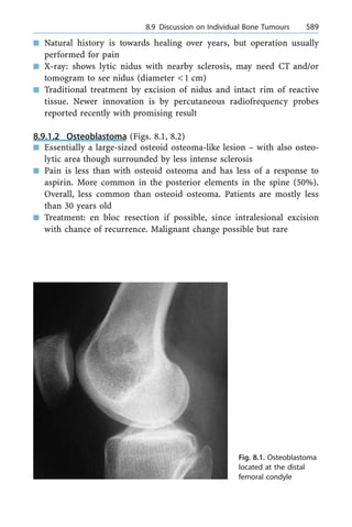
a 1.4 Osteochondritis Dessicans 33](https://image.slidesharecdn.com/aresidentsguideorthopaedics-250108225802-331d4fe3/85/A-Residents-Guide-orthopaedics-surgery-pdf-51-320.jpg)
![n Age: affects also the healing potential
n Others: size of lesion, location (especially weight-bearing)
n Diagnosis: e.g. OCD versus trauma versus hereditary epiphyseal
anomalies
1.4.13 Management Schemes
n Group 1/Juvenile: trial of conservative Rn appropriate for skeletal im-
mature cases, success rates from 50% to 90% reported
n Group 2/Adolescent: assessment of remaining growth potential is most
important
n Group 3/Adult: management can be quite similar to adult osteochon-
dral fracture
1.4.14 Management
n Pure chondral small lesion/small osteochondral defects can initially
be treated in the immature conservatively
n Success of conservative Rn may depend on adequate subchondral
bone attachment (theoretically, if no subchondral bone attached to
the fragment, there is little chance for the articular lesion to heal)
1.4.15 Operative Rn
n Arthroscopic drilling and fixation
± Based on the theory that the lesion is regarded as a fracture non-
union, works by penetration of the subchondral bone to initiate in-
flammatory cascade
± Types: antegrade versus retrograde drilling, an undesirable conse-
quence of antegrade drilling, is the creation of permanent drill
holes in the articular surface that fill with fibrocartilage [retrograde
drilling with bone graft (BG) sometimes advocated for in situ le-
sions with intact overlying articular cartilage]
± Fixation of osteochondral lesions increase the likelihood of main-
taining joint congruity during healing and potential to allow early
range of motion (ROM; by means of K-wires, Herbert, Acutrak) ±
important to bury screw head
n Abrasion chondroplasty and microfracture
± Again, stimulation of cartilage regeneration by penetration of the
subchondral bone to release pluripotential stem cells
a 1.4 Osteochondritis Dessicans 35](https://image.slidesharecdn.com/aresidentsguideorthopaedics-250108225802-331d4fe3/85/A-Residents-Guide-orthopaedics-surgery-pdf-53-320.jpg)
![(The low metabolic rate enables it to remain under tension for long
periods without risk of ischaemia and necrosis)
1.5.7 Blood Supply
n Perimysial at MTJ
n Periosteal at OTJ
n Paratenon ± major supply
Paratenon vessels enter the tendon substance and passing longitudi-
nally within the endotenon sheath form a capillary loop network
n Tendons enclosed in synovial sheaths are supplied by vinculae
n Vascularity compromised at junctional zones and areas of friction/tor-
sion/compression ± no capillary anastomosis: [e.g. (1) supraspinatus
near its insertion, (2) Tendo-achilles (TA) ± where the combined ten-
don of Gastrosoleus undergoes a twist that raises stresses across this
site]
1.5.8 Nerve Supply
n Innervation mainly afferent
n Nerve endings mostly at MTJ
n Four types of nerve endings:
± Free ± pain reception
± Golgi ± mechano-reception
± Paccinian ± pressure sensors
± Ruffini
1.5.9 Biomechanics
n Tendon is the strongest component in the muscle-tendon-bone unit
n Tensile strength is one-half that of stainless steel (e.g. 1 cm2
cross-sec-
tion can bear weight of 500±1000 kg)
1.5.10 Force±Elongation Curve
n Less useful than the stress±strain curve because, unlike the stress±
strain curve, not only depends on the mechanical behaviour of the
tissue; shape of curve also depends on the length and cross-sectional
area (more cross-sectional area, larger loads can be applied; longer
the tissue fibres, greater the elongation before failure)
a 1.5 Tendon Basic Science 39](https://image.slidesharecdn.com/aresidentsguideorthopaedics-250108225802-331d4fe3/85/A-Residents-Guide-orthopaedics-surgery-pdf-57-320.jpg)
![Motor Unit: Number of Muscle Cells Innervated
by a Single Motor Neuron, from 10±2000
n All or none
n In big, two-jointed muscles: ratio high
n Fine co-ordination, e.g. eye muscles: ratio is small
Motor End Plate
n Wasted away after too long denervation (2 years)
NMJ: Release of Acetylcholine (ACh)
Across Synapse on Arrival of Impulse
n Negative inhibition either competitive, e.g. curare that binds ACh re-
ceptors, or non-competitive, e.g. depolarising agent such as suxa-
methonium
n Reversal agents include, for example, neostigmine which prevents
ACh breakdown and reverses non-depolarising agents
Muscle Structure Hierarchy
n Start with sacromere (between two Z lines)
n ? Myofibril ? muscle fibre ? fasicle ? muscle
n A/`Skeleton' of each muscle fibre is endomysium ± the sacroplasmic
reticulum, rather like endoplasmic reticulum, is calcium rich
n T-tubes penetrate, help spread action potential
n B/Exo-`skeleton'; with perimysium around each fascicle and epimy-
sium around each bundle of muscle
MTJ
n Weak link between muscle and tendon
n Especially injured in eccentric exercises
n Although sometimes it is either the muscle proper which is partial or
complete tear, or sometimes tendon itself (tendon has stronger tensile
strength than muscle)
[P.S. Tendon more likely injured with greater muscle force (eccentric);
also depends on any weakness of the tendon and ratio of cross-sec-
tion of muscle versus tendon]
68 1 Orthopaedic Basic Science and Commmon Injuries](https://image.slidesharecdn.com/aresidentsguideorthopaedics-250108225802-331d4fe3/85/A-Residents-Guide-orthopaedics-surgery-pdf-86-320.jpg)
![1.9.4 Outcome
n Sprouts make distal connection then nerve fibre matures (increased
axon and myelin thickness)
n Neurites that fail to make distal connection die back and are lost; if
the perineurium is not disrupted, then the axons will be guided along
the original path at 1 mm/day
1.9.5 Seddon Classes of Nerve Injury
n Neuropraxia: most are compressive in aetiology ? local conduction
block/demyelination ± heal by repair of demyelination, especially of
the thick myelin nerves
n Axonotmesis: mostly traction Ô severe compression cases ? Waller-
ian degeneration, prognosis not bad since will regenerate and not
mis-wire (sensory recovers better since sensory receptors live longer,
especially in more proximal injuries)
n Neurotmesis ± complete cut, no recovery unless repaired ± yet can
mis-wire, hence reduced mass of innervation
1.9.6 Sunderland Classification (Six Types)
n Neuropraxia: no Tinel sign
n Axon: both epi- and perineurium intact, Tinel +, progresses distally
n Axon: only epineurium injured, Tinel +, progresses distally
n Axon: perineurium injured, Tinel + but Tinel not progressing distally
n Neurotemesis
n Neuroma in continuity (i.e. partly cut nerve, the remainder can be
first/second/third/fourth degree of injury)
1.9.7 Feature of the Sunderland Classification
n Accounts for injuries between axonotmesis and neurotemesis ± based
on involvement of perineurium
1.9.8 Assessing after a nerve injury
n Motor: assess power and diagnose level of injury
n Sensory: mapping and pattern recognition
n Autonomic: e.g. wrinkle test
± [Reflex sympathetic dystrophy (RSD) in 3%, featured by swelling,
porosis, sweating, pain, etc.]
± Tinel sign may be present
a 1.9 Neural Injury 71](https://image.slidesharecdn.com/aresidentsguideorthopaedics-250108225802-331d4fe3/85/A-Residents-Guide-orthopaedics-surgery-pdf-89-320.jpg)
![± [Note: motor end plates waste away in 1 year, muscle atrophied by
2±3 months, sensory function (erratic) can return in up to 2 years]
1.9.17.1 Assessing Return of Motor Power
n M0: nil
n M1: see contraction proximally
n M2: see contraction proximally and distally
n M3: muscle against resistance
n M4: some independent movements possible
n M5: normal
1.9.17.2 Assessing Return of Sensation (General Recovery Sequence:
Pain > Touch > Vibration > Two-Point/Stereognosis)
n S0: nil in nerve autonomous area
n S1: deep pain in auto area
n S2: some pain and touch in autonomic area
n S3: pain and touch over whole autonomic area
n S4: two-point returns
n S5: normal
(Two point discrimination best correlation to final function)
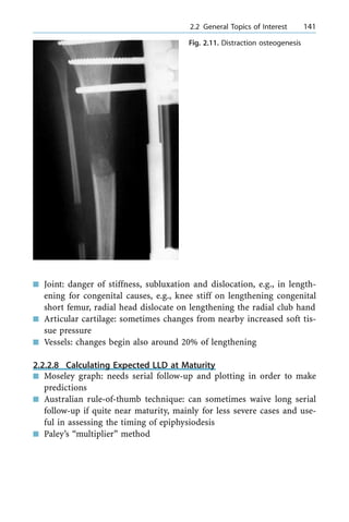
1.10 Growth Factors in Orthopaedics
1.10.1 Clinical Relevance and Introduction
n Most experiments done in animals, interactions complex. Clinical
trials mostly not yet quite available, with some exceptions
n Applications:
± Medicine: e.g. GH defects
± Orthopaedics:
Enhancement in fracture healing and role in normal bony healing
Enhancement of fusion OTs and Rn of bone defects
Possible future use in osteoporosis
Distraction osteogenesis
BMP-related clinical diseases, possible future role of inhibition of
heterotopic ossification (HO) by antagonists
Future role in management of osteolysis around total joint im-
plants
a 1.10 Growth Factors in Orthopaedics 75](https://image.slidesharecdn.com/aresidentsguideorthopaedics-250108225802-331d4fe3/85/A-Residents-Guide-orthopaedics-surgery-pdf-93-320.jpg)
![1.11.13.5 Individual Agents
1.11.13.5.1 Calcium and Vitamin D
n All Rn regimes rendered futile if there is not adequate calcium
n Helps prevent bone loss, especially in elderly ± especially in institu-
tions
± Most studies have not shown fracture reduction, but reduction or
prevention of bone loss
± Though one study says it does by Dawson-Hughes in NEJM
1.11.13.5.2 HRT as a Preventive Agent
n Two very important studies (on fracture risks)
± HERS [oral oestrogen and progestin ± no definite decrease in frac-
ture (if not more fracture) ± do not reduce myocardial infarction
[MI]/cardiovacular system (CVS) morbidity]
± WHI [women's health initiative]
Important large RCT with more than 16,000 women shows a hip
fracture risk after 5.2 years
Trial stopped early due to an increase in cardiovascular events in
the HRT group
n HRT increases in bone density are accepted
n New advice from some experts
± Better taper off HRT, especially in cases of CHD and breast cancer
± Oestrogen sometimes used to decrease menopausal symptom, but
not in osteoporosis reduction
Latest Results of the WHI Study
n It took three decades to get this formal RCT done, which involved
more than 16,000 patients and compared oestrogen/progestin with
placebo
n The study ended prematurely in May 2001 because HRT caused a
higher incidence of: heart disease (30%), breast cancer (26%), cere-
brovascular accident (CVA) (40%) and pulmonary embolism (110%)
n Morbidity far greater than the benefits of less hip fracture (±34%)
and colon cancer (±37%)
84 1 Orthopaedic Basic Science and Commmon Injuries](https://image.slidesharecdn.com/aresidentsguideorthopaedics-250108225802-331d4fe3/85/A-Residents-Guide-orthopaedics-surgery-pdf-102-320.jpg)
![Details of Bisphosphonates
n Contraindications:
± Oesophageal problems (not with risedronate) sometimes less with
re-challenge
± Patient cannot stand/sit-up for various reasons
± Drug sensitivity
± Low [Ca] with renal failure
n Uses:
± Prevention and Rn of osteoporosis
± Possible role (animal model) in total joint osteolysis
± IV administration, e.g. pamidronate in malignant hypercalcaemia
n Dose:
± As prevention: 5 mg q.d., or 35 mg per week
± As Rn: 10 mg q.d., or 70 mg per week
n Administration: glass of water on empty stomach in morning before
eating and drinking, remain upright for 0.5 h
n Potential disadvantage: decreased micro-repair of bone, and bones
may be more brittle with chronic use
1.11.13.5.6 Combination Therapy
n HRT and alendronate: with 20% additive effect
n Other combinations: minimal effect
n Experimental: intermittent PTH and bisphosphonates
1.11.13.5.7 Experimental Agents
PTH: Two Main Actions
n Helps calcium homeostasis: maintains serum calcium for proper cell
function ± across three areas: gut, bone and kidney
n Controls and modulates cells active in the remodelling cycle ? increases
the size, activity and working life of osteoblasts and strengthens the tra-
beculae ± sometimes even bypassing local mechanostatic limits
[Given continuously ± bone is lost; given intermittently ± works via
TGF-b, decreasing apotosis of osteoblasts]
± Action stops on withdraw ± but this might be blocked by alendro-
nate; said to be good for steroid cases
± Some initial reports of associations with osteosarcoma in animals,
but now thought to be dose and species related; trials are resumed
86 1 Orthopaedic Basic Science and Commmon Injuries](https://image.slidesharecdn.com/aresidentsguideorthopaedics-250108225802-331d4fe3/85/A-Residents-Guide-orthopaedics-surgery-pdf-104-320.jpg)
![4. Finkelstein JS, Klibanski A et al. (1998) Prevention of estrogen deficiency related
bone loss with human parathyroid hormone: a randomized controlled trial. JAMA
280:1067±1073
5. Boyan BD, Schwartz LD, et al. (1992) Effects of bone morphogenic protein on the
expression of glycosaminoglycans, collagen, and alkaline phosphatase in nonunion
cell cultures. CORR 278:286±304
6. Salter RB, Simmonds DF et al. (1980) The protective effect of continuous passive
motion on the healing of full thickness defects in articular cartilage. An experimen-
tal investigation in the rabbit. J Bone Joint Surg Am 62:1232±1251
7. Shelbourne KD, Jari S et al. (2003) Outcome of untreated traumatic articular de-
fects of the knee. A natural history study. J Bone Joint Surg Am 85A[Suppl 2]:8±16
8. Laros GS, Cooper RR et al. (1971) Influence of physical activity on ligament inser-
tions in the knees of dogs. J Bone Joint Surg Am 53:275±286
90 1 Orthopaedic Basic Science and Commmon Injuries](https://image.slidesharecdn.com/aresidentsguideorthopaedics-250108225802-331d4fe3/85/A-Residents-Guide-orthopaedics-surgery-pdf-108-320.jpg)
![2.1 Generalised Disorders
2.1.1 Skeletal Dysplasia
2.1.1.1 Definition of Terms
n Dysplasia: The term implies a generalised abnormality in growth and
development
n Dysostosis: Denotes maldevelopment of a single bone or body seg-
ment
n Dystrophy: Defined as a disorder, usually congenital, of the structure
or function of an organ or tissue due to its ªperverted nutritionº. It
includes agenesis, atrophy, hypertrophy, hyperplasia and metaplasia
2.1.1.2 Other Terminology
n Of limb shortening:
± Rhizomelic, i.e. proximal short
± Mesomelic, i.e. middle short
± Acromelic, i.e. distal segment short
n Descriptive terms:
± Diatrophic: Twisted
± Campomelic: Bent limb
± Metatrophic: Changing
± Hyphomelic: Bend forward
2.1.1.3 Classification (Rubin)
n Epiphysis: hyperplasia (e.g. Trevor)
± Hypoplasia [spondylo-epiphyseal dysplasia (SED), multiple epiphy-
seal dysplasia (MED), pseudo-achondroplasia]
n Physis: hyperplasia (e.g. endochondromatosis)
± Hypoplasia (e.g. achondroplasia)
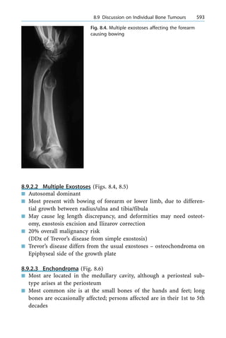
n Metaphysis: hyperplasia (e.g. multiple exostoses)
± Hypoplasia (e.g. osteopetrosis)
n Diaphysis: hyperplasia (e.g. diaphyseal dysplasia)
n Hypoplasia [e.g. osteogenesis imperfecta (OI)]
Others: e.g. osteopathia striata (Fig. 2.1)
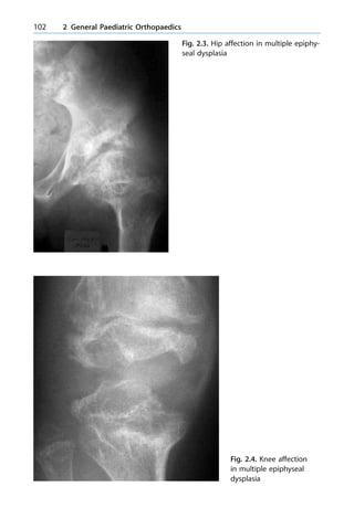
a 2.1 Generalised Disorders 95](https://image.slidesharecdn.com/aresidentsguideorthopaedics-250108225802-331d4fe3/85/A-Residents-Guide-orthopaedics-surgery-pdf-113-320.jpg)
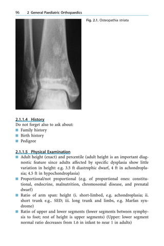
![n Dysmorphic features
n Clinical deformity
[distal upper limb (UL) to proximal 75% usually; distal lower limb
(LL) to proximal 82%]
2.1.1.6 Investigation
n X-ray (XR): skull, pelvis, lumbar spine, hands/wrists, knees
n Laboratory work: e.g. endocrine tests, genetic tests
n Pathology/histological anomalies: e.g. some dysplasia have special mi-
croscopic abnormalities ± pseudoachondroplasia, diastrophic dwarf-
ism, kniest
2.1.1.7 General Goals of Treatment
n Accurate diagnosis (Dx)
n Genetic counsel
n Psychological support
n Symptomatic treatment:
± Orthopaedic: e.g. prevent deformity, stabilise lax joints, support
brittle bones, correction of leg length discrepancy (LLD), joint re-
construction
± Others: dental, eye consult, etc.
n Hormonal
n ÔGene therapy
2.1.1.8 Specific Dysplasias
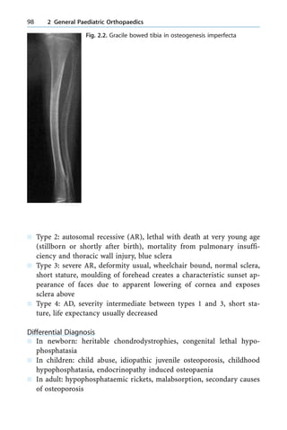
2.1.1.8.1 Osteogenesis Imperfecta (Fig. 2.2)
n A hereditary condition featured by tendency to have multiple frac-
tures from mutation and changed structure of type-1 collagen. Asso-
ciated with involvement of other body tissues, such as poor dentition,
laxity of ligaments, eye (cornea thinned), etc.
n The fracture callus in OI ± heals and appears early; is identical to the
rest of the skeleton, i.e. easily deformed with weight-bearing and mus-
cle actions
Silence Classes
n Type 1: mildest, autosomal dominant (AD), blue sclera, normal life
expectancy
a 2.1 Generalised Disorders 97](https://image.slidesharecdn.com/aresidentsguideorthopaedics-250108225802-331d4fe3/85/A-Residents-Guide-orthopaedics-surgery-pdf-115-320.jpg)
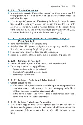
![2.1.2 Cerebral Palsy
2.1.2.1 Definition
n Group of non-progressive, motor (mainly) impairment syndromes
secondary to lesions or anomalies of the brain from foetal to around
age 2 years
± [But its manifestations can change over time with growth, develop-
ment and maturation]
n Can have defects in sensation, cognition, seizure, gastrointestinal/gen-
itourinary problems, etc.
2.1.2.2 Aetiology
n Intrinsic abnormal central nervous system (CNS) structure (e.g. chro-
mosomal, metabolic disease)
n External insult (e.g. infection, ischaemia)
a 2.1 Generalised Disorders 105
Fig. 2.7. Multiple exostoses affecting
the ankle
Fig. 2.8. Ollier's disease](https://image.slidesharecdn.com/aresidentsguideorthopaedics-250108225802-331d4fe3/85/A-Residents-Guide-orthopaedics-surgery-pdf-123-320.jpg)
![n Implication
± Prone to recurrence of deformity
± Bony surgeries on both sides usually may be required
± Adequate release of adductor on one side and very minimal only
release of adductor on the side with active abductor tone ± if not,
one ends up with abduction deformity
± If varus derotational osteotomy (VDRO) is planned ± one side IR,
the other side external rotation (ER)
2.1.2.10.3 Problem 3: Impending Hip Subluxation
n Frequently for these cases, there is a roughly 2-year window of oppor-
tunity to tackle the impending dislocation
n Parents must be told there is a 30±40% chance of being symptomatic
with pain if left untreated
n Treatment mostly involves both femoral varus derotational osteotomy
and pelvic surgery: most of these lack posterolateral coverage, so-
called triplane posterolateral actabuloplasty may be needed. Subluxa-
tion cases usually no degeneration yet. But acetabulum sometimes
does wear away quickly, especially posteriorly, and may need 3D com-
puted tomography (CT) scan preoperatively for assessment
[Note: Acetabulum widely believed to remodel up to age 4 years and
much less remodelling after age 8 years]
2.1.2.10.4 Chronic Hip Dislocation with No Pain
n If chronic and no pain, and does not interfere with transfers, etc. ±
can be observed (especially if bilateral)
2.1.2.10.5 Painful Hip Dislocation with Symptomatic OA
of Femoral Head
n Open capsule and look at head
n If good ± reconstruction of hip on both sides
n If bad ± resect femoral head, can consider a hypervalgus osteotomy
(the previous Shanz operation described by Rang is less used nowa-
days)
n Other options for late OA. Not with good results and seldom recom-
mended (hip arthroplasty unlikely to be good with this osteoporotic
bone in wheelchair bound patients and uneven muscle pull; resection
arthroplasty causes much post-op pain and proximal migration, even
if post-op traction given.)
108 2 General Paediatric Orthopaedics](https://image.slidesharecdn.com/aresidentsguideorthopaedics-250108225802-331d4fe3/85/A-Residents-Guide-orthopaedics-surgery-pdf-126-320.jpg)
![2.1.3.3 Cause of Deformity
n Muscle imbalance
n Example: Quadriceps weakness, the antagonists hamstring also slowed
growth since intermittent stretching is the stimulus by opposition to
growth
n As the femur grows, the relatively short hamstring causes a contrac-
ture
n It follows that deformities are less commonly seen in polio adults
whose paralysis increased in adulthood (e.g. post-polio syndrome)
n The child frequently ambulates by crawling
n Scoliosis is common (sometimes no intercostal muscles to even hold
the rib cage)
n Deformities usually do not occur in the totally paralysed
n Careful document with muscle charts [Medical Research Council
(MRC) grading] of each and every muscle group
2.1.3.4 Key to Testing Different Muscle Groups
n Against gravity if weak
n If not that weak, against resistance
n Beware of trick movements (e.g. weak TA power with some plantar
flexion still made possible by toe flexors)
2.1.3.5 Chronic Phase
n This is the phase that may need surgery
n Greater than 18 months
n Earlier neglect is a frequent cause that makes later repeated surgeries
necessary
n XR: Osteoporosis and thinned cortices common
2.1.3.6 Options of Treatment
n Physiotherapy
n Casting: Milder ones Ô wedging
n Surgery ? e.g. TAL, or lengthen hamstrings, knee extension osteot-
omy (In more supple cases, tendon transfers may be considered)
n All three methods combined to treat severe deformities
a 2.1 Generalised Disorders 115](https://image.slidesharecdn.com/aresidentsguideorthopaedics-250108225802-331d4fe3/85/A-Residents-Guide-orthopaedics-surgery-pdf-133-320.jpg)
![Elbow [Biceps Paralysis]
n Biceps if paralysed are disabling, cannot bring food to mouth
n Three options:
± Steindler flexorplasty (transfer origin of forearm flexors)
± Pectoralis major transfer to the biceps
± If deltoid also paralysed, may need fusion if scapular muscles func-
tional (But avoid surgery if no hand function present)
Elbow (Triceps Paralysis)
n Cannot extend elbow with triceps paralysed
n Triceps is needed for Calipers walking
n No tendon transfer for triceps
n But not too disabling if the LL are functioning well, however
Forearm Contractures
n May need tendon transfers to restore muscle balance
n Pronation/supination contracture: may need to release the interosse-
ous membrane (IOM), sometimes reattach site of insertion of biceps
Hand Problems
n Intrinsic hand muscle
n Extrinsic flexors and extensors
± Differentiate between intrinsic versus extrinsic weakness or both
and treat accordingly
Thumb Opposition Weakness
n Difficult to pick up objects
n May be forced to use the clumsy key pinch
n Example of operation: Camitz, flexor digitorum superficialis 4th; but
test the extrinsics first
Interossei Weakness
n Clawing results (intrinsic minus hand)
n But much lesser than, say, leprosy or the usual ulna nerve palsy
n EPB ? 1st dorsal plus tendon transfers as in ulna nerve palsy (to cor-
rect the MCPJ hyperextension)
a 2.1 Generalised Disorders 117](https://image.slidesharecdn.com/aresidentsguideorthopaedics-250108225802-331d4fe3/85/A-Residents-Guide-orthopaedics-surgery-pdf-135-320.jpg)
![2.1.5 Malformations
2.1.5.1 Malformations with Chromosomal Abnormalities
n Examples include Turner's and Klinefelter's syndrome, which will not
be discussed in this book
2.1.5.2 Malformations without Chromosomal Abnormalities
[Only neurofibromatosis (NF) will be discussed]
2.1.5.2.1 Introduction
n Multisystem
n Affects cell growth of neural tissue
n AD for types 1 and 2, 50% as new mutants in NF 1: Identification of
mutation by the ªprotein truncation testº. Detects a shortened p prod-
uct; this test only can detect 70% of NF 1 mutations
n Not rare ± 1 in 4000, NF 2 very rare
n Protein product of NF 1 is neurofibromin, of NF 2 is schwannomin
n Two peaks for severe clinical problems, age 5±10 years and age 35±50
years, the latter peak related to CA
2.1.5.2.2 Types
n NF 1: Protean manifestations, every patient with NF 1 will eventually
show some features of the disease (sometimes more prominent after
adolescence); categories include neurocutaneous stigmata, eyes, tu-
mours, tibia dysplasia and orthopaedic problems ± scoliosis, kyphos-
coliosis, lordoscoliosis, etc.
n NF 2: not important orthopaedically, associated with acoustic neuro-
ma, gene on long arm of chromosome 22
2.1.5.2.3 Dx Criteria (NIH Consensus Development Conference)
n Greater than six caf au lait (5 mm diameter in child, and 15 mm di-
ameter in adults)
n Greater than two neurofibromata (or 1 plexiform)
n Freckling axilla/groin
n Optic glioma
n Greater than two iris hamatomata (Lisch nodules)
n First degree relative (e.g. parent, sibling)
n Characteristic bone lesion, e.g. thinned long bone cortex Ô pseudoar-
throsis or spenoid dysplasia
124 2 General Paediatric Orthopaedics](https://image.slidesharecdn.com/aresidentsguideorthopaedics-250108225802-331d4fe3/85/A-Residents-Guide-orthopaedics-surgery-pdf-142-320.jpg)
![2.3.2.10 Alternative: Mose Circle Criteria
n Compares sphericity between AP and lateral views
n [good 0 mm, fair 1±2 mm, poor greater than 3 mm]
2.3.2.11 History and Examination
n Hx: painless/painful limp, acute onset rare, may have minor recent
trauma
n P/E: limp, ; ROM (IR, abduction), thigh atrophy, (LLD)
(clinical at-risk signs ? hip stiff, obese, ?female)
2.3.2.12 Investigation
n XR: classify into stages, check bone age
(Early stage XR: medial joint space widens, decreased size of ossific
nucleus, subchondral fracture) (in fragmentation, area of radiodensity
and lucency)
n Use of arthrogram: hinge abduction detection, check proper contain-
ment and head shape
n USS: synovitis, head shape
n Others, e.g. MRI
2.3.2.13 Factors Affecting the Development
of Femoral Head Deformity
n Growth disturbance of epiphysis and physis
n Repair
n Disease process
n Type of treatment being given
2.3.2.14 Possible Residual Deformity
n Coxa Magna
n Premature physeal arrest
n Deformed femoral head
n Overgrowth of trochanter
n Osteochondritis dessicans
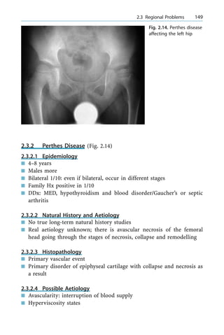
2.3.2.15 Prognostic Factors
n Age most important (cut off age 6 years): reflects remodel potential
n Other important areas:
± Extent of involvement (Herring class)
a 2.3 Regional Problems 151](https://image.slidesharecdn.com/aresidentsguideorthopaedics-250108225802-331d4fe3/85/A-Residents-Guide-orthopaedics-surgery-pdf-169-320.jpg)

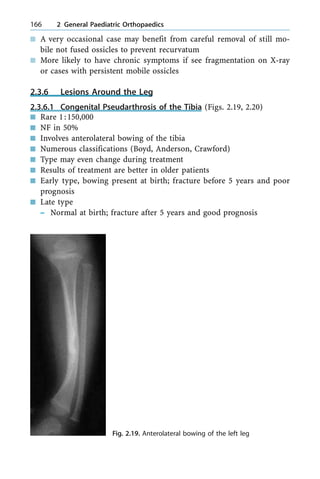
![2.3.6.2.2 Summary of Possible Associations
n Foot: equinovalgus, valgus hind-foot, sometimes absence of one or
more lateral rays
n Ankle: absent lateral malleolus, or hypoplastic. Ball-and-socket ankle
joint
n Fibula: hypoplastic or absent
n Tibia: can be shortened (proximal/mid/distal with valgus)
n Knee: ligament lax sometimes PFJ problem or foot instability
n Femur: acetabular dysplasia, femur varus/valgus, femur sometimes
short, lateral femoral condyle hypoplastic
2.3.6.2.3 Kalamchi Classification
n Types IA, IB: hypoplasia with a portion of fibula present
n Type II: complete absence of the fibula ? hip, knee anomalies com-
mon as are cruciates and knee valgus and maldeveloped lateral femo-
ral condyle. The distal tibial plate/epiphysis can be abnormal. Some-
times tarsal bones anomalies
[More severe forms there can be ankle valgus (stable or unstable); tar-
sal coalition that will eventuate in a ball-and-socket ankle joint. No-
tice tibial bow uncommon with partial fibula deficiency]
172 2 General Paediatric Orthopaedics
Fig. 2.22. A child with
fibula hemimelia](https://image.slidesharecdn.com/aresidentsguideorthopaedics-250108225802-331d4fe3/85/A-Residents-Guide-orthopaedics-surgery-pdf-190-320.jpg)
![± (Persistent varus after casting). Can be from varus deformity of the
calcaneus and/or cuboid or varus of the MT
2.3.7.1.4 Differential Diagnosis
n MT adductus: lacks hind-foot varus and equinus
n Vertical talus: does have hind-foot equinus but with hind-foot valgus
and fore-foot valgus
2.3.7.1.5 XR Assessment: Pearls and Pitfalls
n Feet held in position of best correction in weight-bearing or in infant,
simulated standing
n Since the AP and lateral talocalcaneal (TC) angles are hind-foot an-
gles, the XR beam be focused on the hind-foot (about 308 from the
vertical for AP; the lateral can be transmalleolar, fibula overlapping
the posterior half of the tibia, to avoid rotational distortion
n Sometimes lateral dorsiflexion/plantarflexion views assess ankle mo-
tion especially if flat topped talus, and assess mid-foot hypermobility.
[In the older child ? can also focus XR on mid-foot to assess the
dorsaolateral subluxation of the talo-navicular joint (TNJ)]
2.3.7.1.6 Management: When Should It Start?
n From day 1 according to Ponseti
n Most need 4±8 casts, in fact seldom greater than 7
n Early recurrence needs a further 3±4 casts
n Later ones, TA transfer not to cuboid (valgus force), but to the third
cuneiform
2.3.7.1.7 Conservative Treatment: Ponseti Versus Kite
n Main difference:
± Kite not use tenotomy, sometimes feet kept in cast for 2 years
± Kite uses calcaneo-cuboid joint (CCJ) as pivot instead of talar head,
which Ponseti says is ªwrongº
2.3.7.1.8 Possible Age Limit to Conservative Treatment
n Ponseti says not too rigid
n Others claim if present late at several months success rate low, but
Ponseti thinks that several months old still possible to use his method
176 2 General Paediatric Orthopaedics](https://image.slidesharecdn.com/aresidentsguideorthopaedics-250108225802-331d4fe3/85/A-Residents-Guide-orthopaedics-surgery-pdf-194-320.jpg)
![n Plantar release
n Posterolateral tether take down
n Lateral extended release
2.3.7.1.15 Surgical Approaches (Pros and Cons)
n Turco's posteromedial (not tackle posterolateral tether stressed by Car-
roll; some cases also need more extensive CCJ/lateral TNJ release)
n Carroll's two incision (main critic is residual varus danger since per-
sistent posteromedial tether)
n Cicinati's cicumferential (difficult to deal with TA, but in practice no
problem; sometimes difficult to do plantar release)
2.3.7.1.16 Elements of Release in Greater Details
During Posterior Release
n TA lengthened
n Posterior capsule need release [exposed by retracting peronei and
flexor hallucis longus (FHL) and excise a piece of fat]
During Medial Release
n Keys are abductor hallucis and neurovascular bundle
n Abductor hallucis may need excision
n Part of posteromedial portion of deltoid ligament may need release
n TNJ capsule most require release
Plantar Release for Cavus
n Retract and protect medial/lateral plantar nerves
n Then eases release of plantar aponeurosis and short plantar muscles
n Sheaths of TP and FHL usually need releases
[Avoid plantar release if already rocker buttom]
Posterolateral Tether Release
n TC and calcaneofibular ligaments released
n Peronei sheath released
During Extended Lateral Release
n May extend to the CCJ and the other side of the subtalar joint
178 2 General Paediatric Orthopaedics](https://image.slidesharecdn.com/aresidentsguideorthopaedics-250108225802-331d4fe3/85/A-Residents-Guide-orthopaedics-surgery-pdf-196-320.jpg)

![n Bilateral complex syndactyly with symphalangism: the digits are fused
distally from the M/P onwards, triangular shaped bones, thumb if in-
volved usually with delta phalanx; polydactyly rare. Vascular/tendon
anomalies are common
The Three Types of Apert
n The I/F, M/F and R/F as ªCentral Digital Massº, with thumb and little
finger (L/F) free
n Above and webbed fourth web, thumb free
n Thumb and digital mass share a common nail
X-ray: Assess number of digits involved, number of MCs, number of
phalanges, any cross-unions, any delta phalanx, sometimes joint sur-
face configurations
Management of Apert
n The aim of treatment: to achieve a functional hand in one or two
stages, e.g.:
± Thumb osteotomy to correct angulation, first web release, second/
fourth web release
± Central digital ªmassº is divided to produce four digits and a
thumb at around age 5 years of age
(In type 3, a preliminary nail separation is needed)
3.1.4.6.5 Acrosyndactyly
n Common association with constriction ring syndrome
n Walsh classes:
± Moderate: two phalanges [and one inter-phalangeal joint (IPJ) per
digit]
± Severe: one phalanx
n Treatment: in general, release distal tethering bands early. This per-
mits better growth; more proximal separation can be done later
3.1.4.6.6 Complex Syndactyly
n In general, sometimes very tricky: accessory phalanges may be pre-
sent between the skeletons of syndactyly digits either in organised
form ± ªconcealed central polydactylyº ± or in an apparent jumble of
bones lying transversely, obliquely and longitudinally
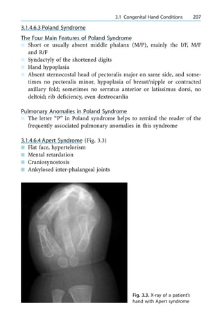
208 3 Principles of Hand Surgery](https://image.slidesharecdn.com/aresidentsguideorthopaedics-250108225802-331d4fe3/85/A-Residents-Guide-orthopaedics-surgery-pdf-226-320.jpg)
![3.1.4.8.2 Other Features
n 1 in 10,000
n More in Asians, most being Unilateral
n Sporadic more common. Ô AD (tri-phalangeal thumb)
n Timing of Operation: 6±9 months
± If equal sized ± can combine both ± ensure articular congruent
± If size unequal ± excise smaller, reconstruct the collateral
± If complex ± add sometimes intrinsic/extrinsic tendon transfers,
Ô osteotomy and surgery on the plate
3.1.4.8.3 Post-axial Polydactyly
n Much more common in Blacks 1/300 versus 1/3000 in non-blacks
n Post-axial most common in Blacks
n Pre-axial most common in Asians and Whites
(Post-axial most common overall)
3.1.4.8.4 Turek (and Stelling) Classification
n Type 1. Extra soft tissue mass, no bone
n Type 2. Normal-looking digit articulates with either phalanx or MC
n Type 3. With MC of its own
`Central polydactyly' ± involves either of the middle three digits; the
extra digit mostly a Turek's type-2 anomaly [autosomal dominant
(AD) and sometimes associated with foot polydactyly/syndactyly]
3.1.4.8.5 Treatment of the Turek's Types
n Type 1. Ligate; watch for bleeding (or by formal excision in theatre)
n Type 2. Preserve important structures such as the ulna collateral, of
MCP of L/F and the abductor digiti quinti insertion
n Type 3. Excise extra digit
(Type 1 with incomplete penetrance; types 2 and 3 AD with marked
penetrance)
3.1.4.9 Macrodactyly (Figs. 3.5, 3.6)
n Pathomechanics: neural, vascular, and hormonal factors have all been
implicated
n More than one digit not uncommon
n Two types ± (1) starts in infancy and (2) starts in adolescence
a 3.1 Congenital Hand Conditions 211](https://image.slidesharecdn.com/aresidentsguideorthopaedics-250108225802-331d4fe3/85/A-Residents-Guide-orthopaedics-surgery-pdf-229-320.jpg)
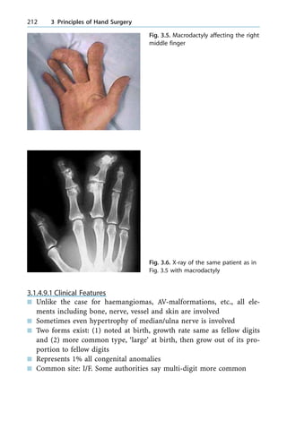
![3.1.4.9.2 Treatment
n Operation needed because function is compromised. The affected fin-
ger can also be stiff, and angulated
n Phased debulking each time on one side more prudent ± remove ex-
cess skin and fat. Sometimes requires osteotomy to correct angula-
tion. Sometimes epiphysiodesis (when size reaches that of the same
digit) and/or bone shortening if digit is already too large, e.g. fusion
of distal and M/P while part of M/P excised)
n Assess need of carpal tunnel syndrome (CTS) release and occasional
case needs excision of the digital nerve. The areas supplied by the ab-
normal nerve are usually with abnormal sensation anyway
n Resistant cases result in amputation ± especially when the rest of the
digits are normal.
3.1.4.10 The Five Types of Hypoplastic Thumbs
n Absent ± most need pollicisation (e.g. radial club hand) before pre-
hensile function is fully developed in children less than 3 years old.
In the case of radial club hand, centralisation ought to be done in 6±
12 months, and pollicisation 6 months thereafter. If there is a delayed
Dx of more than 3 years, the child with radial club hand sometimes
would have adapted to the use of ulna 2 rays and there is a possibility
that pollicisation might not be absolutely necessary. Also, never cen-
tralise in radial clubs if the elbow cannot be flexed
n Short thumbs [i.e. cannot reach level of proximal interphalangeal joint
(PIPJ) of I/F]
n Adducted thumbs ± associated 1st web contracture, needs opponens-
plasty
n Abducted thumbs ± abnormal flexor pollicis longus (FPL) attachment
n Floating thumbs ± possibility of reconstruction reported in some
studies; others prefer pollicisation
3.1.4.10.1 Absent Thumb
n Many are associated with radial club hand
n Most will require pollicisation of the index
3.1.4.10.2 Floating Type
n Treatment: many favour amputation and pollicisation. In Japanese lit-
erature, there are attempts at reconstruction with variable success
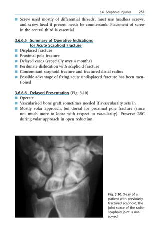
a 3.1 Congenital Hand Conditions 213](https://image.slidesharecdn.com/aresidentsguideorthopaedics-250108225802-331d4fe3/85/A-Residents-Guide-orthopaedics-surgery-pdf-231-320.jpg)
![n Most of these thumbs are connected by flimsy pedicle, and the two
phalanges are hypoplastic. The MC may be rudimentary or absent
n The position of the floating thumb is usually more distal and radial
than the normal thumb.
3.1.4.10.3 Short Thumb
n Most have little functional compromise
n Association with Holt-Oram Syndrome, and Fanconi's Anaemia; MC
short and slender
n If there is short broad MC ± think of myositis ossificans progressiva
(dystrophic dwarfism)
n If there is short broad distal phalanx (D/P) ± think of brachydactyly,
Apert syndrome
n Operation: only if excess short: (i) deepen web space and (ii) distrac-
tion lengthening
3.1.4.10.4 Adducted Thumb
n Pathogenesis: web contracture, and poor thenar muscles
n Principle of treatment includes Z-plasties and/or fascial release (dor-
sal flap/SG), and opponensplasty
[Opponenplasty options are transfer of abductor digiti minimi, flexor
digitorum superficialis (FDS) and even the extensor digiti minimi]
3.1.4.10.5 Abducted Thumb
n Two main types according to Manske: (a) stable CMCJ ± reconstruct;
correct web release adduction, release abnormal slip, and provide sta-
bility and (b) unstable CMCJ ± pollicise
n Pathogenesis ± abnormal insertion of FPL (1 slip at volar D/P, 2nd
slip passes dorsally and radially to join EPL); causes abduction with
action of FPL
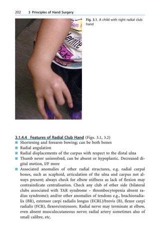
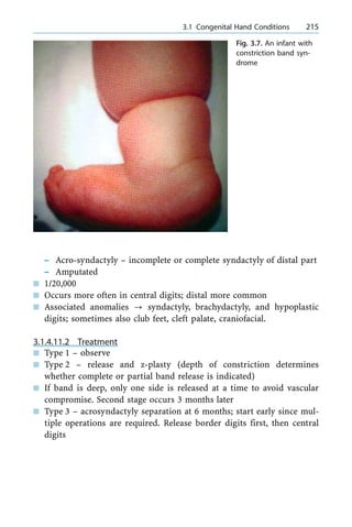
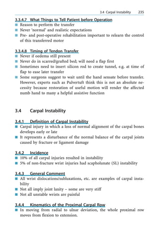
3.1.4.11 Constriction Band Syndrome (Fig. 3.7)
3.1.4.11.1 Introduction
n Definition: deep skin crease encircling a digit, thus causes varying de-
gree of vascular or lymphatic compromise
n Four types (Patterson)
± Mild groove
± Deep groove, abnormal distal
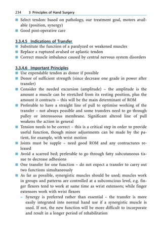
214 3 Principles of Hand Surgery](https://image.slidesharecdn.com/aresidentsguideorthopaedics-250108225802-331d4fe3/85/A-Residents-Guide-orthopaedics-surgery-pdf-232-320.jpg)

![n Tendon gapping is regarded as the hallmark of tendon failure ± a gap
of 2 mm or more is significant
3.3.1.5 Rationale of the Active Extension
and Passive Flexion Programme
3.3.1.6 Basic Biomechanics
n Chicks tendon studies claim that motion and tension promote the
greatest increase in cellular activity, but the absence of both compo-
nents generates least activity
n Tensile strength of common repair technique may not be enough to
reliably support active tendon mobilisation
n Too strong a repeated tensile stress too early will increase gap formation
n Tendons subjected to tensile stress in an active motion post-operative
regimen maintained their strength to a far greater extent than immo-
bile tendons
3.3.1.7 Partial Tendon Laceration
n Many recommend no repair if tendon is more than 70% intact, based
on the reason that repair may delay healing
n Rehabilitation of these unrepaired tendons can follow Strickland's
frayed tendon protocol
3.3.1.8 Zone 2 Flexor Tendon Injuries
n The reader is assumed to know the Kleinert zones
n Zone 2, or no man's land, has the special anatomy of Champer's
chiasm as follows:
± [Flexor digitorum profundus (FDP) travels through the decussation
of FDS Champer's chiasm ± during digit flexion, the two slips of
FDS move forward toward the midline to compress the FDP, like a
bat's tendon-locking mechanism]
n Although adhesion is more likely to form in zone-2 injuries, the mod-
ern trend is to repair both FDS and FDP in such injuries
3.3.1.9 Physical Assessment
n Resting posture sometimes typical, even diagnostic
n Proximal end of cut flexor tendon can retract to variable extent proxi-
mally. When the flexor tendon is under tension while being cut, it can
retract significantly sometimes even to the carpal tunnel
a 3.3 Tendon Injuries 227](https://image.slidesharecdn.com/aresidentsguideorthopaedics-250108225802-331d4fe3/85/A-Residents-Guide-orthopaedics-surgery-pdf-245-320.jpg)
![don at the level of proximal phalanx (P/P), proceed down the digit
and insert to the dorsum of D/P]
n The reader is assumed to know the different zones
3.3.3.3 General Comments
n Few studies on vascularity ± (1) major=synovial diffusion and (2)
muscle branches under the extensor retinaculum and distal to it
n Flat shape and varying thickness and contour ± placement of sutures
is determined more by anatomy than by its vascular supply
n All suture methods weaker then flexors; the Kleinert modification of
the Bunnell technique is strongest, followed by Kessler; others include
mattress, figure-of-8, etc.
n Because of the flat shape, there is marked propensity for them to
bunch during tenorraphy; this shortening can restrict PIP/MCP mo-
tions, and result in more loss of flexion
n The ideal suture technique with maximum tensile strength and mini-
mum shortening has not yet been devised
n It is important to note that it is the post-operative splinting or some-
times k-wire that may help prevent post-operative rupture
n Zone-8 cases ± epimysial invagination should be performed without
strangulation of muscles, which could cause further necrosis. Forearm
fascia repair may prevent muscle herniation
3.3.3.4 Complex Injuries
n Bone grafts, flaps or skin grafts should preferably be done before ex-
tensor tendon reconstruction in complex injuries
n Tissue equilibrium and full mobility of joints achieved before tendon
transfer and grafting
n Refer to the section on rheumatoid arthritis (RA) for treatment of
rheumatoid related tendon ruptures
3.3.3.4.1 Zone 1
n Closed mallet ± nonstop splint for 6±8 weeks (recognised alternative
is trans-articular k-wire. Cx: breakage of wire, chondrolysis, sepsis,
etc.). Open repair of extensor if failed closed splinting treatment
n Open mallet ± open repair
n Closed mallet with fracture and/or DP subluxation ± consider open if
it involves 30±50% articular area or more. If less than 30% and no
subluxation proceed with closed treatment
230 3 Principles of Hand Surgery](https://image.slidesharecdn.com/aresidentsguideorthopaedics-250108225802-331d4fe3/85/A-Residents-Guide-orthopaedics-surgery-pdf-248-320.jpg)

![3.5.5.5.3 Scenario 3: Ulna Styloid Fracture
n Need to treat any nonunion or malunion
3.5.5.5.4 Scenario 4: Chronic TFCC Injury
n Some study showed that one can still intervene with reasonable re-
sults up to 3±4 months
n DRUJ can be made unstable due to malunited distal radius. These
cases may need osteotomy, especially if the distal radius is shortened
with ulna impaction. In these situations, can consider a joint levelling
procedure, which frequently involves osteotomy of the distal radius
n If Sigmoid notch area is degenerated, may need to salvage, e.g. Sauve-
Kapanji
3.5.6 Example of DRUJ Reconstruction [Linshield Procedure]
n Use half of FCU as a sling
n Make in a strip
n Sling ulna head back
3.5.7 Examples of Procedures to Tackle Length Discrepancies
n Ulna alone ± shortening, wafer operation, and Sauve-Kapanji
n Radius alone ± osteotomy
n Both ± rarely mentioned in the literature
3.5.8 Role of Joint Levelling
n Reconstruction procedure alone in the presence of significant length
differences (between distal radius and ulna) with no joint levelling
may not work
n Restoration of joint congruency is important
3.5.9 In the Setting of Distal Radius Injury
n Checking for clinical DRUJ instability is most important, since relying
on X-ray assessment of ulna styloid is not good enough
3.5.9.1 Category I: Acute Situations
n Associated fractured distal radius: assessing integrity of the sigmoid
notch, good restoration of radial shortening and prevention of the
dorsal tilt are all very important. Hence, first ensure the adequate an-
atomical restoration of distal radius anatomy, then check DRUJ insta-
a 3.5 Injuries and Instability of the Distal Radioulna Joint 247](https://image.slidesharecdn.com/aresidentsguideorthopaedics-250108225802-331d4fe3/85/A-Residents-Guide-orthopaedics-surgery-pdf-265-320.jpg)
![n Underlying pathology
n If there is associated dissociated loss of motor or sensory function,
prognosis is sometimes better
3.7.1.6 Median Nerve
3.7.1.6.1 Carpal Tunnel Syndrome
n Symptom ± numbness, but can be nonspecific
n Nocturnal symptom
n Reduced motor control of thumb [only hand muscle control by med-
ian ± `LOAF': radial two lumbricals, opponens pollicis, abductor polli-
cis brevis (APB), flexor pollicis brevis (FPB)]
n Precipitating factors ± pregnancy, thyroid disorder, : anatomic con-
tents (fractured carpus, fractured hamate hook, lipoma, abnormal
muscle, median artery, synovitis/RA)
n Association ± perhaps Dupuytren
n Beware of acute CTS secondary to trauma and fracture dislocation
n DDx ± cervical cause, thoracic outlet syndrome, peripheral neuropathy
3.7.1.6.2 Boundaries of Carpal Tunnel
n Floor ± carpus
n Roof ± transverse retinaculum
n Radial border ± scaphoid tubercle and trapezium
n Ulna border ± hook of hamate and pisiform
n Contents: nine flexor tendons (the FDS of 3rd/4th fingers more super-
ficial) and median nerve
3.7.1.6.3 Diagnosis
n Clinical:
± Phalen
± (Reversed phalen) ± 60% sensitive, 85% specific, numbness in 60
seconds
± Tinel ± 75% sensitive, 90% specific
± Gelberman median nerve compression test ± elbow extend, forearm
supinate, wrist flex 608, then press on the nerve
n Investigation
n Exact role of NCT:
n Not needed if Dx certain
254 3 Principles of Hand Surgery](https://image.slidesharecdn.com/aresidentsguideorthopaedics-250108225802-331d4fe3/85/A-Residents-Guide-orthopaedics-surgery-pdf-272-320.jpg)

![Differential diagnosis of negative stress test:
± Really negative stress test
± Not proper technique
± Subtalar unstable
(Other investigations ± e.g. MRI can see ligaments)
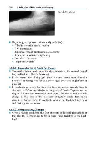
4.2.2.3 Conservative Treatment
n Strengthen peroneals
n Proprioception rehabilitation
n Brace/tapping/use of orthotics
n If successful, continue; later training is sports specific
4.2.2.4 Indication for Surgery
n Failed conservative treatment
n Not all cases need to have positive test to perform surgery
4.2.2.5 Selection of Arthroscopic versus Open Surgery
n ATFL intracapsular, arthroscopic intervention is possible
n CFL extracapsular, cannot use scope to intervene
n Sometimes ligaments are too attenuated and only scar remains; may
need open surgery, especially in chronic cases
n (P.S. CFL contributes most to subtalar instability, if any)
4.2.2.6 Good Indications for Arthroscopic Interventions
n Lateral gutter impingement
n Anterior impingement
n Cartilage injury, e.g. flaps
n Loose bodies and OCD
n Chronic synovitis
4.2.2.7 Types of Open Surgery
n Anatomical ± brostrum repair ± direct ligament repair of ATFL/CFL
± Advantages ± anatomical; spares peroneal; good track record
± [Post-operative: 2 weeks non-weight bearing (NWB); 2 weeks
weight bearing (WB) cast; 8 weeks brace]
n Peroneal used/tenodesis procedure (e.g. Watson Jones)
± Advantages ± good if ligament replaced by scar; in cases with de-
generative joint disease (DJD), gives more sense of stability
± Disadvantage ± sacrifice peroneal, not anatomical
a 4.2 Acute Ankle Ligament Injuries 301](https://image.slidesharecdn.com/aresidentsguideorthopaedics-250108225802-331d4fe3/85/A-Residents-Guide-orthopaedics-surgery-pdf-318-320.jpg)
![Result is fore-foot abduction, mid-foot pronation and hind-foot val-
gus
Cxs if untreated: impingement laterally at subtalar level and osteoar-
thritis (OA) of the subtalar joint
4.4.1.4 XR Assessment
n Anteroposterior (AP): may see fore-foot abduction or TNJ subluxation
n Lateral: may see mid-foot sagging
n Tenography: does not correlate well with operative findings
n MRI: only if the diagnosis is in question; not in every patient
n Ultrasound: may see size of tendon and changes in echogenicity in-
side the tendon; cheaper than MRI
4.4.1.5 Clinical Stages
n Stage 1: tendinitis, no deformity
n Stage 2: tendon degeneration with elongation, flexible deformity
n Stage 3: fixed deformity, hind-foot degeneration
4.4.1.6 Management According to Stages
n Stage 1: mostly conservative [rest, non-steroidal anti-inflammatory
drugs (NSAIDs), TA stretching, PTT strengthening, arch support, even
custom ankle foot orthosis or short leg cast]; operate if fails to re-
spond by debridement, augment or repair and/or correct predisposing
bony anomalies
n Stage 2: conservative means similar; most require tendon augmenta-
tion with flexor digitorum longus (FDL)/flexor hallucis longis (FHL),
limited fusion (e.g. subtalar or double), and/or calcaneal medial dis-
placement osteotomy/Evans calcaneal lengthening
n Stage 3: conservative as above with or without custom-made shoe; OT
usually involves a triple fusion
4.4.2 Peroneal Tendon Injuries
4.4.2.1 Pertinent Anatomy
n The peroneal groove is, in fact, the fibula groove and there is a rim of
cartilage at the lateral border. The depth of the groove is variable
n The superior peroneal retinaculum is the primary lateral restraint
against peroneal instabilty; it is parallel to the CFL, hence, those in-
version injuries affecting the CFL may injure the retinaculum
306 4 Principles of Foot and Ankle Surgery](https://image.slidesharecdn.com/aresidentsguideorthopaedics-250108225802-331d4fe3/85/A-Residents-Guide-orthopaedics-surgery-pdf-323-320.jpg)
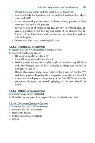
![4.5.1.1 Introduction
n Hallux valgus (HV) is not a single disorder. It represents a complex
deformity of the first ray that is frequently accompanied by deformity
and symptoms of the lesser toes
n Elements of HV that may occur:
± First MT varus deformity
± Big toe in valgus
± Bunion
± OA of the first MTPJ, occasionally
± Hammer toe, usually the second toe
± Corns, calluses and transfer metatarsalgia
4.5.1.2 Predisposing Factors for the Production of HV
n Genu valgum with pronation of feet
n Pes planus
n Increased obliquity of the first MT-cuneiform joint
n Long first ray
n Excess valgus tilt of the articular surface of the first MT head [and
proximal phalanx (P/P) articular surface]
n External factors: most important is shoe wear
(Note: among the different factors, lateral deviation of the big toe is
found to be most significant, although metatarsus primus varus may
be an important factor in adolescents)
4.5.1.3 Pathomechanics of HV
n Increased HV angle at first MTPJ (especially greater than 358) ? pro-
nation of first ray ? abductor hallucis moves in plantar direction, i.e.
medial capsular ligament as the only medial restraint ? HV increases
from unopposed adductor hallucis ? MT head drifts medially from
the sesamoid. The lateral sesamoid is displaced into the first MT
space
n Valgus of big toe usually creates hammer of the second toe
4.5.1.4 Clinical Assessment
n Assessing the presence of symptoms (usually pain) and the exact lo-
cation of pain is important, since not every patient with HV needs
surgery
n Physical assessment is very important
312 4 Principles of Foot and Ankle Surgery](https://image.slidesharecdn.com/aresidentsguideorthopaedics-250108225802-331d4fe3/85/A-Residents-Guide-orthopaedics-surgery-pdf-329-320.jpg)
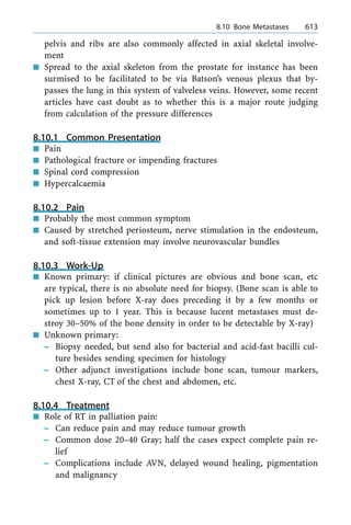
![5.1.4.1.10 Summary of Different Management in Different Ages
In Children
n Possibility of successful pars repair for very symptomatic spondyloly-
sis since there is a normal disc
n Possibility of higher chance of further slip from ligamentous laxity,
especially in girls and despite fusion in-situ [posterolateral (PL) inter-
transverse is the gold standard]
n Possibility of (seemingly solid) `fusion mass' to slip further (especially
if decompression procedures done) in children
Adolescent Spondylolysis
n Many think that pars defect (lytic) type does not occur at birth, and
is hence developmental
n Direct repair can be considered in symptomatic cases with normal
disc, especially if no slip
n Juvenile sportsmen/women are predisposed to develop this condition
Other Features of Adolescent Spondylolysis/Spondylolisthesis
n Disc mostly normal
n Even for high grades, may still get away with posterior in-situ fusion
Adults and Above
n The spondylolytic variety of isthmic-type spondylolisthesis is less
likely to be associated with neurological deficit, since the posterior
arch separates from both pars defects.; hence with significant neurol-
ogy and/or acute/fast progressive neurology, we can rule out disc as
the cause (especially in cauda equina, for example)
n Degenerative as the adult male/female ages; laxity is less and, here,
despite an intact posterior arch (unlike isthmic), slip progression is
mostly not too severe
n Dysplastic: real potential danger of neural injury, since there is a com-
bined effect of intact post arch and a tendency for L-S kyphosis ± e.g.
rounding off of the anterior sacrum with loss of the anterior buttress
a 5.1 Common Spinal Deformities 355](https://image.slidesharecdn.com/aresidentsguideorthopaedics-250108225802-331d4fe3/85/A-Residents-Guide-orthopaedics-surgery-pdf-371-320.jpg)
![Low-Grade Middle-Aged Spondylolisthesis
n Pain ± carefully diagnose where the pain is from, especially in work-
er's compensation cases, e.g. nerve blocks, and discogram is some-
times needed
n Neurology ± the risk of cauda equina and severe neurology is much
less than with the dysplastic type, since there is no intact neural arch
in such cases
n Both pain and neurology ± symptom/grade can worsen as disc subse-
quently degenerates. Beware, pain coming from nearby disc is also
possible
Middle-Age High-Grade Spondylolisthesis
n In-situ fusion with or without decompression; versus instrumented fu-
sion
n Fusion is more advisable to put in front [anterior spinal fusion (ASF)/
posterior lumbar inter-vertebral fusion (PLIF)] especially for high-
grades to increase fusion success, since the chance of fusion is higher
with graft in compression
Spondyloptosis
n Clinical features: vertical buttock, typical gait sometimes deep loin
crease present, marked hamstring spasm
n Choice of operation needs individual assessment. For severe cases,
may consider Gaine's procedure; an alternative will be the use of fibu-
la grafting
5.1.4.1.11 Prognosis in Severe Slip
n Age (worse in the young)
n Sex (more often in females)
n Slip degree >308
n Slip angle >458
n Trapezoidal L5 and dome shaped sacrum
n Spina bifida (possible weakened posterior tension band)
5.1.4.1.12 Rn Pearls ± High-Grade Spondylolisthesis
n Special In/views
± Long film standing anteroposterior (AP)/lateral, especially lateral to
assess overall sagittal balance
356 5 Spine Surgery Principles](https://image.slidesharecdn.com/aresidentsguideorthopaedics-250108225802-331d4fe3/85/A-Residents-Guide-orthopaedics-surgery-pdf-372-320.jpg)
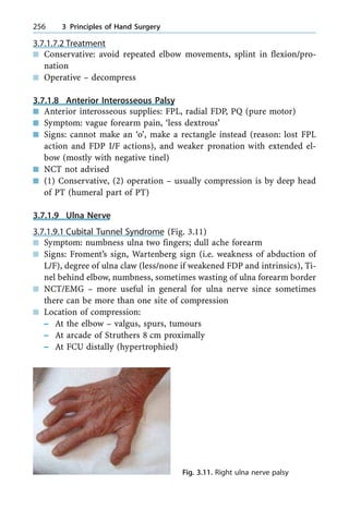
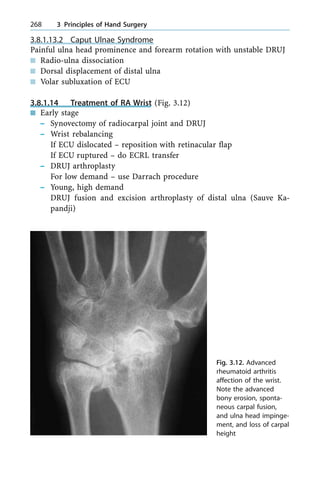
![5.1.5.10 King's Classification
n Type I ± double curve, lumbar major
n Type II ± double curve, thoracic major
n Type III ± single thoracic
n Type IV ± long C curve with L4 in curve
n Type V ± double thoracic
5.1.5.11 Aims of Treatment
n To obtain a stable balanced spine, centred over the pelvis in the coro-
nal/sagittal planes
n Stop curve progression
n Minimise patient morbidity and fuse as few segments as possible
5.1.5.12 Indications for Bracing
n Early Dx
n Mild curve (<308)
n Skeletally immature, Risser 3 or less
(Refer to landmark paper: 1995 SRS Brace Study [5])
5.1.5.13 Brace Wearing
n Dose-dependent effect at more than 20 h (preferably 23 h)
n Examples: Boston underarm (apex below T7); Milwaukee (apex above
T7)
n When to consider weaning: when skeletal maturity is reached
± Menarche over 2 years
± Height assessment
± Risser sign assessment
± Hand (bone age)
5.1.5.14 Indications for Surgery
n Late Dx of significant curves
n Severe curves (>458)
n Truncal imbalance
n Failed brace treatment, can be non-compliance or progression despite
bracing
362 5 Spine Surgery Principles](https://image.slidesharecdn.com/aresidentsguideorthopaedics-250108225802-331d4fe3/85/A-Residents-Guide-orthopaedics-surgery-pdf-378-320.jpg)

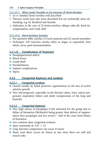
![5.3 The Degenerative Spine
5.3.1 Cervical Myelopathy
5.3.1.1 Definition
Refers to spinal cord dysfunction located at the cervical level arising
from mechanical compression and vascular compromise
5.3.1.2 Mechanism of Injury to Cord
n Direct mechanical compression, e.g. hard disc, soft disc prolapse, liga-
mentum flavum, ossification of posterior longitudinal ligament (OPLL),
etc.
n Dynamic compression and stretching, e.g. pincer action of osteophyte
spurs produced on F/E
n Possible vascular factors
5.3.1.3 Symptoms
n Neck pain and/or weakness, numbness, wasting, and clumsy hands,
difficulty in walking and unsteadiness, tendency to fall
n Sphincter disturbance rather uncommon
5.3.1.4 Signs
n Upper motor neurone ± brisk jerks, Babinski [depending on level(s)
of compression, can have combination of both upper motor neurone/
lower motor neurone signs in the UL]
n Inverted supinator reflex
n Lhermitte signs
n Hoffman sign ± one of the myelopathy hand sign
n Scapulohumeral reflex (Shimuzu 1993) ± if positive, indicative of high
lesion level around C3/4
n Myelopathic hand signs (10-s test, finger-escape test) ± if positive, in-
dicative of cervical myelopathy
5.3.1.5 Occasional Atypical Presentation
n If multiple level disease
n Needs to know the discrepancy between the bony level and the corre-
sponding level of the neural tissue that can be affected, i.e. discrepan-
cy between neurological and bone segment
a 5.3 The Degenerative Spine 381](https://image.slidesharecdn.com/aresidentsguideorthopaedics-250108225802-331d4fe3/85/A-Residents-Guide-orthopaedics-surgery-pdf-397-320.jpg)
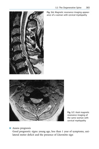
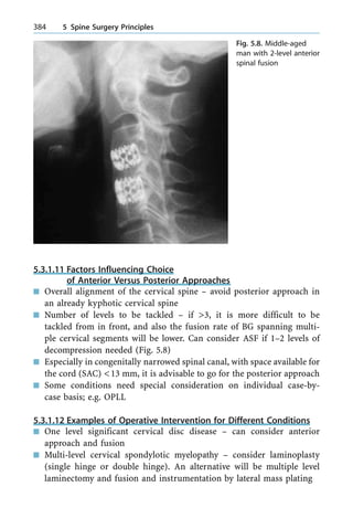
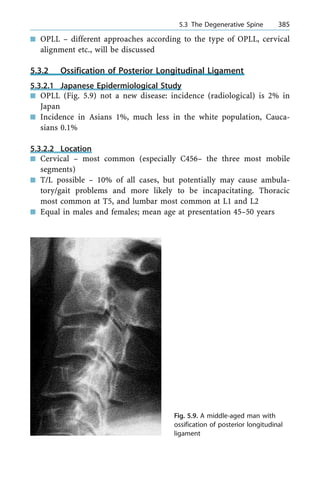
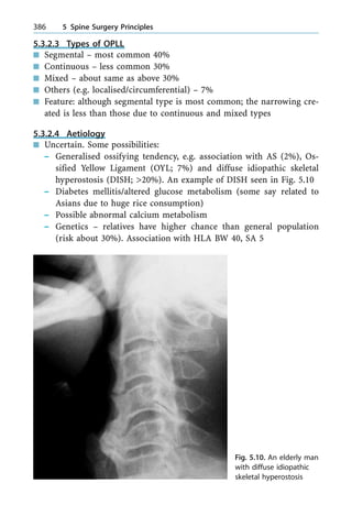
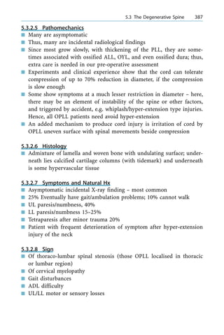
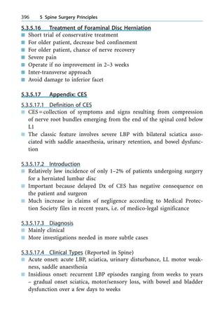
![n Bladder may be palpable, late cases can be painless retention (chart
residual urine after Foley insertion)
n Findings can be quite asymmetrical
5.3.5.17.12 Summary of `Red Flags' Signs
n Severe LBP with bilateral or unilateral sciatica
n Bladder and bowel dysfunction
n Anaesthesia or paraesthesia in perineal region or buttocks
n Significant LL weakness
n Gait disturbances
n Sexual dysfunction
5.3.5.17.13 Investigations
n Pre-operative X-ray
n MRI (urgent) should be ordered if suspect cauda equina compression
n Other investigations ± e.g. urodynamic studies but should not delay
emergent surgery
5.3.5.17.14 Other Possible Investigations
n Spinal angiography if suspect arteriovenous malformation
n Cystometry and/or fluoroscopy to assess detrusor muscle function
n Anorectal manometry and defaecography
n Some cases may use transurethral PG E1 application to rule out a vas-
cular cause of erectile dysfunction
n Motor and sensory evoked potentials
5.3.5.17.15 Timing of Surgery
n Many retrospective studies ± but most are small series giving widely
different conclusions
± Studies for urgent decompression have been reported previously by
Shapiro and by Ahn
± Studies with ªnoº correlation between timing of surgery and end
result have previously been reported by Buchner and Kostuik and
Delamarter
± Studies for urgent decompression in incomplete CES only were re-
cently reported by Gleave (Br J Neurosurgery 2002 [4])
(No role of steroids)
a 5.3 The Degenerative Spine 399](https://image.slidesharecdn.com/aresidentsguideorthopaedics-250108225802-331d4fe3/85/A-Residents-Guide-orthopaedics-surgery-pdf-415-320.jpg)
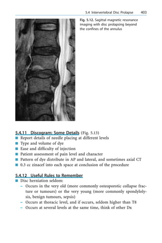
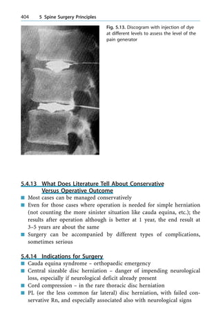
![n Intractable discogenic pain (need attention to Dx and pre-operative
work-up as in general surgery for relief of sciatica more effective than
back pain. The same holds for surgery for disc herniation besides dis-
cogenic pain)
5.4.15 Types of Operation for Disc Disease
n Fenestration and diskectomy
n Laminotomy and diskectomy
n Laminectomy ± mostly for the large central herniation (as in causing
cauda equina syndrome) may even need bilateral laminectomy
n Role of spinal fusion ± (PL or even anterior) for discogenic pain
cases. Seldom needed in the simple straight forward surgery for a disc
herniation
5.4.16 Operative Complications
n Wrong level
n Persistent pain despite surgery (see the section on failed back surgery
for management)
n Retained disc material
n Infection (superficial or deep)
n Injury to the dura and CSF leakage
n Complications peculiar to the surgery performed, e.g. if posterior in-
strumentation performed in an attempt to `improve' fusion, then
hardware complications, etc.
5.5 Spinal Tumours
5.5.1 Primary Spinal Tumours
5.5.1.1 Location of Spine Primaries
n Tumours with predilection for posterior elements (and/or spread to
anterior): osteoblastoma, osteoid osteoma
n Tumours with predilection for sacrum: chordoma, giant cell tumour
n Tumours with predilection for anterior body: most of the rest
n Haemic tumours/Ewing: most are part of more generalised affection,
and hence systemic Rn important
[P.S. aneurysmal bone cyst (ABC) can be located at either anterior or
posterior elements]
a 5.5 Spinal Tumours 405](https://image.slidesharecdn.com/aresidentsguideorthopaedics-250108225802-331d4fe3/85/A-Residents-Guide-orthopaedics-surgery-pdf-421-320.jpg)
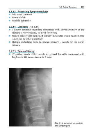
![± AAI plus cranial settling: use MRI to assess (whether traction is
(1) reducible ± post fusion or (2) irreducible ± C1 laminectomy or
anterior decompression and C0/C1 fusion)
± Conservative for cranial settling if: isolated, stable, asymptomatic,
without cord compression
± SAS: canal diameter on X-ray <14 mm; MRI: F/E for true space ±
if <13 mm post fusion
5.6.1.16 Other Special Circumstances
n Case for transoral resection of the dens:
± Irreducible ventral extradural compression of the cervicomedullary
junction
± Irreducible cranial settling
± Odontoid fracture with complex compression fracture fragments
with large fibrous pannus
[N.B. 80% of rheumatoid arthritis patients with cranial settling
with reducible lesion that require only post fusion]
n Conservative treatment of subaxial spine
n Traction and halo vest for 4 months
5.6.1.17 The Operative Option
n Pre-operative traction ± up to 3 weeks ± leads to correction of deformity,
reduction of translocation and might abolish need of anterior surgery
n Approach: anterior approach for anterior compression and anterior
decompression usually also requires posterior fixation
n Bone quality: in osteoporotic bone, lamina may be strongest ± sub-
laminar wiring
n (Fixation with no fusion reported by Crockard)
5.6.1.18 Types of Surgery
n Gallie (and Brooks) fusion
n C1/2 transarticular screw; contraindications
± Collapsed lateral atlantal mass
± C1/2 comminuted fracture
± Marked osteoporosis
± Anomalous vertebral artery (20% cases according to Crockard)
n Co-C2 fusion (Fig. 5.15) in: cranial settling, fixed anterior subluxation,
with or without resection of posterior ring of C1
n Technique of C0/C1: Swiss and Ranford's loop
a 5.6 Inflammatory Diseases Affecting the Spine 415](https://image.slidesharecdn.com/aresidentsguideorthopaedics-250108225802-331d4fe3/85/A-Residents-Guide-orthopaedics-surgery-pdf-431-320.jpg)
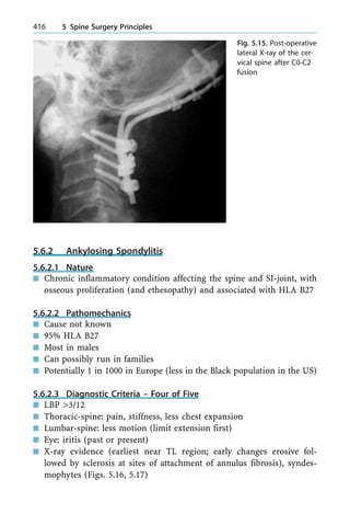
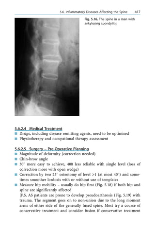
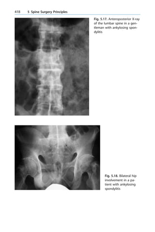
![fails. Recurrence is not uncommon, sometimes at another level that
can escape solid spontaneous fusion]
5.6.2.6 Surgical ± Consider Spinal Osteotomy
if Creates Functional Deficits
5.6.2.6.1 Lumbar Osteotomy
n Smith-Peterson method: later modified by McMaster in '85
± Many Cxs: aortic rupture, gastric dilation, mesenteric artery
thrombosis
n Thomasen: modified it± by instrumenting the spine before destabilis-
ing it. Close the wedge:
± Close wedge with decreased Cxs and deemed better
n Others: e.g. multiple pedicular closing wedges, fewer complications
5.6.2.6.2 Cervical Osteotomy
n Sitting position
n Decompress first (posterior laminectomy) and close defect after de-
compression
a 5.6 Inflammatory Diseases Affecting the Spine 419
Fig. 5.19. Pseudoarthro-
sis in a patient with an-
kylosing spondylitis](https://image.slidesharecdn.com/aresidentsguideorthopaedics-250108225802-331d4fe3/85/A-Residents-Guide-orthopaedics-surgery-pdf-435-320.jpg)
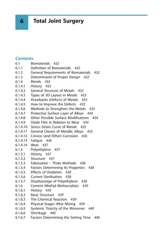
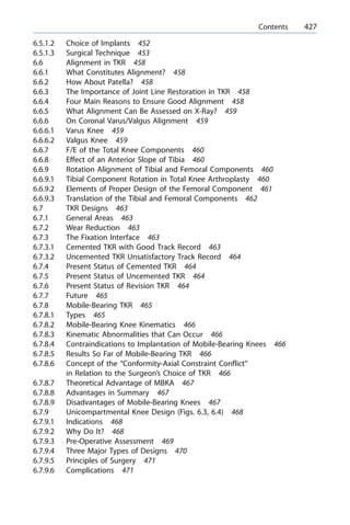
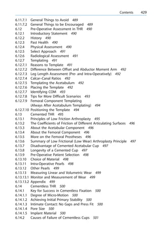
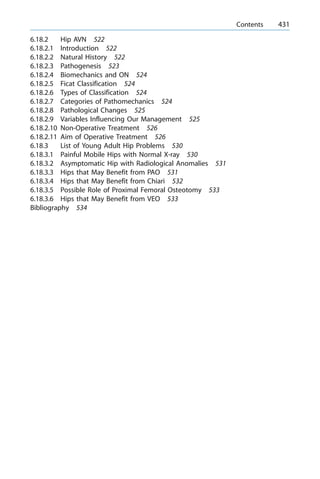
![6.1.4.3 Types of 3D Layout in Metals
n FCC, e.g. 316L S Steel
n BCC, e.g. beta Ti
n HCP, e.g. CoCr, alpha Ti
The above are the three most common ways, though there are 14 pos-
sible patterns
6.1.4.4 Drawbacks (Defects) of Metals
n Point ± a lattice site not occupied by metal atom
n Line defect ± dislocation. Improvements in metal design ought to pre-
vent dislocations ± a dislocation is the result of an extra half plane of
atoms in a crystal ± locally distorts the crystal structure (when a
stress of sufficient magnitude is applied, these dislocations can move
through the lattice ± permanent change in shape of the crystal, and
metal is plastically deformed)
n Area defects ± at grain boundaries
n Volume defects ± voids or cracks (even superficial scratches can lower
the fatigue strength)
6.1.4.5 How to Improve the Defects
n Those processes that tend to prevent the motion of dislocation will
act to strengthen the metal
n Grain boundary sliding is rarer ± will not be discussed here
6.1.4.6 Methods to Strengthen the Metals
n Make an alloy: (solid solution strengthening) both interstitial or sub-
stitutional (if added atom is small, e.g. C/O/N/B) when we add one or
more elements to the `solvent' metal
[Example of institutional ± adding 0.1% carbon to iron=steel (´10
increased strength)]
n Cold working: the principle is to increase strength by increasing dislo-
cation density, which entangles these dislocations into tight bundles ±
the resultant structure is much more difficult to move within the lattice
Cold working is often performed on 316L stainless steel as strength-
ening ± the metal can be cold worked by rolling, compression and
other deformation mechanisms
A process of annealing (heating) sometimes follows cold working and
causes the alloy to return to pre-cold worked structure. The reason
a 6.1 Biomaterials 433](https://image.slidesharecdn.com/aresidentsguideorthopaedics-250108225802-331d4fe3/85/A-Residents-Guide-orthopaedics-surgery-pdf-448-320.jpg)
![n HCP and/or FCC
n Used in total joint since it has high strength, hardness (resists surface
deformation) and corrosion resistance (and/or hardened by precipi-
tate of carbides, less wear and more surface hardness)
6.1.4.11.3 Ti-Based Alloys
n Advantages ± high strength, low modulus, excellent corrosion resis-
tance, biocompatible
n Drawback: notch sensitivity (sensitive to surface flaws) ? can acceler-
ate fatigue process and avoid sharp edges of geometry, e.g. porous-
coating junctions, both of which raise the stress
n Type commonly used in total joint: Ti-6Al-4V (V is vanadium, not vi-
tallium) ? high strength and high fatigue resistance (due to the bi-
modal microstructure ± with alpha and beta phases)
[New: beta Ti (under investigation) ± lesser E (elastic modulus)]
n HCP
n Four grades that vary in oxygen content (function as institutional
strengthening element)
n Why is pure Ti not used in total joint? ± not enough strength to carry
the large stresses
6.1.4.12 Crevice (and Other) Corrosion
n Corrosion process ± limited by the passive oxide film; ± if the rate is
low enough for the body to eliminate the generated corrosion debris,
then this ionic and particle release may not be clinically significant
n There are situations, as in the modular junctions, where mechanically
assisted crevice corrosion (fretting) has been reported; this corrosion
is a conjoint effect of mechanical abrasion of the oxide film that cov-
ers the surface of the alloy in the taper and the restricted crevice en-
vironment
n Besides a local effect, this corrosion can cause systemic levels of Co
and Cr
n Modular taper corrosion can sometimes cause fatigue failure of Co/Cr
stems
6.1.4.13 Fatigue
n Fatigue in orthopaedic implants is now less common with improved
materials and processing
436 6 Total Joint Surgery](https://image.slidesharecdn.com/aresidentsguideorthopaedics-250108225802-331d4fe3/85/A-Residents-Guide-orthopaedics-surgery-pdf-451-320.jpg)
![± Oxidative degradation may increase the modulus and brittleness of
the polyethylene
6.1.6 Cement (Methyl Methacrylate)
6.1.6.1 History
n In olden days, as cranioplast, plexiglass, judet hip, etc.
n Previous examples were all transparent; now they can be seen on X-
ray with barium sulphate
6.1.6.2 Basic Structure
n Methyl methacrylate (MMA) is one member of a group of polymers
widely known as polyethylene, polyvinyl chloride, polystyrene ± re-
peating carbon-based units of variable chain length
n Monomer with MW of only 100
n Longer chain length and greater viscosity; ultimate chain length of
the polymer depends on the manufacturing process ± initiators and
activators
6.1.6.3 The Chemical Reaction
n Powder: pre-polymerised polymethyl methacrylate (PMMA) beads
and initiator (dibenzoyl peroxide)
n Liquid: monomer and activator (N/N dimethyl-p-toludine)
[Main reaction is benzoyl peroxide radical reacting with monomer to
begin the polymerisation process]
(Note: 478C bone necrosis, 568C protein denaturation; modern ce-
ment with 1-mm to 4-mm-thick mantle does not however appear to
produce excess temperature enough to cause necrosis)
6.1.6.4 Physical Stages After Mixing
n Stage 1 ± lumpy after mixing (monomer±polymer conglomerates)
n Stage 2 ± liquid stage; can then be transferred to cement gun
n Stage 3 ± dough; less viscous, further swelling and polymerisation
(time where cement no longer sticks to gloves; prone to produce lami-
nations at this stage); average time for doughy state formation is 5±
8 min
n Hardened and exothermic (on average 2600 calories/20 g) ± a combi-
nation of the bulk polymerisation of the monomer MMA and matrix
a 6.1 Biomaterials 439](https://image.slidesharecdn.com/aresidentsguideorthopaedics-250108225802-331d4fe3/85/A-Residents-Guide-orthopaedics-surgery-pdf-454-320.jpg)
![n Insulation
n As bioactive coats (bone on-growth)
6.1.7.6 Advantages of Ceramics Used as Biological Coating
n More normal in physiological environments, e.g. C/Ca/P/O
n Elastic modulus more similar to nearby tissues (decrease the magni-
tude of interfacial microstrain differences)
n Very biocompatible
n In dentistry, off white/intraoral application
n An interfacial barrier to transfer of elements, heat or electrons from
the substrates
If direct contact and integration with bone is attained, there is im-
proved force transfer
[If used as coatings, the initial active response with bone is short
term (less than 1 month), and most of the active biomaterial (other
than the surface zone) is intended to remain as an implant over the
long term (years). For example, porous and particulate material is
used in bone grafts (BGs) intended for long-term augmentations, and
implant coatings are intended for attachment to bone and soft tissue]
[Some are degradable ± as scaffold to deliver growth factors, for ex-
ample]
6.1.7.7 Summary of Advantages of Ceramics
n Inert, stable
n Biocompatible
n Low friction
n High strength ± especially fully dense ceramic biomaterials such as
aluminium and zirconium oxides in total joints
6.1.7.8 Summary of Disadvantages of Ceramics
n Brittle (better to use larger bulk/component size) ± they are brittle
(non-ductile) because of their atomic bond responses to mechanical
forces
n Prone to fracture, since they are mechanically weak when loaded cy-
clically under high magnitude tension and shear-stress conditions.
(Hence often used in larger bulk structural forms, where mechanical
stresses will be lower)
n Sensitive to hoop stress
444 6 Total Joint Surgery](https://image.slidesharecdn.com/aresidentsguideorthopaedics-250108225802-331d4fe3/85/A-Residents-Guide-orthopaedics-surgery-pdf-459-320.jpg)
![6.2.3 Third Body Wear
n Can be thought of as a form of abrasive wear when a free particle gets
entrapped onto a pair of articulating couple, or sometimes the parti-
cle gets embedded onto the softer surface
6.2.4 Fatigue Wear
n Occurs when subsurface/surface cyclic shear stress in a material ex-
ceeds the fatigue limit of that material
n Cases with significant wear can result in delamination, not just micro-
scopic or macroscopic pits. In the case of polyethylene, it is believed
that oxidative degradation (if present) can be an added predisposing
factor
6.2.5 Corrosion Wear
n Can be thought of as a form of third body wear, the liberated corro-
sive debris acting as an abrasive free body. Since the motion of an ar-
ticulating couple removes corrosive products, liberation of such prod-
ucts exposes a greater surface (less resistant to corrosion) to even
further corrosion
6.2.6 Effect of the Wear Particles
n With the millions of cycles we walk each year, the plentiful submicron
particles generated can induce periprosthetic osteolysis (discussed in
the next section), which is a significant causation of aseptic loosening
of the total joint replacement
n Arthroplasty surgeons have also discovered the concept of effective
joint space. For example, wear particles can find their way between
the bone±implant interface in the non-circumferentially porous-coated
femoral component, and osteolysis can occur in spaces far beyond the
joint itself, aided in part by the continuous pumping or fluctuating
hydrostatic forces between the bone and the implant.
[Note: the major modes of wear in both THR (Fig. 6.1, showing ec-
centric wear of acetabular polyethylene) and total knee replacement
(TKR) include adhesion and abrasion. However, in TKR, the fatigue
mode of wear is important, and delamination can occur]
446 6 Total Joint Surgery](https://image.slidesharecdn.com/aresidentsguideorthopaedics-250108225802-331d4fe3/85/A-Residents-Guide-orthopaedics-surgery-pdf-461-320.jpg)

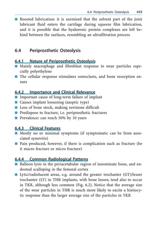
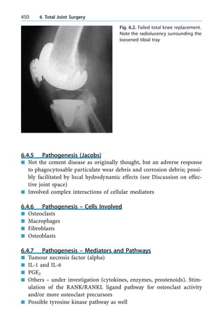
![± Proper restoration of soft-tissue balancing and assessment of the
need for lateral release
Patella-clunk syndrome is commonly associated with posterior-sta-
bilised designs. Its occurrence can be minimised by careful de-
bridement of the peripatella retinaculum, avoidance of proximal
placement of the patella button, and use more modern PS knee de-
signs with a more posteriorly situated intercondylar box (Ip D, Int
Orthop 2002 [2])
6.6 Alignment in TKR
6.6.1 What Constitutes Alignment
n Most people talk mainly about varus/valgus alignment in TKR
n This is over-simplified; the knee has three axes with many degrees of
freedom
n Alignment involves not only varus/valgus, but any F/E of component,
rotational alignment and also translational alignment along each axis
6.6.2 How About Patella?
n The 6 degrees of freedom apply to the patella as well, but since patella
oriented in the coronal plane, the nomenclature is different
6.6.3 The Importance of Joint Line Restoration in TKR
n Proper restoration of joint line needed for proper collaterals function-
ing
n Effect of patella baja ± needs to be prevented in TKR. Patella baja not
only predisposes to patella-clunk, but also anterior knee pain and/or
motion restriction
6.6.4 Four Main Reasons to Ensure Good Alignment
n Biomechanics ± more symmetric bone loading
n Prevent premature failure of polyethylene (note catastrophic failure
can occur when too much force on one compartment)
n Prevent premature loosening ± bone prosthesis and bone±cement
prosthesis interfaces
n Prevent undue strain on ligaments that can cause gross instability
458 6 Total Joint Surgery](https://image.slidesharecdn.com/aresidentsguideorthopaedics-250108225802-331d4fe3/85/A-Residents-Guide-orthopaedics-surgery-pdf-473-320.jpg)
![6.7.8.7 Theoretical Advantage of MBKA
n Less linear wear since more conformity
n The dreaded increase in axial torque with increased conformity de-
creased with ability to rotate; some designs allow AP translation,
others even multidirectional, retaining the cruciates
n CoCr used as the tibial base-plate decreases wear from the rotating
forces (compared with Ti implants), as it is stiffer
n Rotating platform. Said to be better tolerant to minor degrees of tibial
base-plate malrotations, but is not a substitute for good surgical tech-
nique
n Theoretically, the self-aligning ability of MBKA might improve PF me-
chanics, but there is no concrete evidence
6.7.8.8 Advantages in Summary
n Self-aligning [especially in cases with anterior cruciate ligament
(ACL) and PCL still intact]
n Less contact stress (more conformity ± at least during the phases of
gait cycle with high loading such as the stance phase)
n Less contact stress means less surface and subsurface wear
n Decreased peak stresses which are better distributed
n Slightly more forgiving with respect to minor degrees of rotational
malalignment
6.7.8.9 Disadvantages of Mobile-Bearing Knees
n Possibility of more backside wear; some series report some osteolysis
possibly from the smaller wear particles produced
n Bearing dislocation ± attention to technique, design and especially
not excessive flexion gap
n Kinematics also not normal
n Overall, possibly less tolerant to instability
n Sometimes soft-tissue impingement especially the polyaxial ones, and
questionable smooth mobility of platforms/bearings
n The best result longer term, e.g. with LCS, matches but does not sur-
pass the best results of fixed-bearing designs. In Callaghan's study,
the ROM attained with LCS in fact is less than that with most good
series of fixed-bearing designs
n Possibility of more backside wear, especially with designs with multi-
directional motion and meniscal-bearing ones ± i.e. despite linear
a 6.7 TKR Designs 467](https://image.slidesharecdn.com/aresidentsguideorthopaedics-250108225802-331d4fe3/85/A-Residents-Guide-orthopaedics-surgery-pdf-482-320.jpg)
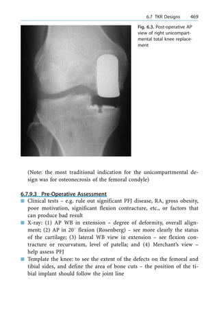
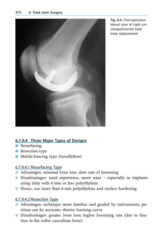

![so-called `teeter effect' of the femur toggling on the intercondylar emi-
nence during gait)
6.8.3.6.10 Reasons Why Distal Femoral Osteotomy
is a Non-Forgiving Operation
n Difficulty cutting the wedge effectively
n Need effective stabilisation of the closed osteotomy
n Need to predict accurately the wedge size necessary to ensure proper
correction of the limb
6.8.3.6.11 Rare Situation for Considering HTO for Valgus Knees
n 12±158 is the upper limit for consideration; exceeding 12±158 of val-
gus correction or 108 of resultant joint line obliquity tends to result
in lateral subluxation of the tibia and gives poor results (Coventry J
Bone Joint Surg Am 1985 [2]; also Maquet CORR 1985 [3])
n MCL laxity can also result from removing a bone wedge from above
MCL insertion
6.8.3.6.12 Pros and Cons and Results of Distal Femoral Osteotomy
n Pros ± conversion to TKR result quite good; may need stem to span
screw holes; union mostly good
n Cons ± not a forgiving operation (done less nowadays), need bulky
blade plate hardware, and sometimes impinges on the vastus medialis
6.8.3.7 Section 3: Unicompartmental Knee
n Advantages
± Mimimal bone resection
± Easy to revise
± More physiological, since cruciates retained (might be able to
dance, golf)
± Rehabilitation much quicker than with osteotomy
± Results in the 1st 10 years better than with HTO (Coventry ± HTO
results decline after 7 years)
± Less blood loss
n Disadvantages:
± Learning curve ± some models (resurfacing type instead of resec-
tion type; no elaborate instrument or blocks that may be unfami-
liar to TKR surgeon)
a 6.8 Treatment of the Young- and Middle-Aged Knee OA Patients 477](https://image.slidesharecdn.com/aresidentsguideorthopaedics-250108225802-331d4fe3/85/A-Residents-Guide-orthopaedics-surgery-pdf-492-320.jpg)
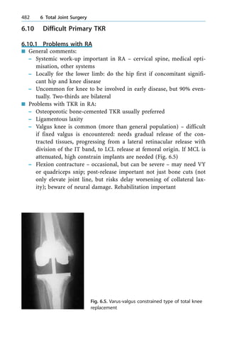
![± Type of implant ± unless ligaments are very poor, too high a con-
straint transmits shear and rotation forces to the bone±cement in-
terface. Scott recommends PCL-retaining some reports of PCL
commonly still intact in 90%
± Other problems ± cysts (sometimes large) commonly need grafts
and stemmed prosthesis for protection
± More complications arise from infection, wound and, since patients
tend to be younger when they need joint replacement, they are
more likely to need revision and undergo component loosening
and wear, etc.
6.10.1.1 TKR in RA ± Long-Term Result
n Several series report 85%+ survivals at 10 years (Ranawart)
n Some argue that this is partly due to the lower functional demand of
most of these patients
n Not much data at more than 10 years
n Laskin reported 50% high late failures with PCL retention (CORR
1997 [4]) ± PCL was found to be absent at revision, although many
other series do not report similar problems (e.g. Whiteside JA and na-
tionwide Swedish studies)
n There have been some reports of common (>60%) incidence of a ti-
bial component in radiolucent lines with PS implants (e.g. Laskin, J
Bone Joint Surg 1990), although a few others claim a good result.
(Currently, it is not settled beyond doubt whether to retain the PCL,
and whether PS implants are good or not)
6.10.2 Fusion Take Down
n Complications ± more infection, wound healing problems, poor ROM
± frequently needs manipulation under anaesthesia (MUA). Incidence
of loosening and revision is higher
n Pearls ± beware, ligaments can be gone (may need increased con-
straints), very scarred, and tibial tubercle osteotomy may be needed.
Post-operative continuous passive motion ensures the motion
achieved intra-operatively is maintained, releasing adhesions under
the quadriceps
n Outcome depends on: degree of pre-operative deformity, aetiology of
ankylosis and patient's age (and likely also on status of ligaments)
a 6.10 Difficult Primary TKR 483](https://image.slidesharecdn.com/aresidentsguideorthopaedics-250108225802-331d4fe3/85/A-Residents-Guide-orthopaedics-surgery-pdf-498-320.jpg)
![n If the component medial edge is just lateral to teardrop, the horizontal
and vertical distances from the teardrop should closely approximate
those of the normal hip ± Muller templates can help
6.12.7.7 Identifying COM
n The ideal position for the acetabular template and component is
achieved by placing the inferomedial edge adjacent to the lateral mar-
gin of the teardrop. In most cases, this will restore the COM and can
be checked using the Muller method
6.12.7.8 Tips for More Difficult Scenarios
n Protrusio ± restore medial wall, achieve component lateralisation ±
added advantage is increase in offset and less impingement; try to get
in a larger component with peripheral rim contact ± get initial stabili-
ty and/or antiprotrusio rings, especially if poor bone
n Lateralized acetabulum common [from large osteophyte especially in
patients with diffuse idiopathic skeletal hyperostosis (DISH)]: proper
removal using teardrop (transverse acetabular ligament marks the in-
ferior border of the true acetabulum) as landmark, ensuring good
coverage for cup. Incomplete removal result is not good, be it a ce-
mented or cementless cup (inadequate host bone cover).
Reaming technique ± straight medial reaming to within a few milli-
metres of planned depth of reaming; this is to avoid superior place-
ment of the component.
How to tackle excess femoral anteversion ± (1) use modular implants
that can adjust version, (2) implants should be custom made; and (3)
if use standard stem ± can even need subtrochanteric osteotomy
n Superolateral defect and dysplastic hips: many hips migrate superolat-
erally ± uncovers lateral margin of the acetabulum
± Options ± (1) High hip centre ± small component 22-mm head;
bone available may not be enough; danger of impingement on the
anterior column and anterior inferior iliac spine with flexion and
IR, also on the ischium in extension and ER, increased loading/
wear. (2) BG favoured by Allan Gross. (3) Barrack sometimes put
cup slightly more vertical and used an offset liner (not too com-
monly used method)
a 6.12 Pre-Operative Assessment in THR 493](https://image.slidesharecdn.com/aresidentsguideorthopaedics-250108225802-331d4fe3/85/A-Residents-Guide-orthopaedics-surgery-pdf-508-320.jpg)
![± Problems with hip dysplasia ± (1) excess anteversion ± use straight
femoral stem and (2) posterior rotated GT ± may need to osteoto-
mise or even de-rotate GT
6.12.7.9 Femoral Component Templating
[Always After Acetabulum Templating]
n Ensure X-ray taken in 158 IR ± needed to most accurately predict
stem size, neck length, offset and neck resection level
(If hip in fixed ER, determine COM of hip then transpose template to
the opposite side hip for femoral templating)
6.12.7.10 Positioning the Template
n The femoral template is moved vertically until the centre of the femo-
ral head template is at the same vertical height as the planned aceta-
bulum COM. No matter what type of stem, the template should be
kept centred along the neutral axis of the femur and not in varus or
valgus
6.12.7.10.1 Various Implant Types
n Proximal-coated components ± proximal fit and fill emphasised
n Extensive-coated cementless ± fixation mainly distal, and a tight fit at
isthmus is sought
n Cemented ± need enough space (of 1±2 mm) for a proper cement
mantle
6.12.7.10.2 Neck Cut
n Ideally, femoral bone stock should be maintained and neck cut usually
planned to be between 1 cm and 2 cm above LT
n Pearl: anatomical stems in particular must have the neck cut made at
a predetermined height to avoid anterior impingement of the tip of
stem distally
What Happens with Improper Neck Cut
n The neck resection should be planned so as to utilise a neck length
without a `skirt' or thickened extension of the head, as this tends to
impinge at extremes of motion; such impingement can cause de-
creased ROM and instability
494 6 Total Joint Surgery](https://image.slidesharecdn.com/aresidentsguideorthopaedics-250108225802-331d4fe3/85/A-Residents-Guide-orthopaedics-surgery-pdf-509-320.jpg)
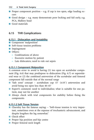
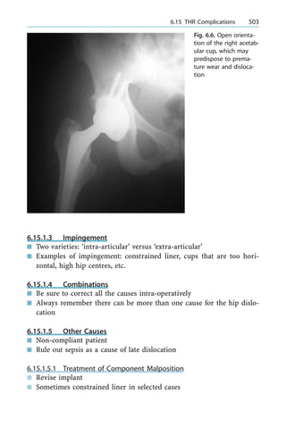
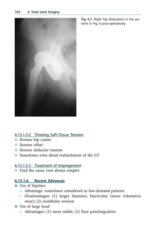
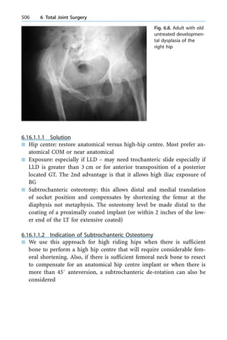
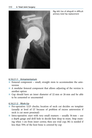
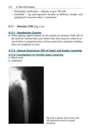
![n Marked low rate for THR performed for primary OA hip: 1.3 per
100,000 versus 60 per 100,000 (Mayo Clinic data), versus Norway's 140
per 100,000
n It was observed in some studies that patients who had THR for pri-
mary OA hip seldom underwent TKR and vice versa
n Other predisposing factors ± long leg OA; and some claims by many
experts that 25±45% of cases are from hip dysplasia, while bearing in
mind studies, for example, from Japan that low CE angle is common
but primary OA uncommon for some uncertain reason
6.18.1.3 Pitfalls
n Hip dysplasia and patients with paediatric hip condition are common
predisposing causes for hip arthritis (especially in countries where
primary OA is uncommon). It is imperative to catch the window of
opportunity to intervene by pelvic and/or femoral osteotomies de-
pending on the underlying pathology if the patient is too young for
THR, like in his or her twenties
n Two causes of acute deterioration of symptoms ± (1) ruptured or torn
labrum and (2) sometimes OA in some rarer case ± can progress rap-
idly in terms of few months
6.18.1.4 Causes of Secondary OA
n AVN ± sclerosis, lucency, flattening of head (will be discussed in de-
tail in the next section)
n Paget's disease (Fig. 6.11) ± bone enlarges, coarse trabeculation, cor-
tex thickened
n Inflammatory arthritis ± diffuse joint space narrowing, osteopenia,
erosion
n Trauma remodelling ± distorted contour with sclerosis and remodelling
n DDH ± shallow acetabulum, increased acetabular index, sometimes
hip subluxation or dislocation
n Slip ± femoral retroversion, convexity of head±neck junction, short
femoral head/short broad neck
n Perthes disease
[Others: Charchot joint is less common in the hip, but sometimes af-
fects the knee (Fig. 6.12) and elbow. Crystal arthropathy, like gout
(Fig. 6.13), mostly spares the hip, but can cause OA in other peripher-
al joints]
520 6 Total Joint Surgery](https://image.slidesharecdn.com/aresidentsguideorthopaedics-250108225802-331d4fe3/85/A-Residents-Guide-orthopaedics-surgery-pdf-535-320.jpg)
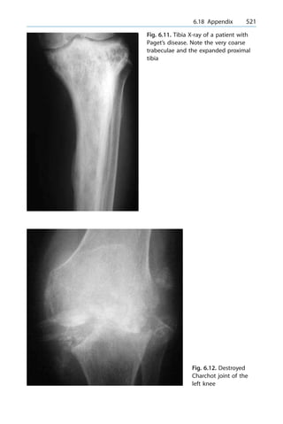
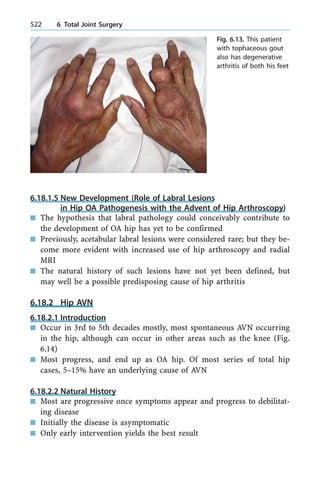
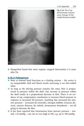
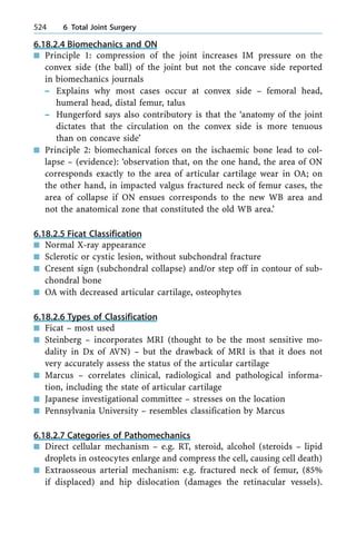
![Study of effect of arterial tamponade ± animal study on hip blood
flow (Launder J Bone Joint Surg 1981 [6]) (some claim, however, that
the adult hip is more resistant to this effect)
n Extraosseous venous mechanism: it is not clear whether this is a
cause or effect. Experiment from Johns Hopkin's Hospital: obstruction
in vein flow causes only temporary decrease in blood flow, then rapid
return to normal
n Intraosseous extravascular: e.g. hypertrophy of fat cells/steroid, Gau-
cher's; and possible role of intraosseous oedema haemorrhage
Mechanism here is that an increase in compartment pressure tends to
collapse the thin-walled vessels and causes decreased blood flow
n Intraosseous intravascular: e.g. sickle cell anaemia, dysbaric, intravas-
cular coagulation (recently proposed ± Schwartzman phenomenon ±
this involves the concept that an immune complex deposition may
lead to arteriolar haemorrhage and ON), possibly fat emboli (hyper-
lipidaemia, steroids, alcohol). and possible role of increased platelet
and hypofibrinolysis
6.18.2.8 Pathological Changes
n The softened necrotic cancellous bone is unable to withstand physio-
logical forces and collapses, signalling usually the onset of symptoms
n A `cleavage plane' occurs between the articular surface ± which is
nourished by the joint fluid ± and the necrotic bone beneath it. (At
this time, the avascular fragment will `ballote' with compression,
much like a tennis ball will compress and then resume its shape)
n Collapse of the articular cartilage is visible when the fragment no
longer assumes its original shape, and the head flattens
n Near the end stage, the articular surface becomes dissociated from
the cancellous bone and cleaves off as a large fragment. This is called
`delamination' representing late advanced stage and OA begins
n Once the articular surface collapses, salvage of congruent hip is al-
most impossible
6.18.2.9 Variables Influencing Our Management
n Stage ± pre-collapse means stage 1 or 2 in most classification systems
n Size ± first stressed by Steinberg; ON angle is useful concept ± mea-
sure the angle subtended in (AP and frog lateral) <1608 small, >2508
large
a 6.18 Appendix 525](https://image.slidesharecdn.com/aresidentsguideorthopaedics-250108225802-331d4fe3/85/A-Residents-Guide-orthopaedics-surgery-pdf-540-320.jpg)
![n Age ± one-quarter to one-third of the patients are very young (<25
years); need to avoid THR and consider other options, e.g. vascularised
BG, trapdoor procedure, surface replacements and/or osteotomy
n Severity of associated underlying disease (in two-thirds of cases)
5th element added by Sugioka ± site of the lesion (see later discus-
sion)
6.18.2.10 Non-Operative Treatment
n Implantable bone stimulators (some studies have been shown to stim-
ulate osteogenesis; reported in 1989) ± this method has not been ap-
proved by the FDA
n Protected weight bear
n Long-term results not good
6.18.2.11 Aim of Operative Treatment
n Relieve pain
n Preserve head
n Prevent further joint deterioration
n Early intervention important
6.18.2.11.1 Method 1: Core Decompression and/or BG
[Ficat's Stage 1, 2]
n Rationale based on an increase in intraosseous pressure in the femo-
ral head resulting from venous congestion and other pathways (Hun-
gerford)
n Immediate pain relief in most series
n Must be done before head collapse or stage 3
n Most agree that it does play an insignificant role in stage 3 (only 20%
success rate)
n Complications in 1%; need protected WB for 6 weeks
n One study in 1996 included 1200 hips of which 741 had clinical suc-
cess at `all' stages ± although much less effective in stage 3
6.18.2.11.2 Method 2: The Two Main Groups of Osteotomies
(Rotational and Angular)
n Varus (F/E) osteotomy and valgus (usu extension) osteotomy
n Sugioka's transtrochanteric rotational osteotomy (in fact a combina-
tion of three osteotomies)
526 6 Total Joint Surgery](https://image.slidesharecdn.com/aresidentsguideorthopaedics-250108225802-331d4fe3/85/A-Residents-Guide-orthopaedics-surgery-pdf-541-320.jpg)
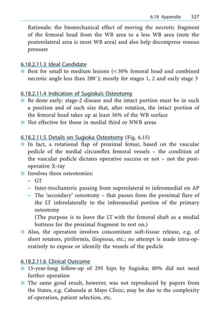
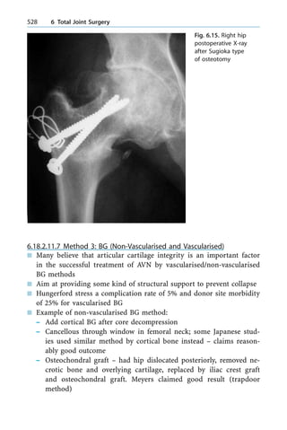
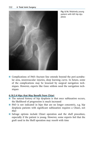
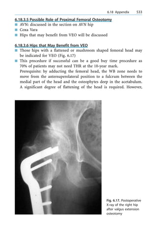
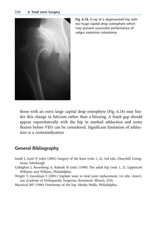
![7.3.2 Pearls 548
7.3.3 Classification of Tibia (and Fibula) Bone Defect [May] 548
7.3.3.1 Cierny-Mader Classes (Here Referring to Osteomyelitis of Tibia) 549
7.3.3.2 Systemic Factors 549
7.3.3.3 Local Factors 549
7.3.4 Diagnosis 549
7.3.5 Treatment 549
7.3.6 Vascularised Bone Graft Results 550
7.3.7 Summary of Key Concepts 550
7.4 Spinal Infections 550
7.4.1 Pyogenic Spinal Infections 550
7.4.2 Non-Pyogenic Granulomatous Infections 551
7.4.2.1 Physical Signs of TB Spine 553
7.4.2.2 Indications for Surgery 554
7.4.2.3 Choice of Graft in Anterior Approach 554
7.4.3 Other Non-Pyogenic Granulomatous Infections 554
7.4.3.1 Other Main Category: Epidural Abscess 554
7.4.3.2 Other Category: Subdural Abscess 555
7.4.3.3 Other Category: IM Abscess 555
7.4.3.4 Other Category: Post-Operative Infections 556
7.4.4 Appendix 556
7.4.4.1 Pitfalls in Dx 556
7.4.4.2 Differential Diagnosis of TB from Pyogenic Spondylitis 557
7.4.4.3 Radiological DDx 557
7.4.4.4 Role of Nuclear Scan in Pyogenic 557
7.4.4.5 Biopsy 557
7.4.4.6 Role of MRI 557
7.4.4.7 Radiological DDx from Non-Infected Causes 558
7.4.4.8 Summary of MRI Assessment 558
7.4.4.9 General Pearls 558
7.4.5 Highlight of Salient Points on TB Spine 558
7.4.5.1 Introduction 558
7.4.5.2 Pathogenesis 559
7.4.5.3 Features in the Affected Child 559
7.4.5.4 General Clinical Features 559
7.4.5.5 Causes of Death 559
7.4.5.6 Special Situations 559
7.4.5.7 Atypical Presentation 560
7.4.5.8 Pathogenesis of Paraplegia 560
7.4.5.9 X-Ray Appearances 560
7.4.5.10 CT Scan 560
7.4.5.11 Role of MRI 561
7.4.5.12 Role of Biopsy 561
7.4.5.14 Adjuncts in Dx 561
7.4.5.15 Main Goals of Rn 561
538 7 Orthopaedic Infections](https://image.slidesharecdn.com/aresidentsguideorthopaedics-250108225802-331d4fe3/85/A-Residents-Guide-orthopaedics-surgery-pdf-552-320.jpg)
![7.1.1.4 Investigations
n Erythrocyte sedimentation rate (ESR) and C-reactive protein (CRP)
are very useful but lack specificity, although sensitive. Seeing a trend
is important. Levels of white blood cells (WBCs) are less so, though
usually on the high end
n Joint aspiration for Gram stain, culture, cell count and biochemistry
is most important for diagnosis (Dx); do not jump to mention bone
scans/magnetic resonance imaging (MRI)
n Ultrasound (USS)-guided aspiration of deeper joints useful, especially
the hip ± USS positive immediately in general after infection, and
many days before X-ray changes
n X-ray: (acute) soft tissue swelling, joint distended/swell capsule (late
changes): joint destruction, osteoporosis, metaphyseal involvement
7.1.1.5 Special Investigation
n Bone scan: pressure inside joint sometimes causes hypo-uptake in
femoral head, may be useful in suspected combined osteomyelitis and
septic arthritis; sometimes if multiple joints are affected (10% of
cases; but this is the figure for adults), and useful in those immuno-
suppressed from whatever cause that may mask symptoms and signs
n MRI: can see not only joint effusion, but see changes if associated os-
teomyelitis
n CT: few indications [but useful in adults in exotic joints such as ste-
noclavicular joint (SCJ)/sacroiliac joint (SIJ) and/or pubic symphysis,
especially in addicts]
7.1.1.6 Differential Diagnosis
n Acute osteomyelitis: can be very similar, but careful examination finds
that joint motion is allowed by the child; however, the two conditions
can co-exist, especially in the hip and shoulder joints
n Juvenile `idiopathic' arthritis: initially can be monoarticular, but there
is little pain and effusion is positive
n Transient synovitis: clinical and aspiration
n Perthes: USS sometimes useful in DDx
n Post-traumatic effusion: Hx of trauma
n Cellulitis and/or bursitis (children: prepatella bursitis more than olec-
ranon)
n Acute rheumatic fever
a 7.1 Septic Arthritis 541](https://image.slidesharecdn.com/aresidentsguideorthopaedics-250108225802-331d4fe3/85/A-Residents-Guide-orthopaedics-surgery-pdf-555-320.jpg)
![n Others: HSP (rare form of hepatorenal dysfunction in children asso-
ciated with purpura), haemophilia, juvenile rheumatoid arthritis
7.1.1.7 Two Main Treatments to Be Given at the Same Time
n IV antibiotics: decrease systemic effect/toxaemia (30±50% blood cul-
ture positive)
n Open drainage and debridement: still the mainstay (in the older child
with knee sepsis, can perform arthroscopic lavage. Hip sepsis often
treated by open drainage)
7.1.1.8 Disadvantage of the Method of Repeated Aspiration
n Pus too thick for adequate aspiration
n Instillation of antibiotics irritant to cartilage
n Tension will recur
n Pain with each procedure
n Uncertain results
7.1.1.9 Open Hip Drainage
n Anterior Smith-Peterson approach used most
n Adequate debridement
n Drain in-situ
n If hip unstable, splint in a partially abducted and/or flexed posture
with or without formal immobilisation if very unstable
n IV then oral antibiotics ± for total of 6 weeks
7.1.1.10 Rare Organisms
n Streptococcus pneumoniae, E. coli, proteus, salmonella (sickle cell pa-
tients), serratia marcescens, clostridia welchii, neisseria, staphylococ-
cus albus, aerobacter, bacteroides, paracolon bacillus.
[Look out for unusual microbes in immunosuppressed patients or
penetration of joint by foreign body (FB), or foreign body inside
joint]
7.1.1.11 DDx Reasons for Negative Culture
n Partial antibiotics treatment (Rn)
n Poor culture technique
n Inadequate anaerobic cultures (some inject the aspirated fluid into
blood culture bottles as well)
542 7 Orthopaedic Infections](https://image.slidesharecdn.com/aresidentsguideorthopaedics-250108225802-331d4fe3/85/A-Residents-Guide-orthopaedics-surgery-pdf-556-320.jpg)
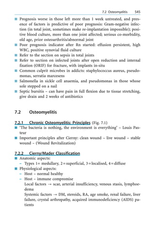
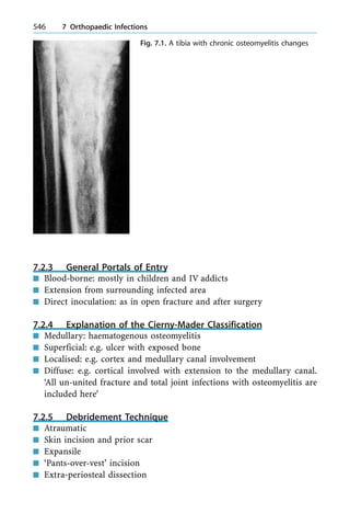
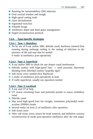
![7.2.6.4 Type 4: Diffuse
n These are infected non-unions or chronic septic joints
n X-ray and CT/MRI all frequently used
n Resection of the infected region frequently involved
n Stabilisation needed [e.g. IM fixation/external fixation (EF)]
n Soft tissue cover mostly required
n 6 weeks of antibiotics post-operatively
7.3 Managing Septic Non-Union
7.3.1 General Principles
n Adequate and thorough debridement to viable and healthy bone.
Sometimes also need to be adequately debrided ± remove non-viable
muscles, debris
n Repeated debridement frequently necessary
n Soft tissue cover brought in early (preferably in less than 1 week if
bed not contaminated, and flap needed). Notice that: (1) muscle flaps
preferable as they help fight infection, are better cover for tibia and
are good base for later split-thickness skin graft (SSG); (2) emergency
soft tissue cover is sometimes needed, e.g. if joint is exposed, bare
tendon, etc.
n Eliminate dead space and IV antibiotics
n Skeletal stabilisation always, EF is useful if large wound in nursing
and raise flaps
n Later Rn of bone defects ± vascular BG considered if bone defect
more than 6 cm and bed not contaminated. Alternative is the Ilizarov
procedure
7.3.2 Pearls
n Soft tissue healing very important or infection persists
n Pasteur: the bug is nothing, the environment is everything
n Especially in trauma open fracture wound, frequently polymicrobial
7.3.3 Classification of Tibia (and Fibula) Bone Defect [May]
n Both T/F intact, less than 3 months rehabilitation expected
n BG needed; 3±6 months rehabilitation expected
n Tibial defect <6 cm, fibula OK; 6±12 months rehabilitation expected
548 7 Orthopaedic Infections](https://image.slidesharecdn.com/aresidentsguideorthopaedics-250108225802-331d4fe3/85/A-Residents-Guide-orthopaedics-surgery-pdf-562-320.jpg)
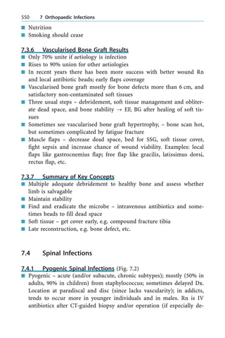
![layed with neural compression, too much destruction and instability,
epidural abscess forming) and treat the cause (DM, steroids)
n Lumbar most common, cervical in IV mainliners injecting their upper
limbs; adults nowadays more gram-negative sepsis ± such as pseudo-
monas, E. coli, and/or anaerobes
n Previous claim of spread from Batson's plexus was doubted since very
high pressure was needed for retrograde flow. Now, most believe it
starts in paradiscal at endplate. As the cartilaginous end plates of
child matures, blind-loops of vascular anastomosis remain ± low-flow
conduits provide a footing for septic emboli and bacterial seeding
n Minimum 4 weeks of IV antibiotics; for those treated conservatively,
need brace, add chin extension in upper thoracic infection and cervi-
cal ones may need halo device. (Brace for 3 months). Overall 75%
success rate with modern Rn
7.4.2 Non-Pyogenic Granulomatous Infections
n Tuberculosis [TB; 10% involves skeleton, and 5% the spine (Fig. 7.3);
other peripheral joints are also affected (Fig. 7.4)]
a 7.4 Spinal Infections 551
Fig. 7.2. An addict with a partially treated
recent pyogenic spondylitis](https://image.slidesharecdn.com/aresidentsguideorthopaedics-250108225802-331d4fe3/85/A-Residents-Guide-orthopaedics-surgery-pdf-565-320.jpg)
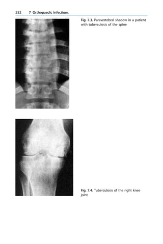

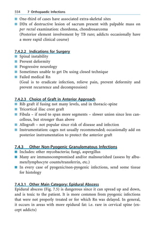
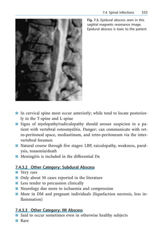
![7.4.5.2 Pathogenesis
n Delayed hypersensitivity (bone destruction by caseous necrosis)
n Selective involvement of the anterior vertebral column and collapse
causes varying degrees of kyphosis
n Early or late paraparesis is another dreaded complication
7.4.5.3 Features in the Affected Child
n Stripping of ALL can cause rapid spread
n Abscesses tend to be larger and amount of bone destruction greater,
but neural complications are comparatively lower than in adults
n Anterior growth apophysis can be destroyed causing affected anterior
growth ± since posterior elements are seldom affected in the usual
case
n Retropharyngeal upper mediastinal paraspinal abscess can cause
upper airway obstruction ± classic inspiratory stridor (Millar asthma)
7.4.5.4 General Clinical Features
n Back pain
n Gibbus (or step)
n Abscess
n Paraparesis
n Constitutional upset
n Look for neurological deficit (in both early and late disease)
7.4.5.5 Causes of Death
n Systemic disease
n Cerviocomedullary disease from abscesses or instability can cause
quadriplegia and even sudden death
n Stridor and respiratory obstruction in the child ± from retropharyn-
geal and upper mediastinal paraspinal masses
7.4.5.6 Special Situations
n Cervical-dorsal region: asthma-like syndrome when the child lies
down at night, from huge soft tissue mass around the bronchus
n Cervical spine: little role of conservative care since paraplegia is com-
mon [prefer anterior spinal fusion (ASF) and debridement]
n T-spine in child: `aneurysmal syndrome' ± abscess lifts up the loose
periosteum; local effects may mimic an aortic aneurysm
a 7.4 Spinal Infections 559](https://image.slidesharecdn.com/aresidentsguideorthopaedics-250108225802-331d4fe3/85/A-Residents-Guide-orthopaedics-surgery-pdf-573-320.jpg)
![7.4.5.7 Atypical Presentation
n Posterior elements (spine) ± disease of the neural arch, picture is like
spinal cord compression rather like cauda equina; not always easy to
diagnose by means of simple X-ray
n Sacoiliac joint ± pain from buttock to knee and/or abscess. Investiga-
tion: CT/MRI ± determine site of abscess and plan accordingly
n Epidural abscess ± pachymeningitis, arachnoiditis; can be lower limb
spastic paresis or picture-like spinal cord tumour. Dx ± MRI (if not
available, cerebrospinal fluid myelogram: shows streaking of contrast)
n Lumbar spine: some authorities prefer conservative; but, if vertebral,
destruction can cause early loss of lordosis or even reverse lordosis,
with LBP in a 20-year long-term study
n Lumbosacral junction disease ± tend to operate in these cases since
kyphosis is likely
n Mutli-level disease is uncommon and has sometimes been mistaken
for metastases in the past
7.4.5.8 Pathogenesis of Paraplegia
n Early onset ± due to pus, disc, subluxation, sequestrum, pachymenin-
gitis (presence of myelitis indicates poorer prognosis)
n Late onset ± kyphus, bone bridge, proximal spinal stenosis
n Interestingly, the deterioration in neurology was more related to canal
compromise than amount of kyphosis
[Note that in many (60%) late cases of paraparesis, there is evidence
of disease reactivation]
7.4.5.9 X-ray Appearances
n Frequently involves two adjacent vertebrae and their adjoining disc
n Anterior part of vertebra may be scalloped
n Fusiform paravertebral abscess shadow can be present, sometimes loss
of psoas shadow
7.4.5.10 CT Scan
n Can see the details of bony destruction and soft tissue calcifications
n (Bone scan is less useful since, although it is sensitive, it is not specific)
560 7 Orthopaedic Infections](https://image.slidesharecdn.com/aresidentsguideorthopaedics-250108225802-331d4fe3/85/A-Residents-Guide-orthopaedics-surgery-pdf-574-320.jpg)
![7.4.5.11 Role of MRI
n Helps in Dx of early disease
n Helps to pick up multicentric lesions before plain X-ray changes get
obvious
n Can show TB arachnoiditis and extradural or intradural spread of the
abscess if present
n Mostly T1 hypodense, T2 hyperintense (gadolinium enhancing)
n Rim enhancement with contrast is common since sequestrum in cen-
tre remains hypointense ± but not specific for TB and can be mi-
micked by pyogenic infection and neoplasm
7.4.5.12 Role of Biopsy
n Neither clinical nor radiological imaging criteria are absolutely patho-
gnomonic ± hence biopsy is usually needed to confirm the Dx
7.4.5.13 Pearls in Definitive Dx
n CT-guided, fine-needle biopsy or video-assisted, thoracoscopic sur-
gery ± less invasive way to get a biopsy or drain an abscess. High cy-
tology yield of 88% reported
n Although the bacterial count is lower than lung lesions (and bacilli
are not seen in 50% of histological lesions), the histological features
of epitheloid granuloma, Langerhan's multinucleated giant cells and
caseation are sufficient in Dx
7.4.5.14 Adjuncts in Dx
n PCR [and enzyme-linked immunosorbent assay (ELISA)] ± amplifies
mycobacterial DNA. May be useful in difficult cases
n MRI ± ring enhancement with contrast, lost cortical margin (show
the compression of neural structures well in, for example, late kyphus,
etc.)
7.4.5.15 Main Goals of Rn
n Eradicate infection
n Preserve and restore neural integrity
n Prevent and correct deformity
a 7.4 Spinal Infections 561](https://image.slidesharecdn.com/aresidentsguideorthopaedics-250108225802-331d4fe3/85/A-Residents-Guide-orthopaedics-surgery-pdf-575-320.jpg)
![7.4.5.22 Comments on Kyphosis Correction
n In multilevel disease, where three or more levels are involved: defor-
mity can reach 1008; to achieve any significant correction of such se-
vere and rigid deformity without neurological loss, the procedure has
to be done gradually
7.4.5.23 Some Recent Trends
n With the advent of spinal instrumentation in the past decade, supple-
menting fusions with anterior or posterior instrumentation has been
tried by some to improve and maintain deformity correction. [Ac-
cording to workers such as Oga, biofilm may be less likely to form
with tuberculous infection even in the presence of an implanted de-
vice]
7.4.5.24 Some Important Bring-Home Messages
n TB spine can only be said to have definitely healed up when solid fu-
sion has been obtained
n This important point was not stressed enough in the MRC report
n Another point not explored enough in MRC trial: with a very pro-
longed follow-up (much greater than 10 years) in 60 patients treated
in Hong Kong University with conservative Rn, nearly half with late
paraparesis and neural loss from progressive kyphus and cord com-
pression, and atrophy and scarring responded poorly despite decom-
pression and BG, and there was no good solution
n The conclusion of MRC that radical surgery ªdid not improve the
chances of a favourable outcome apart from less late deformityº
should be taken with a grain of salt. (Also, the lack of solid fusion
can cause continued deterioration and neural loss)
n Chemotherapy is the most effective Rn, but radical operation causes
earlier fusion, and less delayed deformity
n For multiple vertebral involvement cases, A+P surgery is advised, as
well as ASF+posterior spinal fusion (PSF) since ASF alone in these
cases work less well for the very young child (<7 years)
n After the work of Oga, some had advocated the use of pedicle posteri-
or instrumentation in the earlier stages of thoracic/thoracic-lumbar
disease
564 7 Orthopaedic Infections](https://image.slidesharecdn.com/aresidentsguideorthopaedics-250108225802-331d4fe3/85/A-Residents-Guide-orthopaedics-surgery-pdf-578-320.jpg)
![7.6.15 Duration of IV Antibiotics
and Minimum Inhibitory Concentration
n 6 weeks minimum (as also originally suggested by Insall)
n Some paper in the past, claimed that patient needs to prolong stay in
hospital and maintain minimum inhibitory concentration at therapeu-
tic level ± not always done in every centre since high cost of pro-
longed hospital stay
7.6.16 Use of Antibiotic-Impregnated Cement
n Shown to have positive effect in success to eradicate infection
n Rate of elution differs with different antibiotics
n Do not use antibiotics that are readily inactivated by the exothermic
reaction during cement setting
n Pelicose said to be better than simplex in re-implant since gets
doughy earlier
7.6.17 Use of Cement Spacer and PROSTALAC
n Refer to Vancouver's experience and papers from Clive Duncan's
group
n Spacers: the original concept comes from the instability after resec-
tion arthroplasty ± functions include:
± Provides local concentration of antibiotics
± Maintains length and range of motion
± Frequently also allows ambulation in these elderly subjects
[PROSTALAC=the prosthesis of antibiotic-loaded acrylic cement]
7.6.18 Pearls During Re-Implantation Process:
Cemented Versus Cementless
n Both cemented and cementless revisions have their proponents The
`best' choice lies in individual assessment of each particular case
n Antibiotic-loaded cement is usually used in revisions
n Type of antibiotics depends on sensitivity; if no organism identified,
a few even use more than one antibiotic
n Be prepared to tackle bone defects
n Use of stemmed prosthesis sometimes needed in TKR. In THR, some-
times need long stem for the same token; possible use of allograft (es-
sentially a piece of dead bone) with caution needed
a 7.6 Infected Total Joint Replacements 571](https://image.slidesharecdn.com/aresidentsguideorthopaedics-250108225802-331d4fe3/85/A-Residents-Guide-orthopaedics-surgery-pdf-585-320.jpg)
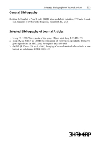
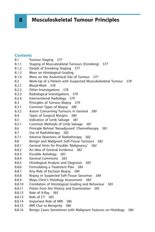
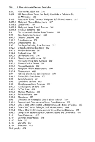
![8.2 Work-Up of a Patient with Suspected Musculoskeletal
Tumour
n History and physical:
± If suspicion of metastases to bone from a distant primary, check
thyroid, breast, prostate, kidney and lung; do a rectal examination
and check for signs of metastases elsewhere
± If suspicion of primary malignant growth from bone, note the age
of the patient [some tumours occur in those of particular age
groups, e.g. giant cell tumour (GCT) mostly occurs after closure of
epiphysis] and the site of the tumour [e.g. different tumours have
predilection for different sites, for instance: (i) in the case of long
bones, Ewing's tumour often occurs in diaphysis, and osteosarco-
mas mostly at metaphysis; while (ii) in the case of the spine, osteo-
blastomas are commonly found in the posterior elements]
8.2.1 Blood-Work
n Blood-work: blood picture (suspected haemic malignancy), erythro-
cyte sedimentation rate (ESR; usually elevated, very high in myeloma),
liver function test (can be deranged if metastases), alkaline phospha-
tase (if bony origin, can be due to bony destruction/increased bone
turnover), Ca/PO4 (some tumours/metastases can have hypercalcae-
mia), tumour markers [e.g. prostate-specific antigen (PSA) if suspect
metastases from prostate] and serum protein electrophoresis (mono-
clonal band in myeloma)
8.2.2 Other Investigations
n Magnetic resonance imaging (MRI): can detect skip lesions, extent of
bony malignancy, any involvement of other compartments and/or
neurovascular structures; helps to plan biopsy tract and definitive re-
section
n Computed tomography (CT): helps assess the extent of bony destruc-
tion by the tumour in areas such as the flat bones of the pelvis or
acetabulum, and the spine, etc, to help plan reconstructive surgery (if
still feasible), e.g. 3D CT reconstruction in planned total hip replace-
ment for metastases involvement of the acetabulum, etc. CT also helps
in the staging of some tumours, e.g. CT thorax
578 8 Musculoskeletal Tumour Principles](https://image.slidesharecdn.com/aresidentsguideorthopaedics-250108225802-331d4fe3/85/A-Residents-Guide-orthopaedics-surgery-pdf-591-320.jpg)