1. TGF-β1 Regulates IAP Expression in Lung Fibroblasts
Madalyn L. Holzapfel*, I.O. Ajayi, Amos E. Dodi M.D., Jeffery C. Horowitz M.D.
ABSTRACT
Idiopathic pulmonary fibrosis (IPF) is a disease that is associated with abnormal
scar tissue within the lung. It does not yet have a known cause; however,
fibroblasts, which are cells responsible for making the abnormal scar tissue, are
key to the pathogenesis of the disease. These fibroblasts accumulate in the lung
and our laboratory has hypothesized that resistance to apoptosis is one
mechanism underlying the abnormal accumulation. To test this hypothesis, we
assessed the effect of TGF-ß1, a pro-fibrotic factor, on fibroblast expression of
XIAP (an inhibitor of apoptosis protein) and the closely related proteins cIAP-1 and
cIAP-2. To do this, fibroblasts were grown in cell culture dishes. Designated
dishes were treated with TGF-ß1 for four hours and other dishes were left
untreated as controls. Protein and RNA was extracted at different time points.
XIAP was assessed by Western blotting and quantitative real time reverse
transcription PCR (qPCR), while cIAP-1 and cIAP-2 were assessed by qPCR. Our
results showed a consistent increase in XIAP protein and mRNA. cIAP-1 mRNA
was similarly increased, while cIAP-2 was not. These results show that TGF-ß1
increases several IAPs in fibroblasts. We conclude that TGF-ß1 regulation of
several IAPs may contribute to fibroblast survival and the development of
abnormal lung scarring. Ongoing studies are evaluating the effects of TGF-ß1 on
IAPs at later time points.
INTRODUCTION
HYPOTHESIS
Fibroblast resistance to apoptosis is mediated
by XIAP and contributes to the abnormal
accumulation of these cells in IPF.
METHODS
RESULTS
To determine if TGF-β1 regulates structurally similar
cIAP-1/cIAP-2, these IAPs and XIAP were treated with
TGF-β1 for four hours and assessed by qRT-PCR.
CONCLUSIONS
• TGF-β1 rapidly increases XIAP at both the mRNA
level and the protein level in normal lung fibroblasts
• TGF-β1 has no significant effect on cIAP-2 and
promotes a small increase in cIAP-1
• Collectively, these findings suggest that regulation of
XIAP and possibly cIAP-1 by TGF-β1 may contribute
to fibroblast resistance to apoptosis in lung fibrosis.
• Ongoing experiments are assessing how TGF-β1
regulates IAPs at later time points
Determine the effect of the pro-fibrotic cytokine TGF-β1
on fibroblast expression of the Inhibitor of Apoptosis
Proteins XIAP, cIAP-1 and cIAP-2.
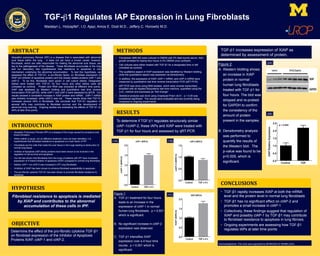
Figure 1:
A. TGF-β1 treatment for four hours
leads to an increase in the
expression of cIAP-1 in normal
human lung fibroblasts. p < 0.001
which is significant.
B. No significant increase in cIAP-2
expression was observed.
C. TGF-β1 intensifies XIAP
expression over a 4 hour time
course. p < 0.001 which is
significant.
TGF-β1 increases expression of XIAP as
determined by assessment of protein.
Figure 2:
A. Western blotting shows
an increase in XIAP
protein in normal
human lung fibroblasts
treated with TGF-β1 for
four hours. The blot was
stripped and re-probed
for GAPDH to confirm
the consistency of the
amount of protein
present in the samples.
B. Densitometry analysis
was performed to
quantify the results of
the Western blot. The
p-value was found to be
p<0.005, which is
significant.
• Fibroblasts (IMR-90) were cultured in DMEM with 5% fetal bovine serum, then
growth arrested for twenty-four hours in 0% DMEM once confluent.
• Cell cultures were either treated with TGF-β1 for a designated time or left
untreated as controls.
• The qualitative aspect of XIAP expression was identified by Western blotting,
while the quantitative aspect was assessed via densitometry.
• In addition, the expression of XIAP, cIAP-1 mRNA, and cIAP-2 mRNA were
measured by quantitative real time reverse transcription PCR (qRT-PCR).
• qRT-PCR was done using RNA isolates, which were reverse transcribed,
amplified with an Applied Biosystems real time machine, quantified using the
ΔΔCT method and expressed as “fold change”.
• Statistical analysis was done using Graphpad Prism v6.01. p < 0.05 was
considered significant. The results were evaluated and are currently being
compared to ongoing experiments.
• Idiopathic Pulmonary Fibrosis (IPF) is a disease of the lungs caused by excessive scar
tissue formation.
• While neither a cause, nor an effective treatment, have yet been identified, it is
hypothesized that fibroblast resistance to apoptosis is a main factor of IPF.
• Fibroblasts are the cells that make the scar tissue in the lungs leading to destruction of
normal lung tissue.
• Inhibitor of Apoptosis (IAP) family proteins have been shown to be involved in the
regulation of cell survival and apoptosis
• Our lab has shown that fibroblasts from the lungs of patients with IPF have increased
expression of X-linked inhibitor of apoptosis (XIAP) compared to normal lung fibroblasts.
• Neither cIAP-1 nor cIAP-2 was increased in IPF lung fibroblasts.
• Inhibition of XIAP has been shown to enhance fibroblast susceptibility to apoptosis.
• The pro-fibrotic cytokine TGF-ß1 has been shown to promote fibroblast resistance to
apoptosis.
Acknowledgments: This work was supported by NIH/NHLBI HL105489 (JCH).
(1A) (1B)
(1C)
OBJECTIVE
(2A)
(2B)