Downloaded 65 times
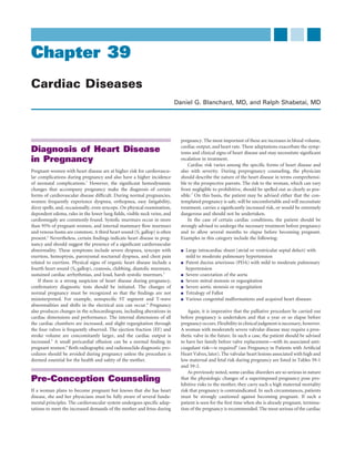
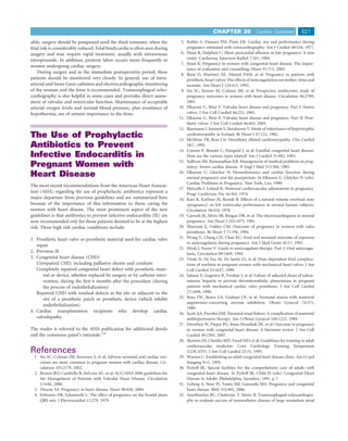
![CHAPTER 39 Cardiac Diseases 807
Aorta LV
LV
Septal
RV defect RV
Pulmonary
artery
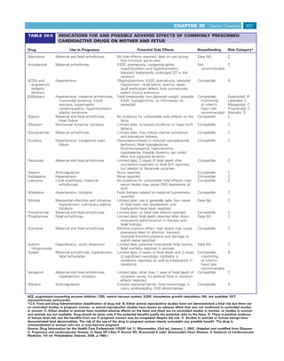
Tetralogy of Fallot Normal
FIGURE 39-8 Tetralogy of Fallot. The anatomic pathology (left)
compared with normal (right). Note the ventricular septal defect, the
aorta (which overrides the defect), the pulmonary stenosis, and the
right ventricular hypertrophy. LV, left ventricle; RV, right ventricle.
(Reprinted by permission of the publisher. From Taussig HB:
Congenital Malformations of the Heart. Cambridge, Mass, Harvard
University Press, 1960. Copyright © 1960 by the Commonwealth
Fund of the President and Fellows of Harvard College.)
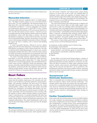
FIGURE 39-7 Pressure tracings in severe pulmonary stenosis.
Pulmonary pressure is extremely low and appears damped. Right
ventricular pressure is suprasystemic. (From Shabetai R, Adolph RJ:
Principles of cardiac catheterization. In Fowler NO [ed]: Cardiac
Diagnosis and Treatment. Hagerstown, MD: Harper & Row, 1980,
p 106.)
RV
Pulmonic stenosis is generally well tolerated so that neither preg-
nancy nor labor poses a significant threat.49 Prophylaxis against infec-
tive endocarditis is necessary. More severe pulmonary stenosis requires LV
AO
treatment. Unlike aortic stenosis, however, critical pulmonary stenosis
does not require valve replacement or open repair. Most cases are
treated successfully with transvenous balloon valvuloplasty.50 Ideally, MV
this should be carried out before pregnancy is undertaken; if a woman
does become pregnant and develops intractable right-sided heart
failure, the procedure can still be safely performed (but at some risk
to the fetus). Extreme pulmonary stenosis (right ventricular systolic
pressure > systemic systolic pressure) is a contraindication to preg- FIGURE 39-9 Transthoracic echocardiographic image of tetralogy
nancy until the lesion is adequately treated. of Fallot. A large ventricular septal defect is present, and the aorta
(AO) overrides the interventricular septum. LV, left ventricle; MV,
mitral valve; RV, right ventricle.
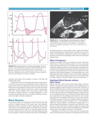
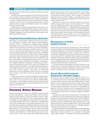
Right-to-Left Shunt without Pulmonary
Hypertension (Tetralogy of Fallot) outflow obstruction that diverts blood flow through the VSD. In the
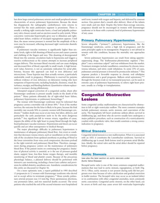
The congenital cyanotic heart diseases discussed so far have been asso- typical case, right and left ventricular systolic pressures are equal but
ciated with a communication between the pulmonary and systemic the pulmonary artery pressure is exceedingly low. A loud, long systolic
circulations and pulmonary vascular resistance sufficiently high to murmur is audible along the left sternal border. The murmur is caused
cause a right-to-left shunt. However, cyanosis occurs in other congeni- by an abnormal flow pattern through the obstructed right ventricular
tal malformations, in which there is a defect between the right and left outflow tract. The pulmonary valve closure sound is usually inaudible.
sides of the heart but also right ventricular outflow obstruction (Figs. Patients are usually cyanotic and often have significant clubbing of the
39-8 and 39-9). Examples include the tetralogy of Fallot and tricuspid fingers and toes. The hematocrit value is greatly elevated because of
atresia. the severe erythrocytosis. Phlebotomy is not indicated to treat the
Tetralogy of Fallot is used to illustrate this class of congenital mal- hematocrit level per se but is indicated if symptoms of hyperviscosity
formation of the heart, because it is by far the most common form of occur. Ignoring this important therapeutic principle leads to a micro-
cyanotic congenital heart disease encountered in pregnancy. Moreover, cytic anemia that further complicates pregnancy. The ECG shows
the offspring of a mother with tetralogy of Fallot has a 2% to 13% severe right ventricular hypertrophy. The chest radiograph is charac-
chance of inheriting the condition.51 The syndrome includes (1) a large terized by a normal-sized heart and a concavity in the region where
defect high in the ventricular septum; (2) pulmonary stenosis, which the pulmonary artery should be (Fig. 39-11). As in all malformations
may be at the valve itself but more commonly is in the infundibulum of this general type, the lung fields are oligemic, showing small vessels
of the right ventricle; (3) dextroposition of the aorta so that the aortic throughout.
orifice sits astride the VSD and overrides, at least in part, the right Most adults born with the tetralogy of Fallot and lesions with
ventricle; and (4) right ventricular hypertrophy (Fig. 39-10). similar pathophysiology have undergone surgical treatment before
A wide spectrum of clinical presentations may be present, depend- reaching young adulthood. Children raised in undeveloped countries
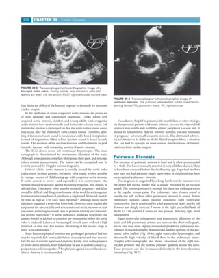
ing on the relative size of the VSD and the degree of right ventricular are an important exception. Many patients have had surgery to close](https://image.slidesharecdn.com/4-u1-0-b978-1-4160-4224-2-50042-9-docpdf-120121090855-phpapp01/85/4-u1-0-b978-1-4160-4224-2-50042-9-docpdf-11-320.jpg)
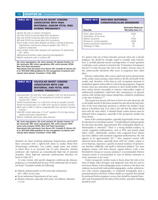
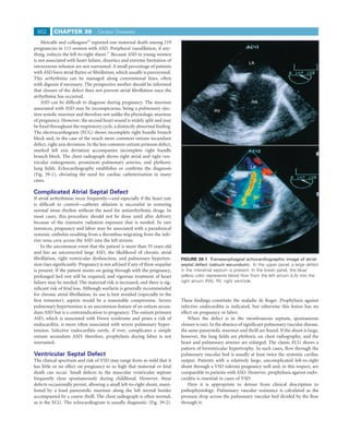
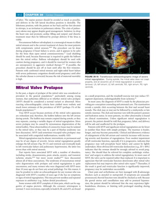
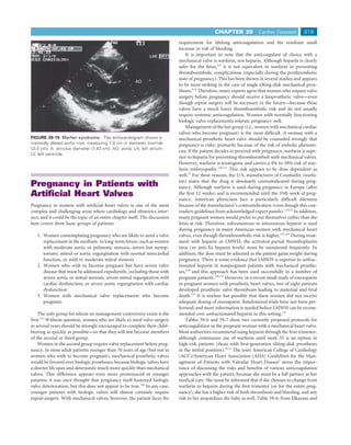
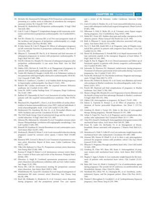
![CHAPTER 39 Cardiac Diseases 809
A B
C D
FIGURE 39-11 Tetralogy of Fallot. A, Chest radiograph. Note concavity in the area of the pulmonary artery, oligemic lungs, and right aortic
arch. B, Right ventriculogram. Note the narrow right ventricular outflow tract. C, Further clarification of the pulmonary arteries. The left ventricle
is slightly opacified via the ventricular septal defect. D, The associated right-sided aortic arch is now visible. (From Shabetai R, Adolph RJ:
Principles of cardiac catheterization. In Fowler NO [ed]: Cardiac Diagnosis and Treatment. Hagerstown, MD: Harper & Row, 1980, p 106.)
require cardiopulmonary bypass and can be carried out with safety for dissection and rupture. A number of centers are now performing
the mother and with less fetal risk than accompanies open heart balloon dilation with stent implantation for adults with unoperated
surgery with cardiopulmonary bypass. Although transvascular balloon aortic coarctation, but large, multicenter studies are currently not
dilation of aortic coarctation is a viable option for children and infants available.56
with coarctation, its use in adults is controversial.55 The procedure If delivery must be undertaken in cases of unoperated coarctation,
is well accepted for treatment of postsurgical renarrowing of the blood pressure can be titrated with β-adrenergic–blocking agents
coarctation, but de novo balloon angioplasty carries a risk of aortic delivered by intravenous drip.](https://image.slidesharecdn.com/4-u1-0-b978-1-4160-4224-2-50042-9-docpdf-120121090855-phpapp01/85/4-u1-0-b978-1-4160-4224-2-50042-9-docpdf-13-320.jpg)
![CHAPTER 39 Cardiac Diseases 813
Mitral Regurgitation Not Caused
by Prolapse
In younger women, mitral regurgitation may be a result of rheumatic
or congenital disease. In older women, mitral regurgitation is more
often a manifestation of hypertension, ischemia, idiopathic myocardial
disease, or infective endocarditis.
Most of the information regarding mitral regurgitation in prolapse
also applies here. In older women, the valve is more likely to be calci-
fied; fewer of the valves are amenable to repair and must be replaced.
The problems posed by prosthetic valves in pregnant women are dis-
cussed later in this chapter; the hemodynamics are illustrated in Figure
39-13, and echocardiography is illustrated in Figure 39-15.
In patients with far-advanced left ventricular dysfunction or failure
who have severe mitral regurgitation, it can be difficult to determine
which is the cause and which the result. In either case, the patient with
a greatly enlarged and hypokinetic ventricle must be advised against
becoming pregnant. Most of the pregnancy would be spent in bed, the
course would be punctuated by episodes of uncompensated congestive
heart failure (any of which could prove fatal or require therapeutic
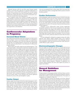
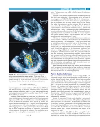
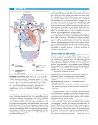
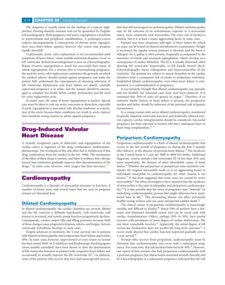
abortion), and the risk to the fetus would exceed 50%. FIGURE 39-16 Hemodynamic data in aortic stenosis. Left
Pregnancy in patients with mild or moderate mitral regurgitation ventricular pressure is 250/40 mm Hg (normal, 120/10 mm Hg). Aortic
systolic pressure is 130 mm Hg lower than the left ventricular
can be managed safely with a conservative regimen of reduced physical
pressure and shows a slow upstroke and vibrations representing the
activity, salt restriction, and low doses of a diuretic agent. Low-dose
systolic thrill. The record above the aortic pressure tracing is a
digoxin may be helpful if atrial fibrillation supervenes. As mentioned phonocardiogram showing the systolic murmur. Also shown is the
previously, severe mitral regurgitation indicates a need for repair or pulmonary wedge pressure (lowest pressure tracing), which is
replacement of the valve when symptoms and/or early evidence of elevated to equal the left ventricular diastolic pressure. The bottom
declining ventricular function appear.2,70 Clearly, surgical treatment is tracing is the electrocardiogram. (From Shabetai R, Adolph RJ:
best undertaken before pregnancy. If the woman is already pregnant, Principles of cardiac catheterization. In Fowler NO [ed]: Cardiac
the physician should make every effort to help her to carry the preg- Diagnosis and Treatment. Hagerstown, MD: Harper & Row, 1980,
nancy to term using strict medical measures. This course is particularly p 106.)
important if clinical, radiologic, and echocardiographic criteria suggest
that the valve is irreparable and would require replacement. phy is more pronounced, the cavity is smaller, and systolic function is
supranormal.
The left ventricle does not dilate until the ventricle fails, and so a
dilated ventricle in aortic stenosis is an ominous sign that calls for
Aortic Valve Disease rapid intervention. In general, aortic valve replacement is preferred to
percutaneous balloon aortic valvuloplasty, but open heart surgery
Aortic Stenosis presents a high risk to the fetus. For this reason, some have advised
balloon aortic valvuloplasty for treatment of aortic stenosis during
(see also the earlier section on aortic stenosis under Congenital
pregnancy,71 but valve replacement will almost certainly have to be
Obstructive Lesions)
done soon after delivery.
The etiologic mechanism of aortic stenosis commonly is degeneration, Hemodynamic monitoring is recommended during labor in
often of a congenitally bicuspid valve. The problem may be encoun- patients with moderate to severe aortic stenosis. Vaginal delivery is
tered in women a decade or more older than those with rheumatic or preferred, with assisted second stage of labor. If cesarean section is
congenital aortic valve disease. The combination of aortic and mitral performed, some have suggested that general anesthesia is preferred.72
stenosis is usually caused by rheumatic heart disease. Critical aortic See Chapter 56 for more details regarding anesthesia management.
stenosis leads to severe left ventricular hypertrophy and, eventually, to Pregnancy in women with a mechanical aortic valve replacement
left ventricular failure. Before overt heart failure develops, syncope or must be undertaken with great caution and meticulous management,
even sudden death may occur. because continuous anticoagulation is necessary (see Pregnancy in
The characteristic findings include an ejection systolic murmur that Patients with Artificial Heart Valves, later).
is harsher and longer and peaks later than the normal ejection murmur
of pregnancy. It is usually loudest at the second right intercostal space.
If aortic stenosis is severe, the pulse upstroke is slow, and left ventricu- Aortic Regurgitation
lar hypertrophy is evident on the ECG. The echocardiogram is a more The etiologic mechanism of aortic regurgitation is commonly rheu-
sensitive and more specific marker of left ventricular hypertrophy. matic heart disease, in which case mitral stenosis often coexists. Other
Doppler echocardiographic measurement of blood flow velocity diseases, such as Marfan syndrome, bicuspid aortic valve, infective
through the aortic valve permits reliable estimation of the systolic endocarditis, and systemic lupus erythematosus, also may cause severe
pressure drop across the valve, as well as calculation of the valve area. aortic regurgitation. This valvular lesion imposes a volume rather than
The hemodynamics are illustrated in Figure 39-16. The left ventricle a pressure overload on the heart and, as such, is usually well tolerated
in women remodels differently than in men. The concentric hypertro- in pregnancy and labor.15](https://image.slidesharecdn.com/4-u1-0-b978-1-4160-4224-2-50042-9-docpdf-120121090855-phpapp01/85/4-u1-0-b978-1-4160-4224-2-50042-9-docpdf-17-320.jpg)
![818 CHAPTER 39 Cardiac Diseases
Disturbances of
Cardiac Rhythm
Isolated supraventricular and ventricular extrasystoles are very
common, and no treatment is necessary. Pre-conception counseling is
simplified by a clear appreciation of several general principles.
Arrhythmia that occurs in the absence of organic heart disease is
almost always benign and is therefore not an indication for pharma-
cologic treatment unless the woman finds the symptoms intolerable.
Reassuring her of the benign nature of this symptom is often all that
is required. Sustained symptomatic arrhythmia, however, requires
treatment, which can be pharmacologic or procedural (e.g., transcath-
eter ablation of an anomalous conduction pathway, insertion of an
implantable cardiac defibrillator).
Pregnancy and labor should be safe except in the group with
sustained ventricular arrhythmia, with its attendant risk of cardiac
arrest and need for vigorous treatment. Pharmacologic treatment for
serious arrhythmia is likely to include newly introduced agents, such
as amiodarone or sotalol, for which there is at best limited knowledge
of potentially unfavorable effects on the fetus. Ideally, pregnancy
should be postponed until the arrhythmia has been eliminated or at
least controlled, preferably by nonpharmacologic means. If antiar-
rhythmic drugs must be used, whenever possible they should be those
that have been used for several decades, allowing prediction of the fetal
risk.
High-grade atrioventricular conduction disturbance, especially if it
is symptomatic, is treated by artificial pacing, which should not influ-
ence pregnancy, labor, or the fetus. Electrical cardioversion or defibril-
lation of the mother’s heart does not disturb or damage the fetal
heart.118
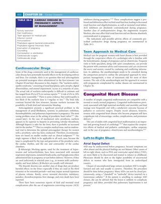
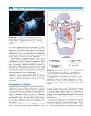
It is clearly desirable to evaluate disturbances of cardiac rhythm and FIGURE 39-18 Aortic aneurysm. Aortogram showing an aneurysm
conduction before pregnancy, proceeding to full electrophysiologic of the ascending aorta (AO) with regurgitation of contrast through an
testing if indicated. This plan avoids exposing a fetus to potentially incompetent aortic valve (arrows) into the left ventricle (LV). (From
Shabetai R, Adolph RJ: Principles of cardiac catheterization. In Fowler
toxic antiarrhythmic agents and the radiation associated with electro-
NO [ed]: Cardiac Diagnosis and Treatment. Hagerstown, MD: Harper
physiologic investigation.
& Row, 1980, p 106.)
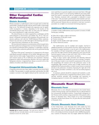
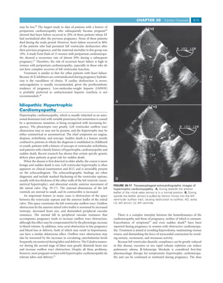
Marfan Syndrome
Marfan syndrome is variably expressed and inherited as an autosomal Deficiency of elastic tissue is the cause of myxomatous degenera-
dominant trait. If it is left untreated, life expectancy is reduced by half tion of the aortic and mitral valves and cystic medial necrosis of the
in those who exhibit the classic syndrome. The basic defect is one of aorta (Figs. 39-18 and 39-19). This abnormality translates to large
connective tissue, particularly fibrillin, and connective tissue weakness aneurysms of the aortic root, multiple aneurysms elsewhere along the
in the aorta causes the dangerous complications, most notably aortic course of the aorta and great vessels, and severe aortic and mitral
dissection.119 regurgitation with resulting heart failure. Surgery is indicated for
Symptoms and signs may include dyspnea and chest pain, an aortic rapidly expanding aneurysm or if dissection is evident. Pregnancy is
diastolic murmur, and a midsystolic click. The best diagnostic test, and poorly tolerated under these conditions, and labor may precipitate
apparently the most critical one for determining the outcome of preg- rupture of an aneurysm or aortic dissection.
nancy, is the echocardiogram. More than 90% of patients have evi- If a woman with Marfan syndrome chooses to become pregnant,
dence of MVP, and 60% have echocardiographic evidence of aortic therapy is directed at markedly limiting physical activity, preventing
root dilatation.120 hypertensive complications, and decreasing the pulsatile forces on the
Pregnancy is particularly dangerous for patients with this syn- aortic wall with the use of a β-blocker. Long-acting β-blockers
drome, because there appears to be a high risk of aortic rupture and are indicated before, during, and after pregnancy in women with
dissection, especially if dilatation of the aortic root is present.47 Women Marfan syndrome.122 Once the aortic root diameter reaches 50 to
with an aortic diameter exceeding 40 mm are at greatest risk of death 55 mm, most authorities recommend prophylactic aortic valve and
during pregnancy.121 The physician should also make sure the woman root replacement because of the high risk of aortic dissection. Abdomi-
understands the 50% risk of genetic transmission of Marfan syndrome nal delivery is recommended to avoid the hemodynamic stress of
to her children. labor.](https://image.slidesharecdn.com/4-u1-0-b978-1-4160-4224-2-50042-9-docpdf-120121090855-phpapp01/85/4-u1-0-b978-1-4160-4224-2-50042-9-docpdf-22-320.jpg)
![822 CHAPTER 39 Cardiac Diseases
septal defects in adults using the buttoned device. Mayo Clin Proc 75:913, 59. Kalra GS, Arora R, Khan JA, et al: Percutaneous mitral commissurotomy
2000. for severe mitral stenosis during pregnancy. Cathet Cardiovasc Diagn
33. Metcalfe J, McAnulty JH, Ueland K: Cardiac Disease and Pregnancy: 33:28, 1994.
Physiology and Management. Boston, Little, Brown, 1986, p 223. 60. Lefevre T, Bonan R, Serra A, et al: Percutaneous mitral valvuloplasty in
34. Rashkind WJ, Mullins CE, Hellenbrand WE, et al: Nonsurgical closure of surgical high risk patients. J Am Coll Cardiol 17:348, 1991.
patent ductus arteriosus: Clinical application of the Rashkind PDA 61. de Souza JAM, Martinez EE, Ambrose JA, et al: Percutaneous balloon
Occluder System. Circulation 75:583, 1987. mitral valvuloplasty in comparison with open mitral valve commissurot-
35. Wood P: The Eisenmenger syndrome or pulmonary hypertension with omy for mitral stenosis during pregnancy. J Am Coll Cardiol 37:900,
reversed central shunt. BMJ 701:755, 1958. 2001.
36. Yentis SM, Steer PJ, Plaat F. Eisenmenger’s syndrome in pregnancy: Mater- 62. Devereux RB, Perloff JK, Reichek N, et al: Mitral valve prolapse. Circula-
nal and fetal mortality in the 1990s. BJOG 105:921, 1998. tion 54:3, 1976.
37. Gleicher N, Midwall J, Hochberger D, et al: Eisenmenger’s syndrome and 63. Levine RA, Handschumacher MD, Sanfilippo AJ, et al: Three-dimensional
pregnancy. Obstet Gynecol Surv 34:721, 1979. echocardiographic reconstruction of the mitral valve, with implications
38. Spinnato JA, Kraynack BJ, Cooper MW: Eisenmenger’s syndrome in preg- for the diagnosis of mitral valve prolapse. Circulation 80:589, 1989.
nancy: Epidural anesthesia for elective cesarean section. N Engl J Med 64. Hartman N, Kramer R, Brown T, et al: Panic disorder in patients with
304:1215, 1981. mitral valve prolapse. Am J Psychiatry 139:669, 1982.
39. Avila WS, Grinberg M, Snitcowsky R, et al: Maternal and fetal outcome in 65. Perloff JK, Child JS, Edwards JE: New guidelines for the clinical diagnosis
pregnant women with Eisenmenger’s syndrome. Eur Heart J 16:460, of mitral valve prolapse. Am J Cardiol 57:1124, 1986.
1995. 66. Freed LA, Levy D, Levine RA, et al: Prevalence and clinical outcome of
40. McCaffrey RM, Dunn LJ: Primary pulmonary hypertension in pregnancy. mitral-valve prolapse. N Engl J Med 341:1, 1999.
Obstet Gynecol Surv 19:567, 1964. 67. Gilon D, Buananno FS, Leavitt M, et al: Lack of evidence of an association
41. Abenhaim L, Moride Y, Brenot F, et al: Appetite suppressant drugs and between mitral-valve prolapse and stroke in young patients. N Engl J Med
the risk of primary pulmonary hypertension. N Engl J Med 335:609, 341:8, 1999.
1996. 68. Butman S, Chandraratna PA, Milne N, et al: Stress myocardial imaging in
42. Barst RJ, Rubin LJ, Long WA, et al: A comparison of continuous intrave- patients with mitral valve prolapse: Evidence of a perfusion abnormality.
nous epoprostenol (prostacyclin) with conventional therapy for primary Cathet Cardiovasc Diagn 8:243, 1982.
pulmonary hypertension: The Primary Pulmonary Hypertension Study 69. Bach DS, Bolling SF: Early improvement in congestive heart failure after
Group [see comments]. N Engl J Med 334:296, 1996. correction of secondary mitral regurgitation in end-stage cardiomyopa-
43. Rothman A, Beltran D, Kriett JM, et al: Graded balloon dilation atrial thy. Am Heart J 129:1165, 1995.
septostomy as a bridge to lung transplantation in pulmonary hyperten- 70. Otto CM: Evaluation and management of chronic mitral regurgitation. N
sion. Am Heart J 125:1763, 1993. Engl J Med 345:740, 2001.
44. Thanopoulos BD, Georgakopoulos D, Tsaousis GS, et al: Percutaneous 71. Banning AP, Pearson JF, Hall RJ: Role of balloon dilatation of the aortic
balloon dilatation of the atrial septum: Immediate and midterm results. valve in pregnant patients with severe aortic stenosis. Br Heart J 70:544,
Heart 76:502, 1996. 1993.
45. Arias F, Pineda J: Aortic stenosis in pregnancy. J Reprod Med 20:229, 72. Silversides CK, Colman JM, Sermer M, et al: Early and intermediate-term
1978. outcomes of pregnancy with congenital aortic stenosis. Am J Cardiol
46. Hameed A, Karaalp IS, Tummala PP, et al: The effect of valvular 91:1386, 2003.
heart disease on maternal and fetal outcome. J Am Coll Cardiol 37:893, 73. Mast ST, Jollis JG, Ryan T, et al: The progression of fenfluramine-
2001. associated valvular heart disease assessed by echocardiography. Ann Intern
47. Elkayam U, Ostrzega E, Shotan A, et al: Cardiovascular problems in Med 134:261, 2001.
pregnant women with the Marfan syndrome. Ann Intern Med 123:117, 74. Connolly HM, Crary JL, McGoon MD, et al: Valvular heart disease associ-
1995. ated with fenfluramine-phentermine. N Engl J Med 337:581, 1997.
48. Purcell IF, Williams DO: Peripartum cardiomyopathy complicating severe 75. Hunt SA, Abraham WT, Chin MH, et al: ACC/AHA guideline update for
aortic stenosis. Int J Cardiol 52:163, 1995. the diagnosis and management of chronic heart failure in the adult. J Am
49. Hameed A, Yuodim K, Mahboob A, et al: Effect of the severity of pulmo- Coll Cardiol 46:1116-43, 2005.
nary stenosis on pregnancy outcome: A case-control study. Am J Obstet 76. Baron O, Hubaut J, Galetta D, et al: Pregnancy and heart-lung transplanta-
Gynecol 191:93, 2004. tion. Heart Lung Transplant 21:914, 2002.
50. Stanger P, Cassidy SC, Girod DA, et al: Balloon pulmonary valvuloplasty: 77. Troche V, Ville Y, Fernandez H: Pregnancy after heart or heart-lung trans-
Results of the valvuloplasty and angioplasty register. Am J Cardiol 65:775, plantation: A series of 10 pregnancies. BJOG 105:454, 1998.
1990. 78. Morini A, Spina V, Aleandri V, et al: Pregnancy after heart transplant:
51. Morris CD, Menashe VD: Recurrence of congenital heart disease in off- Update and case report. Hum Reprod 13:749, 1998.
spring of parents with surgical correction. Clin Res 33:68A, 1985. 79. Pearson GD, Veille JC, Rahimtoola S, et al: Peripartum cardiomyopathy:
52. Meijer JM, Pieper PG, Drenthen W, et al: Pregnancy, fertility, and recur- NHLBI/NIH workshop recommendations and review. JAMA 283:1183,
rence risk in corrected tetralogy of Fallot. Heart 91:801, 2005. 2000.
53. Barash PG, Hobbins JC, Hook R, et al: Management of coarctation of the 80. Hibbard JU, Lindheimer M, Lang RM: A modified definition for peripar-
aorta during pregnancy. J Thorac Cardiovasc Surg 69:781, 1975. tum cardiomyopathy and prognosis based on echocardiography. Obstet
54. Beauchesne LM, Connolly HM, Ammash NM, et al: Coarctation of the Gynecol 94:311, 1999.
aorta: Outcome of pregnancy. J Am Coll Cardiol 38:1728, 2001. 81. Sliwa K, Fett J, Elkayam U. Peripartum cardiomyopathy. Lancet 368:687,
55. Ovaert C, McCrindle BW, Nykanen D, et al: Balloon angioplasty of native 2006.
coarctation: Clinical outcomes and predictors for success. J Am Coll 82. Van Hoeven KH, Kitsis RN, Katz SD, et al: Peripartum versus idiopathic
Cardiol 35:988, 2000. dilated cardiomyopathy in young women: A comparison of clinical,
56. Inglessis I, Landzberg MJ: Interventional catheterization in adult congeni- pathologic and prognostic features. Int J Cardiol 40:57, 1993.
tal heart disease. Circulation 115:1622, 2007. 83. Rezeq MN, Rickenbacher PR, Fowler MB, et al: Incidence of myocarditis
57. Connolly HM, Warnes CR: Ebstein’s anomaly: Outcome of pregnancy. J in peripartum cardiomyopathy. Am J Cardiol 74:474, 1994.
Am Coll Cardiol 23:1194, 1994. 84. Lampert MB, Weinert L, Hibbard J, et al: Contractile reserve in patients
58. Reid JM, Coleman EN, Doig W: Complete congenital heart block: Report with peripartum cardiomyopathy and recovered left ventricular function.
of 35 cases. Br Heart J 48:236, 1982. Am J Obstet Gynecol 176:189, 1997.](https://image.slidesharecdn.com/4-u1-0-b978-1-4160-4224-2-50042-9-docpdf-120121090855-phpapp01/85/4-u1-0-b978-1-4160-4224-2-50042-9-docpdf-26-320.jpg)


This document discusses heart disease in pregnancy. It notes that pregnant women with heart disease are at higher risk for complications. However, the changes of pregnancy can make diagnosing heart conditions difficult. Key signs of heart disease include severe dyspnea, syncope with exertion, and chest pain. If heart disease is suspected, confirmatory tests should be done while accounting for normal pregnancy changes. Pre-conception counseling is important to advise patients on risk levels and necessary pre-pregnancy treatment. Certain high-risk conditions like severe valvular disease may require terminating the pregnancy due to prohibitive maternal mortality risks.

![Cardiovascular disease students [compatibility mode]](https://cdn.slidesharecdn.com/ss_thumbnails/cardiovasculardisease-studentscompatibilitymode-130509054709-phpapp02-thumbnail.jpg?width=640&height=640&fit=bounds)


