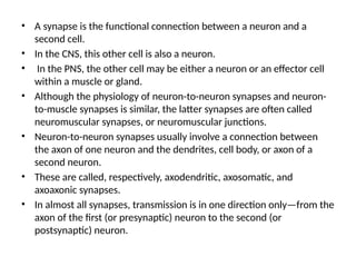
• A synapseis the functional connection between a neuron and a
second cell.
• In the CNS, this other cell is also a neuron.
• In the PNS, the other cell may be either a neuron or an effector cell
within a muscle or gland.
• Although the physiology of neuron-to-neuron synapses and neuron-
to-muscle synapses is similar, the latter synapses are often called
neuromuscular synapses, or neuromuscular junctions.
• Neuron-to-neuron synapses usually involve a connection between
the axon of one neuron and the dendrites, cell body, or axon of a
second neuron.
• These are called, respectively, axodendritic, axosomatic, and
axoaxonic synapses.
• In almost all synapses, transmission is in one direction only—from the
axon of the first (or presynaptic) neuron to the second (or
postsynaptic) neuron.
3.
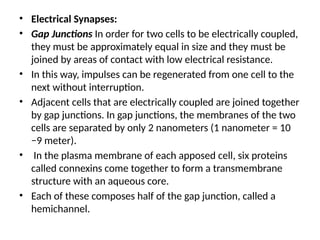
• Electrical Synapses:
•Gap Junctions In order for two cells to be electrically coupled,
they must be approximately equal in size and they must be
joined by areas of contact with low electrical resistance.
• In this way, impulses can be regenerated from one cell to the
next without interruption.
• Adjacent cells that are electrically coupled are joined together
by gap junctions. In gap junctions, the membranes of the two
cells are separated by only 2 nanometers (1 nanometer = 10
−9 meter).
• In the plasma membrane of each apposed cell, six proteins
called connexins come together to form a transmembrane
structure with an aqueous core.
• Each of these composes half of the gap junction, called a
hemichannel.
4.
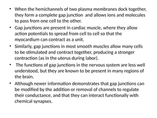
• When thehemichannels of two plasma membranes dock together,
they form a complete gap junction and allows ions and molecules
to pass from one cell to the other.
• Gap junctions are present in cardiac muscle, where they allow
action potentials to spread from cell to cell so that the
myocardium can contract as a unit.
• Similarly, gap junctions in most smooth muscles allow many cells
to be stimulated and contract together, producing a stronger
contraction (as in the uterus during labor).
• The functions of gap junctions in the nervous system are less well
understood, but they are known to be present in many regions of
the brain.
• Although newer information demonstrates that gap junctions can
be modified by the addition or removal of channels to regulate
their conductance, and that they can interact functionally with
chemical synapses.
5.
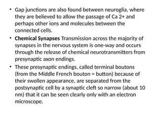
• Gap junctionsare also found between neuroglia, where
they are believed to allow the passage of Ca 2+ and
perhaps other ions and molecules between the
connected cells.
• Chemical Synapses Transmission across the majority of
synapses in the nervous system is one-way and occurs
through the release of chemical neurotransmitters from
presynaptic axon endings.
• These presynaptic endings, called terminal boutons
(from the Middle French bouton = button) because of
their swollen appearance, are separated from the
postsynaptic cell by a synaptic cleft so narrow (about 10
nm) that it can be seen clearly only with an electron
microscope.
6.
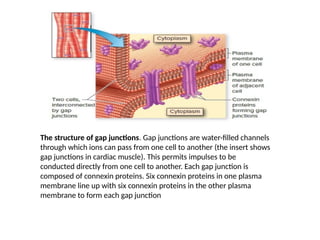
The structure ofgap junctions. Gap junctions are water-filled channels
through which ions can pass from one cell to another (the insert shows
gap junctions in cardiac muscle). This permits impulses to be
conducted directly from one cell to another. Each gap junction is
composed of connexin proteins. Six connexin proteins in one plasma
membrane line up with six connexin proteins in the other plasma
membrane to form each gap junction
7.
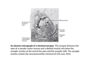
An electron micrographof a chemical synapse. This synapse between the
axon of a somatic motor neuron and a skeletal muscle cell shows the
synaptic vesicles at the end of the axon and the synaptic cleft. The synaptic
vesicles contain the neurotransmitter chemical (in this case, ACh).
8.
• Chemical transmissionrequires that the synaptic cleft stay very
narrow and that neurotransmitter molecules are released near
their receptor proteins in the postsynaptic membrane.
• The physical association of the pre- and postsynaptic membranes
at the chemical synapse is stabilized by the action of particular
membrane proteins.
• Cell adhesion molecules (CAMs) are proteins in the pre and
postsynaptic membranes that project from these membranes into
the synaptic cleft, where they bond to each other.
• Release of Neurotransmitter
• Neurotransmitter molecules within the presynaptic neuron
endings are contained within many small, membrane-enclosed
synaptic vesicles.
• In order for the neurotransmitter within these vesicles to be
released into the synaptic cleft, the vesicle membrane must fuse
with the axon membrane in the process of exocytosis .
9.
• Exocytosis ofsynaptic vesicles, and the consequent release of
neurotransmitter molecules into the synaptic cleft, is triggered by
action potentials that stimulate the entry of Ca 2+ into the
terminal bouton through voltage-gated Ca 2+ channels.
• When there is a greater frequency of action potentials at the
terminal bouton, there is a greater entry of Ca 2+ , and thus a
larger number of synaptic vesicles undergoing exocytosis and
releasing neurotransmitter molecules.
• As a result, a greater frequency of action potentials by the
presynaptic axon will result in greater stimulation of the
postsynaptic neuron.
• Before an action potential arrives at the terminal bouton, many
synaptic vesicles are already attached, or docked, to specialized
sites of the presynaptic plasma membrane.
• Docking involves a SNARE complex of proteins that bridge the
vesicle membrane and the plasma membrane.
10.
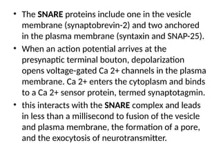
• The SNAREproteins include one in the vesicle
membrane (synaptobrevin-2) and two anchored
in the plasma membrane (syntaxin and SNAP-25).
• When an action potential arrives at the
presynaptic terminal bouton, depolarization
opens voltage-gated Ca 2+ channels in the plasma
membrane. Ca 2+ enters the cytoplasm and binds
to a Ca 2+ sensor protein, termed synaptotagmin.
• this interacts with the SNARE complex and leads
in less than a millisecond to fusion of the vesicle
and plasma membrane, the formation of a pore,
and the exocytosis of neurotransmitter.
11.
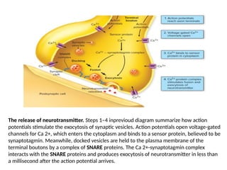
The release ofneurotransmitter. Steps 1–4 inprevioud diagram summarize how action
potentials stimulate the exocytosis of synaptic vesicles. Action potentials open voltage-gated
channels for Ca 2+, which enters the cytoplasm and binds to a sensor protein, believed to be
synaptotagmin. Meanwhile, docked vesicles are held to the plasma membrane of the
terminal boutons by a complex of SNARE proteins. The Ca 2+-synaptotagmin complex
interacts with the SNARE proteins and produces exocytosis of neurotransmitter in less than
a millisecond after the action potential arrives.
12.
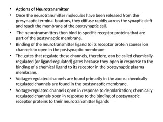
• Actions ofNeurotransmitter
• Once the neurotransmitter molecules have been released from the
presynaptic terminal boutons, they diffuse rapidly across the synaptic cleft
and reach the membrane of the postsynaptic cell.
• The neurotransmitters then bind to specific receptor proteins that are
part of the postsynaptic membrane.
• Binding of the neurotransmitter ligand to its receptor protein causes ion
channels to open in the postsynaptic membrane.
• The gates that regulate these channels, therefore, can be called chemically
regulated (or ligand-regulated) gates because they open in response to the
binding of a chemical ligand to its receptor in the postsynaptic plasma
membrane.
• Voltage-regulated channels are found primarily in the axons; chemically
regulated channels are found in the postsynaptic membrane.
• Voltage-regulated channels open in response to depolarization; chemically
regulated channels open in response to the binding of postsynaptic
receptor proteins to their neurotransmitter ligands
13.
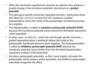
• When thechemically regulated ion channels are opened, they produce a
graded change in the membrane potential, also known as a graded
potential.
• The opening of specific chemically regulated channels—particularly those
that allow Na + or Ca 2+ to enter the cell—produces a graded
depolarization, where the inside of the postsynaptic membrane becomes
less negative.
• This depolarization is called an excitatory postsynaptic potential (EPSP)
because the membrane potential moves toward the threshold required for
action potentials.
• In other cases, as when CI − enters the cell through specific channels, a
graded hyperpolarization is produced (where the inside of the
postsynaptic membrane becomes more negative). This hyperpolarization
is called an inhibitory postsynaptic potential (IPSP) because the
membrane potential moves farther from the threshold depolarization
required to produce action potentials
• Excitatory postsynaptic potentials, as their name implies, stimulate the
postsynaptic cell to produce action potentials, and inhibitory postsynaptic
potentials antagonize this effect.
Definition
• A chemicalsubstance, which is released at the
end of nerve fiber ,by the arrival of nerve
impulse and,by diffusing across the synapse or
junction, effects the transfer of impulse to
another nerve fiber/muscle fiber/or some
other structure.
20.
1)ACETYLCHOLINE
• Acetylcholine (ACh)is used as an excitatory
neurotransmitter by some neurons in the CNS and by
somatic motor neurons at the neuromuscular junction.
• At autonomic nerve endings, ACh may be either excitatory
or inhibitory, depending on the organ involved. • The
varying responses of postsynaptic cells to the same
chemical can be explained by the fact that different
postsynaptic cells have different subtypes of ACh receptors.
• These receptor subtypes can be specifically stimulated by
particular toxins, and they are named for these toxins.
21.
• 1. NicotinicACh receptors.
• They are so named because they can also be activated by
nicotine.
• These are found in specific regions of the brain, in autonomic
ganglia, and in skeletal muscle fibers.
• The release of ACh from somatic motor neurons and its binding
to nicotinic receptors, for example, stimulates muscle
contraction.
• 2. Muscarinic ACh receptors.
• They are so named because they can also be activated by
muscarine (a drug derived from certain poisonous mushrooms).
• These are found in the plasma membrane of smooth muscle
cells, cardiac muscle cells, and the cells of particular glands.
• Thus, the activation of muscarinic ACh receptors over there is
required for the regulation of the cardiovascular system,
digestive system, and others.
23.
2)Monoamines as Neurotransmitters
•They contains catechol ring of 6 carbon with 1 amino
group and 2 hydroxyl group.
• They are broken by MAO (mono amine oxidase).
• Inhibators of MAO cause increase amount of
norepinephrine and dopamine and are use in mood
disorders.
• Their neurons are present in brain and hypothalamus
because of this they have function like
conciousness,mood,motivation,direction,attention,mo
vement,BP,hormone release.
24.
• 1)Epinephrine, 2)norepinephrine,3)serotonin, and
4)dopamine are in the chemical family known as monoamines.
• Serotonin is derived from the amino acid tryptophan.
• Epinephrine, norepinephrine, and dopamine are derived from
the amino acid tyrosine and form a subfamily of monoamines
called the catecholamines
• Like ACh, monoamine neurotransmitters are released by
exocytosis from presynaptic vesicles, diffuse across the
synaptic cleft, and interact with specific receptors in the
membrane of the postsynaptic cell.
• The inhibition of monoamine action is due to (1) reuptake of
monoamines into the presynaptic neuron endings, (2)
enzymatic degradation of monoamines in the presynaptic
neuron endings by monoamine oxidase (MAO), and (3) the
enzymatic degradation of catecholamines in the postsynaptic
neuron by catechol-O-methyltransferase (COMT).
25.
• 1)Epinephrine(also calledadrenaline) is a hormone
secreted by the adrenal gland, not a neurotransmitter,
while nor epinephrine functions both as a hormone
and a neurotransmitter.
• Receptors are called adrenergic receptors.
• Epinephrine have 2 receptor name alpha 1 and alpha 2
• Alpha 1 ,post synaptically inhibit/stimulate activity of
potassium ion.
• Alpha 2,pre synaptically inhibit the release of nor
epinephrine.
• 2)Nor epinephrine have 3 receptors name beta 1 ,beta
2 and beta 3
• All these act via stimulating G protein .
26.
• 3)Serotonin (5-HT)have slow onset, consider as
neuromodulators.
• Serotonergic neurons virtually innervate in brain and spinal cord
• Work with 16 different receptor subtypes
• Excitatory effect on control of muscles
• Inhibatory effect on sensations
• Its activity decrese/slow at night and highest at awakefulness.
• Involves in regulation of food intake,reproductive
behaviour,emotional states for example mood anxiety.
• SSRI(paxil) use in treatment of depression cause inactivation of
pre synaptic serotonin transporter
• These transporter mediate reuptake of serotonin into pre
synaptic cell.
• SSRI cause increase synaptic concentration of serotonin.
27.
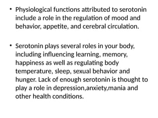
• Physiological functionsattributed to serotonin
include a role in the regulation of mood and
behavior, appetite, and cerebral circulation.
• Serotonin plays several roles in your body,
including influencing learning, memory,
happiness as well as regulating body
temperature, sleep, sexual behavior and
hunger. Lack of enough serotonin is thought to
play a role in depression,anxiety,mania and
other health conditions.
28.
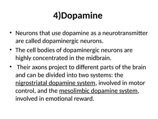
4)Dopamine
• Neurons thatuse dopamine as a neurotransmitter
are called dopaminergic neurons.
• The cell bodies of dopaminergic neurons are
highly concentrated in the midbrain.
• Their axons project to different parts of the brain
and can be divided into two systems: the
nigrostriatal dopamine system, involved in motor
control, and the mesolimbic dopamine system,
involved in emotional reward.
29.
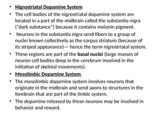
• Nigrostriatal DopamineSystem
• The cell bodies of the nigrostriatal dopamine system are
located in a part of the midbrain called the substantia nigra
(“dark substance”) because it contains melanin pigment.
• Neurons in the substantia nigra send fibers to a group of
nuclei known collectively as the corpus striatum (because of
its striped appearance)— hence the term nigrostriatal system.
• These regions are part of the basal nuclei (large masses of
neuron cell bodies deep in the cerebrum involved in the
initiation of skeletal movements).
• Mesolimbic Dopamine System
• The mesolimbic dopamine system involves neurons that
originate in the midbrain and send axons to structures in the
forebrain that are part of the limbic system.
• The dopamine released by these neurons may be involved in
behavior and reward.
30.
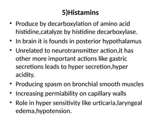
5)Histamins
• Produce bydecarboxylation of amino acid
histidine,catalyze by histidine decarboxylase.
• In brain it is founds in posterior hypothalamus
• Unrelated to neurotransmitter action,it has
other more important actions like gastric
secretions leads to hyper secretion,hyper
acidity.
• Producing spasm on bronchial smooth muscles
• Increasing permiability on capillary walls
• Role in hyper sensitivity like urticaria,laryngeal
edema,hypotension.
31.
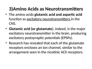
3)Amino Acids asNeurotransmitters
• The amino acids glutamic acid and aspartic acid
function as excitatory neurotransmitters in the
CNS.
• Glutamic acid (or glutamate), indeed, is the major
excitatory neurotransmitter in the brain, producing
excitatory postsynaptic potentials (EPSPs).
• Research has revealed that each of the glutamate
receptors encloses an ion channel, similar to the
arrangement seen in the nicotinic ACh receptors.
32.
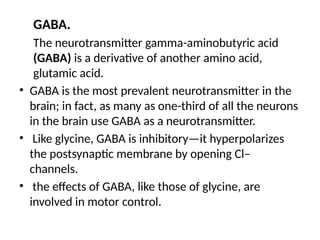
GABA.
The neurotransmitter gamma-aminobutyricacid
(GABA) is a derivative of another amino acid,
glutamic acid.
• GABA is the most prevalent neurotransmitter in the
brain; in fact, as many as one-third of all the neurons
in the brain use GABA as a neurotransmitter.
• Like glycine, GABA is inhibitory—it hyperpolarizes
the postsynaptic membrane by opening Cl–
channels.
• the effects of GABA, like those of glycine, are
involved in motor control.
33.
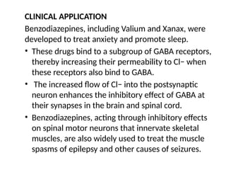
CLINICAL APPLICATION
Benzodiazepines, includingValium and Xanax, were
developed to treat anxiety and promote sleep.
• These drugs bind to a subgroup of GABA receptors,
thereby increasing their permeability to Cl− when
these receptors also bind to GABA.
• The increased flow of Cl− into the postsynaptic
neuron enhances the inhibitory effect of GABA at
their synapses in the brain and spinal cord.
• Benzodiazepines, acting through inhibitory effects
on spinal motor neurons that innervate skeletal
muscles, are also widely used to treat the muscle
spasms of epilepsy and other causes of seizures.
34.
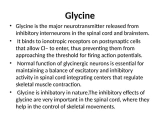
Glycine
• Glycine isthe major neurotransmitter released from
inhibitory interneurons in the spinal cord and brainstem.
• It binds to ionotropic receptors on postsynaptic cells
that allow Cl− to enter, thus preventing them from
approaching the threshold for firing action potentials.
• Normal function of glycinergic neurons is essential for
maintaining a balance of excitatory and inhibitory
activity in spinal cord integrating centers that regulate
skeletal muscle contraction.
• Glycine is inhibatory in nature.The inhibitory effects of
glycine are very important in the spinal cord, where they
help in the control of skeletal movements.
35.
4)Neuropeptides
• The neuropeptides,in contrast, are derived from
large precursor proteins, which in themselves
have little, if any, inherent biological activity.
• The synthesis of these precursors, directed by
mRNA, occurs on ribosomes, which exist only in
the cell body and large dendrites of the neuron,
often a considerable distance from axon
terminals or varicosities where the peptides are
released.
36.
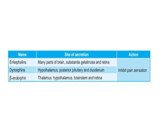
1)Endogenous opioids
• Endogenousopioids—a group of neuropeptides that includes
beta-endorphin, the dynorphins, and the enkephalins— have
attracted much interest because their receptors are the sites
of action of opiate drugs such as morphine and codeine.
• The opiate drugs are powerful analgesics (that is, they relieve
pain without loss of consciousness), and the endogenous
opioids undoubtedly have a function in regulating pain.
• There is also evidence that the opioids function in regulating
eating and drinking behavior, circulatory system function, and
mood and emotion.
37.
2)Neuropeptide Y
• NeuropeptideY has been shown to have a variety of
physiological effects, including a role in the response
to stress, in the regulation of circadiac arhythmias,
and in the control of the cardiovascular system.
• Neuropeptide Y has been shown to inhibit the release
of the excitatory neurotransmitter glutamate in a part
of the brain called the hippocampus.
• Neuropeptide Y is a powerful stimulator of appetite.
Conversely, inhibitors of neuropeptide Y that are
injected into the brain inhibit eating.
38.
5)Gases
• Certain veryshort-lived gases also serve as
neurotransmitters.
• Nitric oxide is the best understood, but recent research
indicates that carbon monoxide and hydrogen sulfide are also
emitted by neurons as signals.
• Gases are not released by exocytosis of presynaptic vesicles,
nor do they bind to postsynaptic plasma membrane
receptors.
• They are produced by enzymes in axon terminals (in
response to Ca2+ entry) and simply diffuse from their sites of
origin in one cell into the intracellular fluid of other neurons
or effector cells, where they bind to and activate proteins.
39.
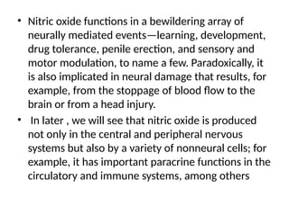
• Nitric oxidefunctions in a bewildering array of
neurally mediated events—learning, development,
drug tolerance, penile erection, and sensory and
motor modulation, to name a few. Paradoxically, it
is also implicated in neural damage that results, for
example, from the stoppage of blood flow to the
brain or from a head injury.
• In later , we will see that nitric oxide is produced
not only in the central and peripheral nervous
systems but also by a variety of nonneural cells; for
example, it has important paracrine functions in the
circulatory and immune systems, among others
40.
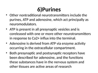
6)Purines
• Other nontraditionalneurotransmitters include the
purines, ATP and adenosine, which act principally as
neuromodulators.
• ATP is present in all presynaptic vesicles and is
coreleased with one or more other neurotransmitters
in response to Ca2+ influx into the terminal.
• Adenosine is derived from ATP via enzyme activity
occurring in the extracellular compartment.
• Both presynaptic and postsynaptic receptors have
been described for adenosine, and the functions
these substances have in the nervous system and
other tissues are active areas of research
41.
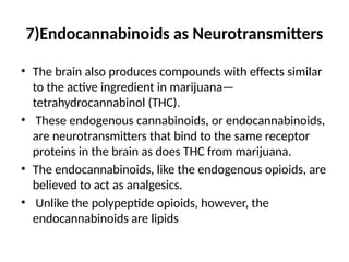
7)Endocannabinoids as Neurotransmitters
•The brain also produces compounds with effects similar
to the active ingredient in marijuana—
tetrahydrocannabinol (THC).
• These endogenous cannabinoids, or endocannabinoids,
are neurotransmitters that bind to the same receptor
proteins in the brain as does THC from marijuana.
• The endocannabinoids, like the endogenous opioids, are
believed to act as analgesics.
• Unlike the polypeptide opioids, however, the
endocannabinoids are lipids