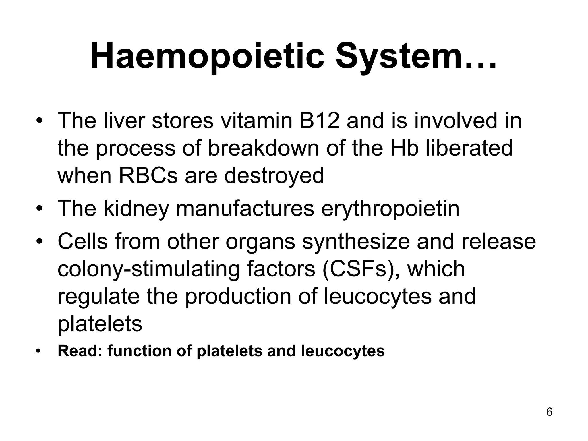
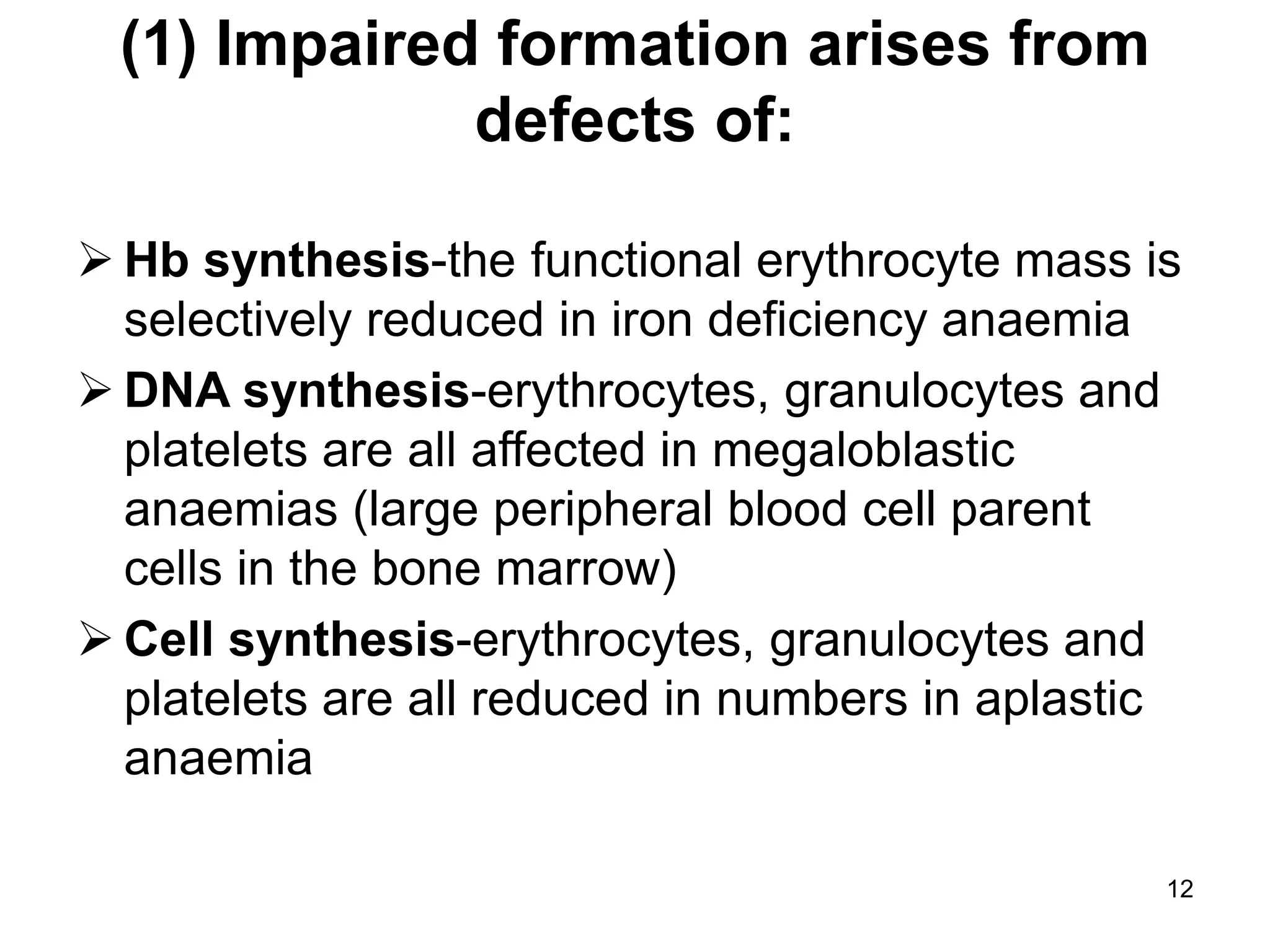
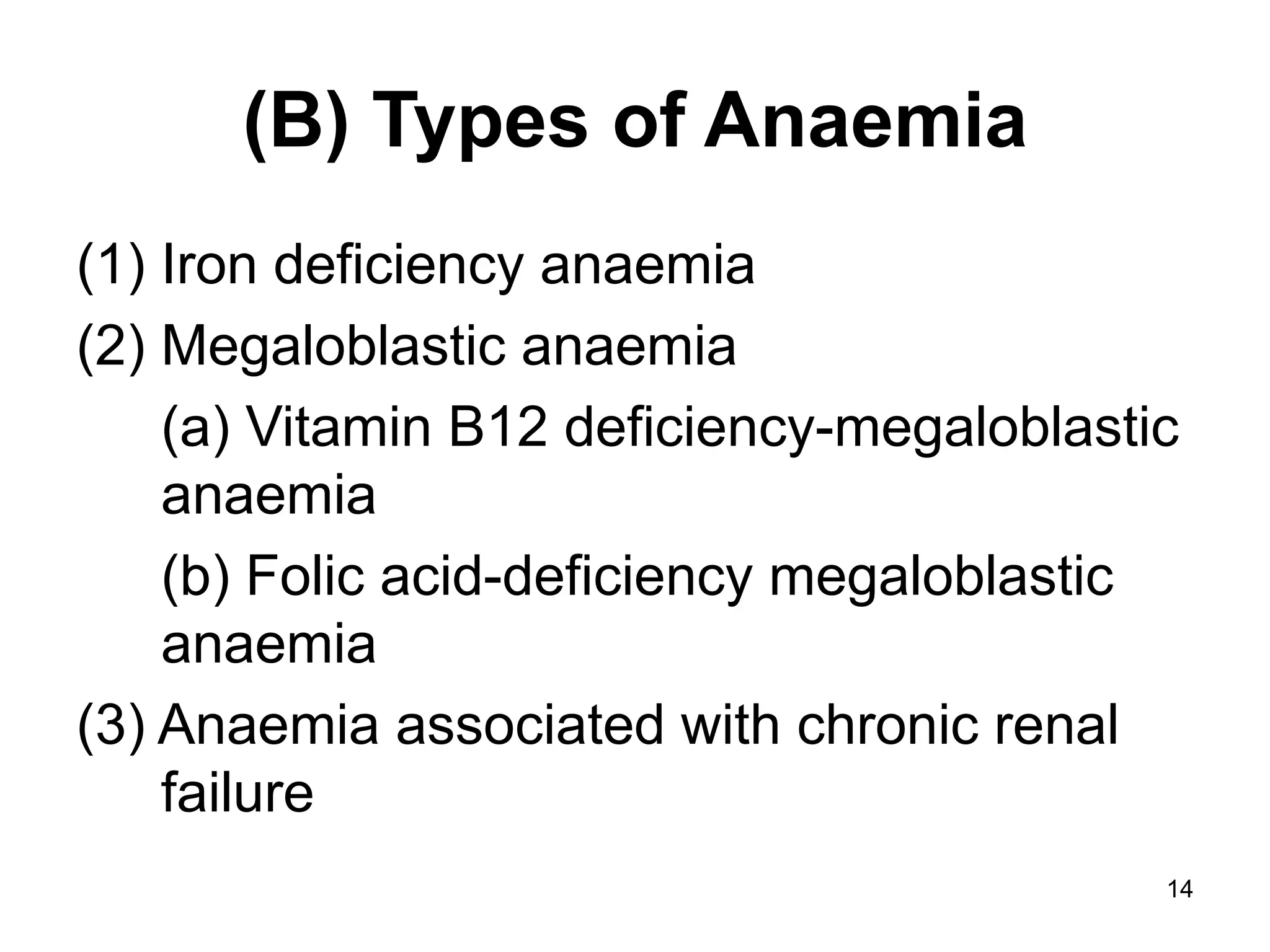
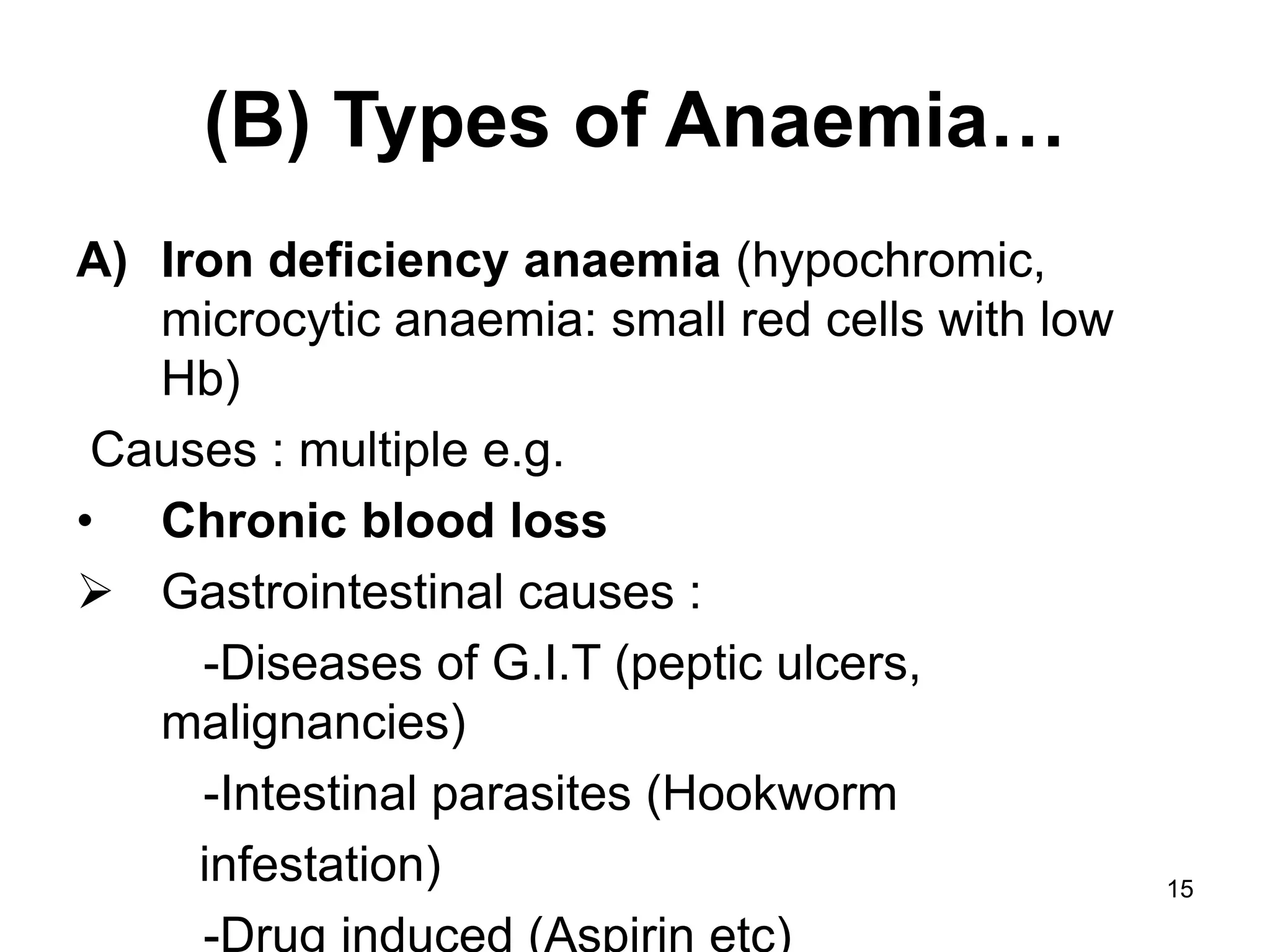
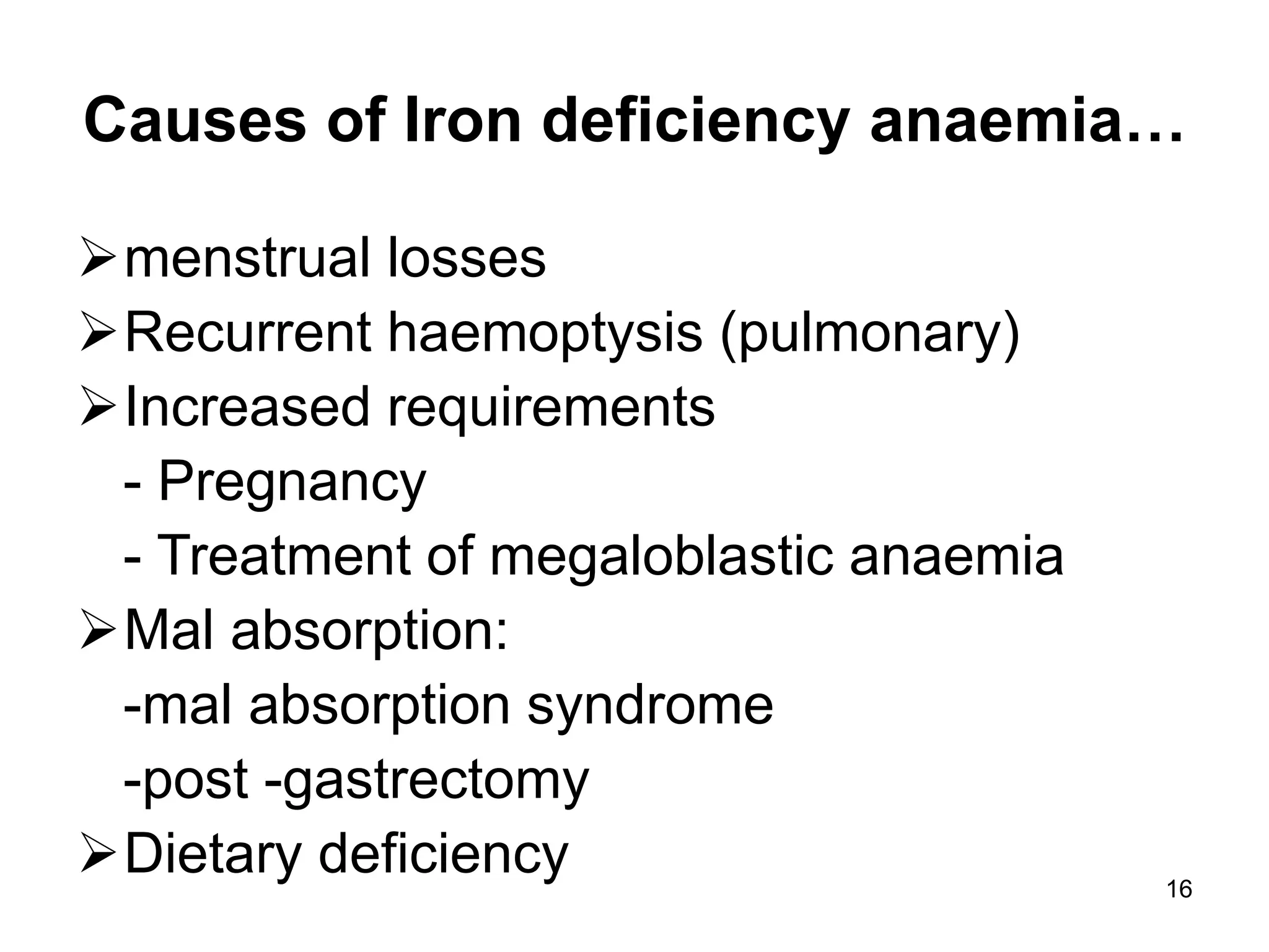
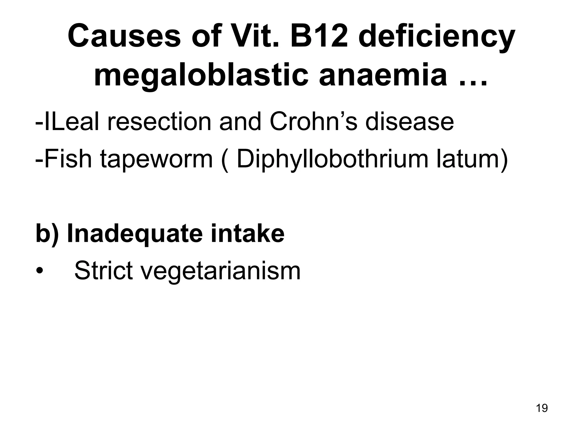
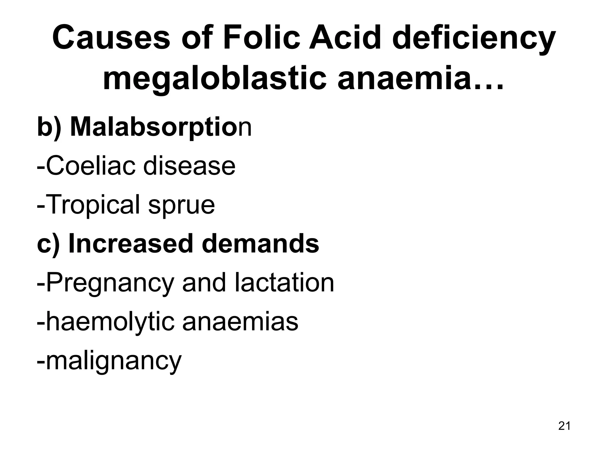
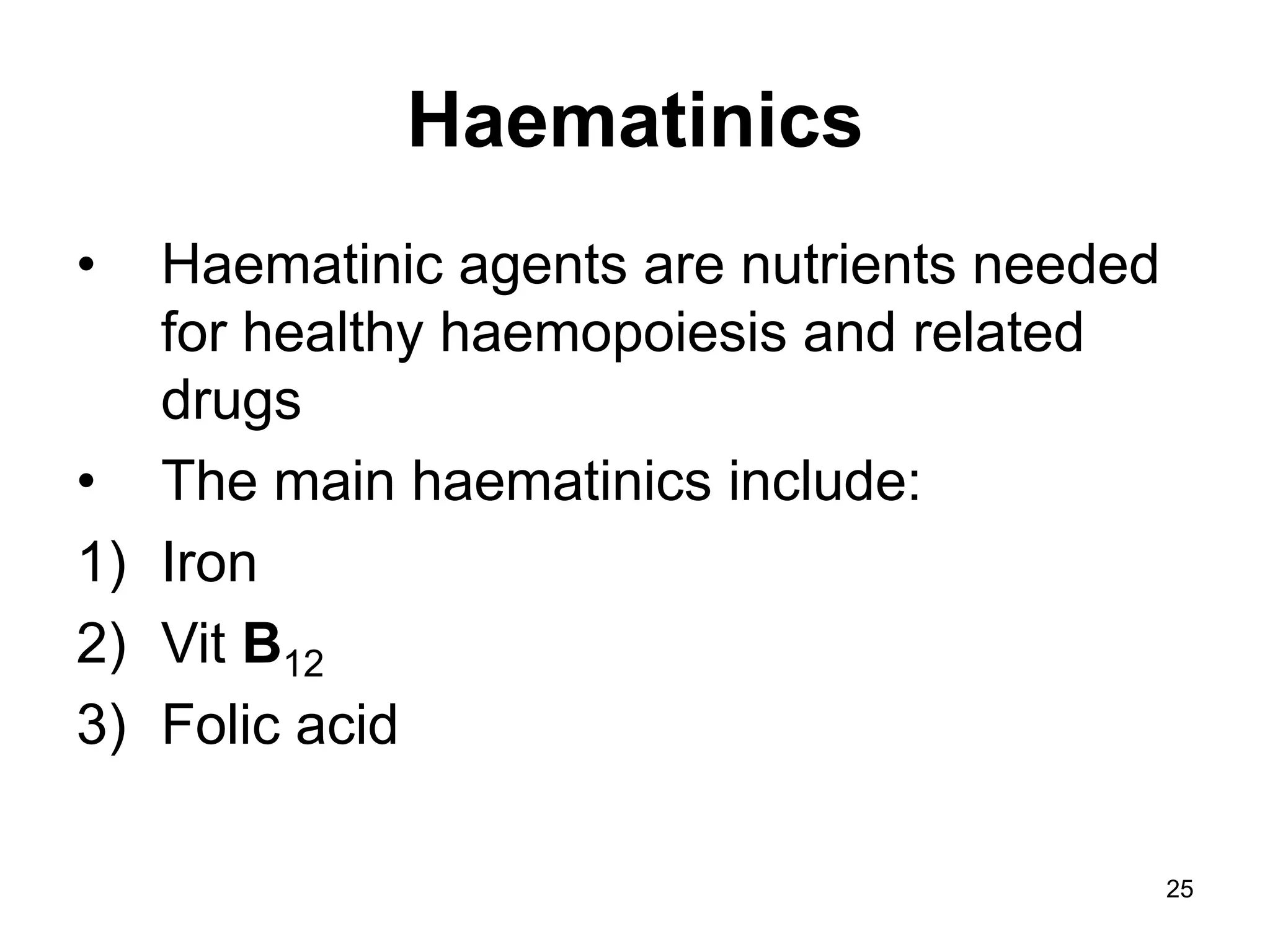
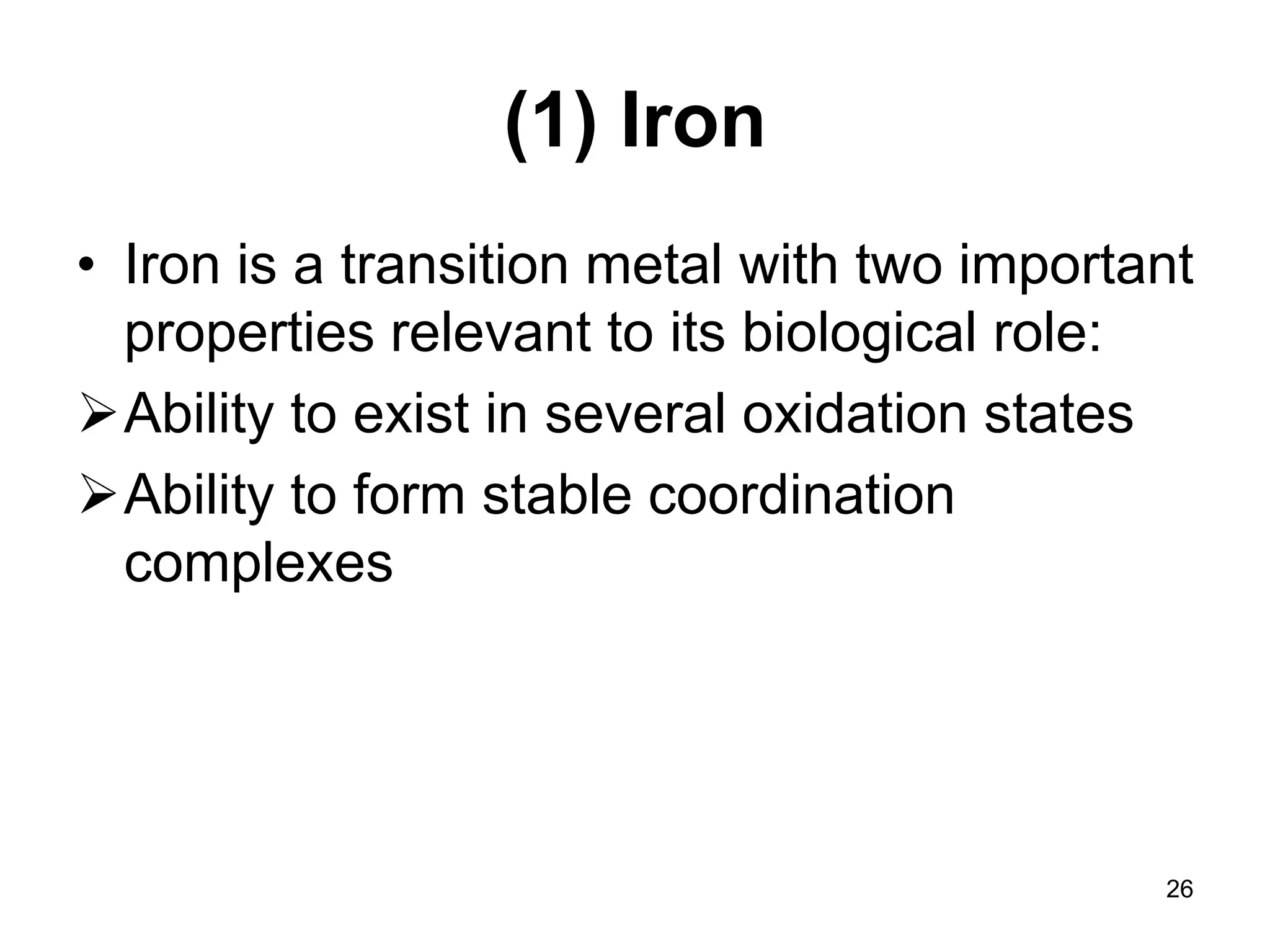
1
HAEMATINICS AND
HAEMOPOIETIC GROWTH
FACTORS
February 15th, 2012
2
Haemopoietic System-
Overview
• This session will summarise the:
 Types of anaemia (due to deficiency of nutrients,
depression of bone marrow or increased
destruction of red cells)
 Main haematinic agents used to treat anaemia
namely iron, folic acid and vitamin B12
 Haemopoietic growth factors-erythropoietin
and colony-stimulating factors (CSFs)
3
Haemopoietic System
• The main components are:
The blood
Bone marrow
Lymph nodes
Thymus
Important accessory organs i.e. spleen,
liver and kidneys
4
Haemopoietic System…
• Blood consists of :
formed elements (red blood cells-RBCs,
white blood cells-WBCs and platelets)
Plasma
• The blood has diverse functions including
key roles in host defence and haemostasis
• In this session we will deal mainly with
RBCs which have the principal function
of carrying oxygen
5
Haemopoietic System…
• The oxygen carrying power of RBCs
(erythrocytes) depends on their
haemoglobin (Hb) content
• The most important site of RBCs formation
in adults is the bone marrow whereas the
spleen acts as their graveyard
• Red cell loss in healthy adults is precisely
balanced by production of new cells
6
Haemopoietic System…
• The liver stores vitamin B12 and is involved in
the process of breakdown of the Hb liberated
when RBCs are destroyed
• The kidney manufactures erythropoietin
• Cells from other organs synthesize and release
colony-stimulating factors (CSFs), which
regulate the production of leucocytes and
platelets
• Read: function of platelets and leucocytes
7
Anaemia-
(A) Introduction
• Definition:
Anaemia is defined as a reduced
concentration of haemoglobin (Hb) in the
blood.
Normal values: male = 13.2 -17.0 g/dL
Normal values:female = 12.0 -16.0 g/dL
• Symptoms of anaemia???
8
Anaemia Symptoms
• Listlessness
• Tiring easily
• Palpitations
• Muscle aches and pains
• Blackouts
• Angina pectoris
• Breathlessness on exertion (high output cardiac
failure)
• Major clinical feature is pallor.
• Others??? etc etc
9
Anaemia…
• Anaemia may give rise to fatigue but,
especially if it is chronic, is often
surprisingly asymptomatic.
• The commonest cause is blood loss
related to menstruation, drug treatment
(e.g. with aspirin or other NSAIDs; or
pathological processes such as colonic
carcinoma or (especially in developing
countries) parasitic infestation.
10
Anaemia…
• Pregnancy and child bearing are other
important physiological drains on iron
reserves. There are several different types
of anaemia, and several different
diagnostic levels.
• Determining indices of red cell size and
haemoglobin content and microscopical
examination of a stained blood smear
allow characterization into these different
types of anaemia.
11
Anaemia…
• The haemoglobin content and red blood
cells (erythrocytes) count of blood are
closely linked.
• There are two general ways in which
erythrocytes are reduced in number:
(1) Impaired formation
(2) Accelerated destruction or loss
12
(1) Impaired formation arises from
defects of:
 Hb synthesis-the functional erythrocyte mass is
selectively reduced in iron deficiency anaemia
 DNA synthesis-erythrocytes, granulocytes and
platelets are all affected in megaloblastic
anaemias (large peripheral blood cell parent
cells in the bone marrow)
 Cell synthesis-erythrocytes, granulocytes and
platelets are all reduced in numbers in aplastic
anaemia
13
(2) Accelerated destruction or
loss arises from:
Chronic blood loss, which leads to iron
deficiency anaemia
Haemolysis, which leads to macrocytic
anaemia (abnormal large erythrocytes in
the peripheral blood)
Drugs used to correct these disorders
are known as HAEMATINICS
14
(B) Types of Anaemia
(1) Iron deficiency anaemia
(2) Megaloblastic anaemia
(a) Vitamin B12 deficiency-megaloblastic
anaemia
(b) Folic acid-deficiency megaloblastic
anaemia
(3) Anaemia associated with chronic renal
failure
15
(B) Types of Anaemia…
A) Iron deficiency anaemia (hypochromic,
microcytic anaemia: small red cells with low
Hb)
Causes : multiple e.g.
• Chronic blood loss
 Gastrointestinal causes :
-Diseases of G.I.T (peptic ulcers,
malignancies)
-Intestinal parasites (Hookworm
infestation)
-Drug induced (Aspirin etc)
16
Causes of Iron deficiency anaemia…
menstrual losses
Recurrent haemoptysis (pulmonary)
Increased requirements
- Pregnancy
- Treatment of megaloblastic anaemia
Mal absorption:
-mal absorption syndrome
-post -gastrectomy
Dietary deficiency
17
Types of Anaemia…
B) Megaloblastic anaemia due to:
-Vit B12 deficiency
-Folic acid deficiency
-or both
Causes of megaloblastic anaemia multiple
e.g.
18
Causes of Vit. B12 deficiency
megaloblastic anaemia
i) Vit. B12 deficiency
a) Malabsorption
• Gastric causes
-Gastrectomy
• Intestinal causes
-Stagnation ( blind loops, ilea-colic fistula )
-Tropical sprue
19
Causes of Vit. B12 deficiency
megaloblastic anaemia …
-ILeal resection and Crohn’s disease
-Fish tapeworm ( Diphyllobothrium latum)
b) Inadequate intake
• Strict vegetarianism
20
Causes of Folic Acid deficiency
megaloblastic anaemia
ii) Folic acid deficiency
a) Inadequate intake
- Dietary deficiency –Old age, alcoholism ,
scurvy
21
Causes of Folic Acid deficiency
megaloblastic anaemia…
b) Malabsorption
-Coeliac disease
-Tropical sprue
c) Increased demands
-Pregnancy and lactation
-haemolytic anaemias
-malignancy
22
Causes of Folic Acid deficiency
megaloblastic anaemia…
d) Drugs
- Anti convulsants (phenytoin and
barbiturates e.g. phenobarbitone) interfere
with absorption
-Dihydrofolate reductase inhibitors
e.g. Pyrimethamine, proguanil and
trimethoprim, methotrexate (interfere with
utilization)
23
• It is important to note that the use of
haematinic agents is often only an adjunct to
treatment of the underlying cause of the
anaemia
• E.g.
 surgery of colon cancer (a common cause of
iron deficiency)
 Anti-helminthic drugs for patients with hookworm
 Sometimes treatment consists of stopping an
offending drug, e.g. a NSAID that causes blood
loss from the stomach
24
HAEMATINIC AGENTS
25
Haematinics
• Haematinic agents are nutrients needed
for healthy haemopoiesis and related
drugs
• The main haematinics include:
1) Iron
2) Vit B12
3) Folic acid
26
(1) Iron
• Iron is a transition metal with two important
properties relevant to its biological role:
Ability to exist in several oxidation states
Ability to form stable coordination
complexes
27
Distribution of Iron in the body of a
healthy 70kg male
Protein Tissue Iron Content (mg)
Haemoglobin Erythrocytes 2600
Myoglobin Muscle 400
Enzymes
(cytochromes,
catalase,
guanylate cyclase
etc
Liver and other
tissues
25
28
IRON-Distribution of iron in the body
of a healthy 70kg male
Protein Tissue Iron content
(mg)
Transferrin Plasma and
extracellular
fluid
8
Ferritin and
haemosiderin
Liver 410
Spleen 48
Bone marrow 300
29
Read on:
• Schematic illustration of iron distribution in
the body :
• Refer Pharmacology by Rang & dale :
 5th edition chapter 21 fig 21.1
 6th edition chapter 22 fig 22.1
 7th edition, Chapter 25, fig 25.1)
30
Aim of therapy in iron – deficiency
anaemia:
-To remove the cause of anaemia by
treating the underlying pathology/or
cause
-To increase the red blood cell mass by
giving Iron.
31
Facts about iron distribution in the
body
• In a 70 kg man there is 4g of iron
- 65% of which circulates in blood as
Haemoglobin
- About one half of the remainder is stored
in the liver, spleen and bone marrow
chiefly as ferritin and haemosiderin
32
Facts about iron distribution in the
body…
• The iron in ferritin and haemosiderin is
available for haemoglobin synthesis
• The rest which is not available for Hb
synthesis is present in myoglobin,
cytochromes and various enzymes.
• Iron is stored in the body in the Fe +++
state
33
Facts about haemoglobin (Revise
Biochemistry)
-Hb is made up of 4 protein chain subunits called
globins
-Each globin contains: one haem moiety
-Haem consists of : Tetrapyrrole porphyrin ring
containing ferrous (Fe 2 +) ion
-Each haem group carry ONE O2 molecule which
is bound reversibly to Fe 2+ and to a histidine
residue in the globin chain
-This reversible binding is the basis of O2 transport
34
Iron turnover and balance
Administration and side effects
• Normal daily requirement for iron is
approx. 5mg for men, and 15mg for
growing kids and child bearing women
(menstruating women).
• Pregnant women require 2-10 times this
amount i.e about 30-150mg daily. Why??
• Because of the demands of the fetus
and increased requirements of the
mother
35
• NB: Each pregnancy ‘costs’ the mother
680 mg iron, equivalent to 1300mls of
blood, owing to the demands of the fetus,
plus requirements of the expanded blood
volume and blood loss at delivery.
36
• Iron in meat for e.g., is generally present
as haem and about 20-40% of haem iron
is available for absorption
• Non-haem iron in food is mainly in the
ferric (Fe +++) state
• Fe +++ has low solubility in intestine
• Fe +++ is converted to Fe ++ iron for
absorption
37
Absorption of Iron
-Fe ++ form
-In stomach iron dissolves and binds to
mucoprotein (carrier)
38
Absorption of Iron…
• The site of iron absorption is the
duodenum and upper jejunum, and
absorption is a two-stage process
involving 1st a rapid uptake across the
brush border and then transfer into the
plasma from the interior of the epithelial
cells.
• The 2nd stage, which is rate limiting, is
energy dependent.
39
Absorption of Iron…
• Haem iron in the diet is absorbed as intact
haem, and the iron is released in the mucosal
cell by the action of haem oxidase.
• Non-haem iron is absorbed in the ferrous
state.
• Within the cell, ferrous iron is oxidised to ferric
iron, which is bound to an intracellular carrier, a
transferrin-like protein; the iron is then either
held in storage in the mucosal cell as ferritin (if
body stores of iron are high) or passed on to the
plasma ( if plasma stores are low)
40
Absorption of Iron…
- In the presence of ascorbic acid, fructose and
various amino acids:
- Iron is detached from mucoprotein ( carrier
molecule) forming soluble low-molecular- weight
complexes that allow it to remain in soluble form
in the intestine.
- Ascorbic acid stimulates iron absorption partly
by reducing Fe +++ iron to the more soluble
Fe ++ form and by forming soluble iron-
ascorbate chelates
41
Administration of Iron
• Iron is given orally or parenterally in
special circumstances.
• Several different preparations of ferrous
iron salts are available for oral
administration
• Mainly one is ferrous sulphate ,with an
elemental iron content of 200ug/mg
42
Administration of Iron…
• Other salts for oral administration are ferrous
succinate, ferrous gluconate, ferrous
fumarate. They are all absorbed to a
comparable extent
• Parenteral iron is rarely given except in patients
who are unable to swallow, or not able to absorb
oral iron because of malabsorption syndromes,
or as a result of surgical procedures or
inflammatory conditions involving the g.i.t
43
Administration of Iron…
• the preparations used are iron-dextran or
iron-sorbitol, both given by deep
intramuscular injection
• Iron dextran (NOT Iron Sorbitol) can be
given by slow intravenous infusion, but this
method of administration should only be
used if absolutely necessary because of
the risk of anaphylactoid reactions
44
Side effects of Iron therapy
• The unwanted effects of oral iron
administration are dose related and
include nausea, abdominal cramps and
diarrhoea
• Acute iron toxicity –common in children
who have swallowed attractively coloured
tablets in mistake for sweets, occur after
ingestion of large quantities of iron salts.
45
Side effects of Iron therapy…
• this can cause:-
: severe necrotizing gastritis with vomiting
: Haemorrhage and diarrhoea
: Circulatory collapse
• Chronic iron toxicity (iron overload)
: usually caused by conditions other than
ingestion of iron salts for e.g.
:chronic haemolytic anaemias or repeated
blood transfusions
46
Side effects of Iron therapy…
• Treatment of acute and chronic iron
toxicity involves the use of iron chelators
such as desferrioxamine
• This is not absorbed from the gut but is
nonetheless given
:intragastrically following acute overdose
( to bind iron in the bowel lumen and
prevent its absorption)
47
Side effects of Iron therapy…
:intramuscularly
:intravenously
:in severe poisoning, it is given by slow
intravenous infusion
• Desferrioxamine forms a complex with
ferric iron, and unlike unbound iron, this is
excreted in the urine
48
Clinical Uses of Iron
• To treat iron deficiency anaemia which can be
caused by:
-chronic blood loss (e.g. menorrhagia,
hookworm, colon cancer)
-increased demand (e.g. in pregnancy and
early infancy)
-Inadequate dietary intake (uncommon in
developed countries)
-Inadequate absorption (e.g. following
gastrectomy)
49
Folic acid and Vitamin B12
• Both are necessary constituents of the
human diet
• Both are essential for DNA Synthesis and
consequently cell division
50
Folic acid and Vitamin B12…
• Deficiency of either vitamin B12 or folic acid
affects tissues with a rapid cell turnover,
particularly bone marrow, but vitamin B12
deficiency also causes important disorders
of nerves, which are not corrected by
treatment with folic acid.
• Deficiency of either vitamin causes
megaloblastic anaemia
51
(2) Vitamin B12
• Is a complex cobalamin compound
• The Vitamin B12 used medically is
hydroxocobalamin
• Dietary sources : Liver, kidney, sea fish,
egg yolk and dairy products.
52
Vitamin B12…
• All cobalamins, dietary and therapeutic,
must be converted to methylcobalamin
(methyl- B12 ) or 5’-
deoxyadenosylcobalamine (ado- B12) for
activity in the body.
• Daily requirements 1-3 mg pregnancy and
lactation 3-5mg
53
Actions
• Actions of Vitamin B12 is required for
conversion of homocysteine to Methionine.
• Vitamin B12 is required for two main
biochemical reactions in humans:
The conversion of methyl-FH4 to FH4
Isomerization of methylmalonyl-CoA to
succinyl-CoA
54
I. The Conversion of Methyl-FH4
to FH4
• The reaction involves conversion of both
methyl-FH4 to FH4 and homocysteine to
methionine.
• The enzyme that accomplishes this is
homocysteine-methionine
methyltransferase
55
See diagram on The role of Vit. B12 in the
synthesis of folate polyglutamate :-
• Read: Rang 7 Dale Pharmacology :-
• 5th edition Chapter 21 fig 21.4
• 6th edition chapter 22 fig 22.4
• 7th edition chapter 25 fig 25.2
56
Role of Vit B12 in the synthesis of
Folate Polyglutamate…
• Methyltetrahydrofolate (Methyl-FH4)
enters cells by active transport.
• The methyl group is transferred to
homocysteine to form methionine via
vitamin B12 , which is bound to the
apoenzyme homocystein-methionine
methyltransferase.
57
Role of Vit B12 in the synthesis of
Folate Polyglutamate…
• Methionine is an important substrate in the
donation of a formate group (shown by the
curved red arrow) for the conversion of
tetrahydrofolate (FH4) to formyl
tetrahydrofolate (formyl FH4), which is the
preferred substrate for the formation of
folate polyglutamates, essential for DNA
synthesis
58
II. Isomerization of Methylmalonyl-
CoA to succinyl-CoA
• B12 is essential / required for
isomerization of methylmalonyl-CoA to
succinyl- CoA an important step in
linking Carbohydrate and lipid
metabolism.
59
Uses of Vit. B12
• Given by injection as Hydroxocobalamin
(I.M)
• Life long treatment in Pernicious anaemia
• Prophylaxis after surgical removal of the
site of production of intrinsic factor (the
stomach) or Vit B12 absorption site (the
terminal ileum )
60
(3) Folic Acid
• Dietary sources include:
Liver,
green vegetables like spinach,
egg,
milk,
synthethised by gut flora
• Dietary requirements: Adult < 0-1 mg/day
pregnancy and lactation 0.8 mg/day
61
Metabolic functions of folic acid
• Folic acid is inactive, so for activity, folate
must be in the FH4 form, in which it is
maintained by dihydrofolate reductase
• This enzyme reduces dietary folic acid to
FH4 and also regenerates FH4 from FH2
produced from FH4 during thymidylate
synthesis
• Folate antagonists act by inhibiting
dihydrofolate reductase
62
• Folates are especially important for the
conversion of deoxyuridylate
monophosphate to deoxythymidylate
monophosphate
• This is rate limiting in mammalian DNA
synthesis and is catalysed by thymidylate
synthetase, with folate acting as methyl
donor
• Note: Folic acid generates thymidylate
from THFA, an essential constituent of
DNA
63
Comparison of the manifestations of
Vit B12 and Folic Acid Deficiency
B12 Folic acid
-megaloblastic anaemia -megaloblastic anaemia
indistinguishable from B12
-Glossitis, git disturbances -Epithelial damage
-Neurological: subacute -Debility, wt loss, sterility
Combined degeneration
of spinal cord, peripheral
neuritis
64
Comparison of the manifestations of
Vit B12 and Folic Acid Deficiency…
B12 Folic Acid
-mental change : poor memory -Neurological symptom DO NOT
hallucinates ,mood OCCUR
• Note:Folate deficiency shows similar clinical
features and blood picture to that of vit B12
deficiency, except there is no neurological
disturbance
• It must be distinguished by history and tests from
Vit. B12 deficiency.
• As an attempt to treat Vit B12 deficiency with folate
can precipitate neuropathy
65
Uses of Folic acid
1) Treatment of megaloblastic anaemia due
to nutritional folate deficiency, which can
be caused by: poor diet (common in
alcoholics), malabsorption syndromes,
drugs e.g. phenytoin
2) Increased demand : pregnancy, lactation,
premature infancy, during treatment of
haemolytic anaemia etc.
66
HAEMOPOIETIC GROWTH
FACTORS
67
HAEMOPOIETIC GROWTH
FACTORS
• Every 60 seconds, a human being must
generate about 120 million granulocytes and
150 million erythrocytes, as well as numerous
mononuclear cells and platelets.
• The cells responsible for this remarkable
productivity are derived from a relatively small
number of self renewing, pluripotent stem cells
laid down during embryogenesis.
68
HAEMOPOIETIC GROWTH
FACTORS…
• Maintenance of haemopoiesis necessitates a
balance between self renewal of the stem cells
on one hand, and differentiation into the various
types of blood cell on the other.
• The factors involved in controlling this balance
are the haemopoietic growth factors, which
direct the division and maturation of the progeny
of these cells down eight possible lines of
development.
69
Haemopoietic Factors
(I) Erythropoietin
(II) Colony-stimulating factors
70
(I) Erythropoietin
• Is a glycoprotein hormone
• It stimulates the production of erythrocytes
(erythropoiesis) in the bone marrow
• In adults, 85% of circulating erythropoietin
is synthesized in the endothelial cells of
the peritubular capillaries in the renal
cortex; the remainder is synthesized in the
liver
71
Erythropoietin…
• Hypoxia is the normal stimulus for
erythropoietin synthesis
• When the plasma concentration of
erythropoietin is low, erythroid stem cells
in the bone marrow exhibit DNA cleavage
followed by apoptosis (programmed cell
death)
• Erythropoietin prevents this process
72
Erythropoietin…
• It increase the number of committed stem
cells that are converted to RBCs
precursors and subsequently to mature
erythrocytes
• In chronic renal failure the ability of the
kidney to produce an adequate supply of
erythropoietin is lost and the deficiency
cannot be made good by synthesis in the
liver.
73
Erythropoietin…
• The erythropoietin deficiency may
result from chronic renal failure and
can result in the development of a
normocytic, hypochromic anaemia
(though this may sometimes be
complicated by other factors as iron
and folate deficiency)
• This may be corrected by epoetin
(recombinant erythropoietin)
74
Clinical Uses of Epoietin
(i) Anaemia of chronic renal failure, treated
by s/c or i/v injection. Response is
fastest after i/v injection
(ii) Anaemia during chemotherapy for
cancer
(iii) Prevention of the anaemia that occurs in
premature infants
(iv) Anaemia of AIDS (exacerbated by
zidovudine treatment)
75
II. Colony-stimulating Factors
• CSFs are called so because they were
found to stimulate the formation of
maturing colonies of leucocytes in semi-
solid medium in vitro
• Are classified as cytokines
• They stimulate particular committed
progenitor cells to proliferate and also
cause irreversible differentiation
76
Colony-stimulating Factors…
• Granulocyte colony-stimulating factors
(G-CSF)
-produced mainly by monocytes,
fibroblasts and endothelial cells,
-controls primarily the development of
neutrophils (stimulates neutrophil
progenitor)
-is available as filgrastim; it is given i/v or
s/c
77
Colony-stimulating Factors…
• Granulocyte-macrophage colony
stimulating factors (GM-CSF)
-stimulates development of many types of
progenitor cell
-is available as molgramostin and is given
i/v, s/c
-Can cause fever, rashes, bone pain,
hypotension, gastrointestinal symptoms
and arterial oxygen desaturation
78
Clinical uses of CSFs
• To reduce the severity and duration of the
neutropenia induced by cytotoxic drug
during anticancer chemotherapy and
following bone marrow transplant
• For persistent neutropenia in advanced
HIV infection
• May have a role in treatment of aplastic
anaemia
79
THE END!
• Read: Pharmacology by Rang & Dale:-
• 5th Edition Chapter 21
• 6th Edition Chapter 22
• 7th Edition Chapter 25

37-anaemia_and_haematinics_notes.ppt

  • 1.
  • 2.
    2 Haemopoietic System- Overview • Thissession will summarise the:  Types of anaemia (due to deficiency of nutrients, depression of bone marrow or increased destruction of red cells)  Main haematinic agents used to treat anaemia namely iron, folic acid and vitamin B12  Haemopoietic growth factors-erythropoietin and colony-stimulating factors (CSFs)
  • 3.
    3 Haemopoietic System • Themain components are: The blood Bone marrow Lymph nodes Thymus Important accessory organs i.e. spleen, liver and kidneys
  • 4.
    4 Haemopoietic System… • Bloodconsists of : formed elements (red blood cells-RBCs, white blood cells-WBCs and platelets) Plasma • The blood has diverse functions including key roles in host defence and haemostasis • In this session we will deal mainly with RBCs which have the principal function of carrying oxygen
  • 5.
    5 Haemopoietic System… • Theoxygen carrying power of RBCs (erythrocytes) depends on their haemoglobin (Hb) content • The most important site of RBCs formation in adults is the bone marrow whereas the spleen acts as their graveyard • Red cell loss in healthy adults is precisely balanced by production of new cells
  • 6.
    6 Haemopoietic System… • Theliver stores vitamin B12 and is involved in the process of breakdown of the Hb liberated when RBCs are destroyed • The kidney manufactures erythropoietin • Cells from other organs synthesize and release colony-stimulating factors (CSFs), which regulate the production of leucocytes and platelets • Read: function of platelets and leucocytes
  • 7.
    7 Anaemia- (A) Introduction • Definition: Anaemiais defined as a reduced concentration of haemoglobin (Hb) in the blood. Normal values: male = 13.2 -17.0 g/dL Normal values:female = 12.0 -16.0 g/dL • Symptoms of anaemia???
  • 8.
    8 Anaemia Symptoms • Listlessness •Tiring easily • Palpitations • Muscle aches and pains • Blackouts • Angina pectoris • Breathlessness on exertion (high output cardiac failure) • Major clinical feature is pallor. • Others??? etc etc
  • 9.
    9 Anaemia… • Anaemia maygive rise to fatigue but, especially if it is chronic, is often surprisingly asymptomatic. • The commonest cause is blood loss related to menstruation, drug treatment (e.g. with aspirin or other NSAIDs; or pathological processes such as colonic carcinoma or (especially in developing countries) parasitic infestation.
  • 10.
    10 Anaemia… • Pregnancy andchild bearing are other important physiological drains on iron reserves. There are several different types of anaemia, and several different diagnostic levels. • Determining indices of red cell size and haemoglobin content and microscopical examination of a stained blood smear allow characterization into these different types of anaemia.
  • 11.
    11 Anaemia… • The haemoglobincontent and red blood cells (erythrocytes) count of blood are closely linked. • There are two general ways in which erythrocytes are reduced in number: (1) Impaired formation (2) Accelerated destruction or loss
  • 12.
    12 (1) Impaired formationarises from defects of:  Hb synthesis-the functional erythrocyte mass is selectively reduced in iron deficiency anaemia  DNA synthesis-erythrocytes, granulocytes and platelets are all affected in megaloblastic anaemias (large peripheral blood cell parent cells in the bone marrow)  Cell synthesis-erythrocytes, granulocytes and platelets are all reduced in numbers in aplastic anaemia
  • 13.
    13 (2) Accelerated destructionor loss arises from: Chronic blood loss, which leads to iron deficiency anaemia Haemolysis, which leads to macrocytic anaemia (abnormal large erythrocytes in the peripheral blood) Drugs used to correct these disorders are known as HAEMATINICS
  • 14.
    14 (B) Types ofAnaemia (1) Iron deficiency anaemia (2) Megaloblastic anaemia (a) Vitamin B12 deficiency-megaloblastic anaemia (b) Folic acid-deficiency megaloblastic anaemia (3) Anaemia associated with chronic renal failure
  • 15.
    15 (B) Types ofAnaemia… A) Iron deficiency anaemia (hypochromic, microcytic anaemia: small red cells with low Hb) Causes : multiple e.g. • Chronic blood loss  Gastrointestinal causes : -Diseases of G.I.T (peptic ulcers, malignancies) -Intestinal parasites (Hookworm infestation) -Drug induced (Aspirin etc)
  • 16.
    16 Causes of Irondeficiency anaemia… menstrual losses Recurrent haemoptysis (pulmonary) Increased requirements - Pregnancy - Treatment of megaloblastic anaemia Mal absorption: -mal absorption syndrome -post -gastrectomy Dietary deficiency
  • 17.
    17 Types of Anaemia… B)Megaloblastic anaemia due to: -Vit B12 deficiency -Folic acid deficiency -or both Causes of megaloblastic anaemia multiple e.g.
  • 18.
    18 Causes of Vit.B12 deficiency megaloblastic anaemia i) Vit. B12 deficiency a) Malabsorption • Gastric causes -Gastrectomy • Intestinal causes -Stagnation ( blind loops, ilea-colic fistula ) -Tropical sprue
  • 19.
    19 Causes of Vit.B12 deficiency megaloblastic anaemia … -ILeal resection and Crohn’s disease -Fish tapeworm ( Diphyllobothrium latum) b) Inadequate intake • Strict vegetarianism
  • 20.
    20 Causes of FolicAcid deficiency megaloblastic anaemia ii) Folic acid deficiency a) Inadequate intake - Dietary deficiency –Old age, alcoholism , scurvy
  • 21.
    21 Causes of FolicAcid deficiency megaloblastic anaemia… b) Malabsorption -Coeliac disease -Tropical sprue c) Increased demands -Pregnancy and lactation -haemolytic anaemias -malignancy
  • 22.
    22 Causes of FolicAcid deficiency megaloblastic anaemia… d) Drugs - Anti convulsants (phenytoin and barbiturates e.g. phenobarbitone) interfere with absorption -Dihydrofolate reductase inhibitors e.g. Pyrimethamine, proguanil and trimethoprim, methotrexate (interfere with utilization)
  • 23.
    23 • It isimportant to note that the use of haematinic agents is often only an adjunct to treatment of the underlying cause of the anaemia • E.g.  surgery of colon cancer (a common cause of iron deficiency)  Anti-helminthic drugs for patients with hookworm  Sometimes treatment consists of stopping an offending drug, e.g. a NSAID that causes blood loss from the stomach
  • 24.
  • 25.
    25 Haematinics • Haematinic agentsare nutrients needed for healthy haemopoiesis and related drugs • The main haematinics include: 1) Iron 2) Vit B12 3) Folic acid
  • 26.
    26 (1) Iron • Ironis a transition metal with two important properties relevant to its biological role: Ability to exist in several oxidation states Ability to form stable coordination complexes
  • 27.
    27 Distribution of Ironin the body of a healthy 70kg male Protein Tissue Iron Content (mg) Haemoglobin Erythrocytes 2600 Myoglobin Muscle 400 Enzymes (cytochromes, catalase, guanylate cyclase etc Liver and other tissues 25
  • 28.
    28 IRON-Distribution of ironin the body of a healthy 70kg male Protein Tissue Iron content (mg) Transferrin Plasma and extracellular fluid 8 Ferritin and haemosiderin Liver 410 Spleen 48 Bone marrow 300
  • 29.
    29 Read on: • Schematicillustration of iron distribution in the body : • Refer Pharmacology by Rang & dale :  5th edition chapter 21 fig 21.1  6th edition chapter 22 fig 22.1  7th edition, Chapter 25, fig 25.1)
  • 30.
    30 Aim of therapyin iron – deficiency anaemia: -To remove the cause of anaemia by treating the underlying pathology/or cause -To increase the red blood cell mass by giving Iron.
  • 31.
    31 Facts about irondistribution in the body • In a 70 kg man there is 4g of iron - 65% of which circulates in blood as Haemoglobin - About one half of the remainder is stored in the liver, spleen and bone marrow chiefly as ferritin and haemosiderin
  • 32.
    32 Facts about irondistribution in the body… • The iron in ferritin and haemosiderin is available for haemoglobin synthesis • The rest which is not available for Hb synthesis is present in myoglobin, cytochromes and various enzymes. • Iron is stored in the body in the Fe +++ state
  • 33.
    33 Facts about haemoglobin(Revise Biochemistry) -Hb is made up of 4 protein chain subunits called globins -Each globin contains: one haem moiety -Haem consists of : Tetrapyrrole porphyrin ring containing ferrous (Fe 2 +) ion -Each haem group carry ONE O2 molecule which is bound reversibly to Fe 2+ and to a histidine residue in the globin chain -This reversible binding is the basis of O2 transport
  • 34.
    34 Iron turnover andbalance Administration and side effects • Normal daily requirement for iron is approx. 5mg for men, and 15mg for growing kids and child bearing women (menstruating women). • Pregnant women require 2-10 times this amount i.e about 30-150mg daily. Why?? • Because of the demands of the fetus and increased requirements of the mother
  • 35.
    35 • NB: Eachpregnancy ‘costs’ the mother 680 mg iron, equivalent to 1300mls of blood, owing to the demands of the fetus, plus requirements of the expanded blood volume and blood loss at delivery.
  • 36.
    36 • Iron inmeat for e.g., is generally present as haem and about 20-40% of haem iron is available for absorption • Non-haem iron in food is mainly in the ferric (Fe +++) state • Fe +++ has low solubility in intestine • Fe +++ is converted to Fe ++ iron for absorption
  • 37.
    37 Absorption of Iron -Fe++ form -In stomach iron dissolves and binds to mucoprotein (carrier)
  • 38.
    38 Absorption of Iron… •The site of iron absorption is the duodenum and upper jejunum, and absorption is a two-stage process involving 1st a rapid uptake across the brush border and then transfer into the plasma from the interior of the epithelial cells. • The 2nd stage, which is rate limiting, is energy dependent.
  • 39.
    39 Absorption of Iron… •Haem iron in the diet is absorbed as intact haem, and the iron is released in the mucosal cell by the action of haem oxidase. • Non-haem iron is absorbed in the ferrous state. • Within the cell, ferrous iron is oxidised to ferric iron, which is bound to an intracellular carrier, a transferrin-like protein; the iron is then either held in storage in the mucosal cell as ferritin (if body stores of iron are high) or passed on to the plasma ( if plasma stores are low)
  • 40.
    40 Absorption of Iron… -In the presence of ascorbic acid, fructose and various amino acids: - Iron is detached from mucoprotein ( carrier molecule) forming soluble low-molecular- weight complexes that allow it to remain in soluble form in the intestine. - Ascorbic acid stimulates iron absorption partly by reducing Fe +++ iron to the more soluble Fe ++ form and by forming soluble iron- ascorbate chelates
  • 41.
    41 Administration of Iron •Iron is given orally or parenterally in special circumstances. • Several different preparations of ferrous iron salts are available for oral administration • Mainly one is ferrous sulphate ,with an elemental iron content of 200ug/mg
  • 42.
    42 Administration of Iron… •Other salts for oral administration are ferrous succinate, ferrous gluconate, ferrous fumarate. They are all absorbed to a comparable extent • Parenteral iron is rarely given except in patients who are unable to swallow, or not able to absorb oral iron because of malabsorption syndromes, or as a result of surgical procedures or inflammatory conditions involving the g.i.t
  • 43.
    43 Administration of Iron… •the preparations used are iron-dextran or iron-sorbitol, both given by deep intramuscular injection • Iron dextran (NOT Iron Sorbitol) can be given by slow intravenous infusion, but this method of administration should only be used if absolutely necessary because of the risk of anaphylactoid reactions
  • 44.
    44 Side effects ofIron therapy • The unwanted effects of oral iron administration are dose related and include nausea, abdominal cramps and diarrhoea • Acute iron toxicity –common in children who have swallowed attractively coloured tablets in mistake for sweets, occur after ingestion of large quantities of iron salts.
  • 45.
    45 Side effects ofIron therapy… • this can cause:- : severe necrotizing gastritis with vomiting : Haemorrhage and diarrhoea : Circulatory collapse • Chronic iron toxicity (iron overload) : usually caused by conditions other than ingestion of iron salts for e.g. :chronic haemolytic anaemias or repeated blood transfusions
  • 46.
    46 Side effects ofIron therapy… • Treatment of acute and chronic iron toxicity involves the use of iron chelators such as desferrioxamine • This is not absorbed from the gut but is nonetheless given :intragastrically following acute overdose ( to bind iron in the bowel lumen and prevent its absorption)
  • 47.
    47 Side effects ofIron therapy… :intramuscularly :intravenously :in severe poisoning, it is given by slow intravenous infusion • Desferrioxamine forms a complex with ferric iron, and unlike unbound iron, this is excreted in the urine
  • 48.
    48 Clinical Uses ofIron • To treat iron deficiency anaemia which can be caused by: -chronic blood loss (e.g. menorrhagia, hookworm, colon cancer) -increased demand (e.g. in pregnancy and early infancy) -Inadequate dietary intake (uncommon in developed countries) -Inadequate absorption (e.g. following gastrectomy)
  • 49.
    49 Folic acid andVitamin B12 • Both are necessary constituents of the human diet • Both are essential for DNA Synthesis and consequently cell division
  • 50.
    50 Folic acid andVitamin B12… • Deficiency of either vitamin B12 or folic acid affects tissues with a rapid cell turnover, particularly bone marrow, but vitamin B12 deficiency also causes important disorders of nerves, which are not corrected by treatment with folic acid. • Deficiency of either vitamin causes megaloblastic anaemia
  • 51.
    51 (2) Vitamin B12 •Is a complex cobalamin compound • The Vitamin B12 used medically is hydroxocobalamin • Dietary sources : Liver, kidney, sea fish, egg yolk and dairy products.
  • 52.
    52 Vitamin B12… • Allcobalamins, dietary and therapeutic, must be converted to methylcobalamin (methyl- B12 ) or 5’- deoxyadenosylcobalamine (ado- B12) for activity in the body. • Daily requirements 1-3 mg pregnancy and lactation 3-5mg
  • 53.
    53 Actions • Actions ofVitamin B12 is required for conversion of homocysteine to Methionine. • Vitamin B12 is required for two main biochemical reactions in humans: The conversion of methyl-FH4 to FH4 Isomerization of methylmalonyl-CoA to succinyl-CoA
  • 54.
    54 I. The Conversionof Methyl-FH4 to FH4 • The reaction involves conversion of both methyl-FH4 to FH4 and homocysteine to methionine. • The enzyme that accomplishes this is homocysteine-methionine methyltransferase
  • 55.
    55 See diagram onThe role of Vit. B12 in the synthesis of folate polyglutamate :- • Read: Rang 7 Dale Pharmacology :- • 5th edition Chapter 21 fig 21.4 • 6th edition chapter 22 fig 22.4 • 7th edition chapter 25 fig 25.2
  • 56.
    56 Role of VitB12 in the synthesis of Folate Polyglutamate… • Methyltetrahydrofolate (Methyl-FH4) enters cells by active transport. • The methyl group is transferred to homocysteine to form methionine via vitamin B12 , which is bound to the apoenzyme homocystein-methionine methyltransferase.
  • 57.
    57 Role of VitB12 in the synthesis of Folate Polyglutamate… • Methionine is an important substrate in the donation of a formate group (shown by the curved red arrow) for the conversion of tetrahydrofolate (FH4) to formyl tetrahydrofolate (formyl FH4), which is the preferred substrate for the formation of folate polyglutamates, essential for DNA synthesis
  • 58.
    58 II. Isomerization ofMethylmalonyl- CoA to succinyl-CoA • B12 is essential / required for isomerization of methylmalonyl-CoA to succinyl- CoA an important step in linking Carbohydrate and lipid metabolism.
  • 59.
    59 Uses of Vit.B12 • Given by injection as Hydroxocobalamin (I.M) • Life long treatment in Pernicious anaemia • Prophylaxis after surgical removal of the site of production of intrinsic factor (the stomach) or Vit B12 absorption site (the terminal ileum )
  • 60.
    60 (3) Folic Acid •Dietary sources include: Liver, green vegetables like spinach, egg, milk, synthethised by gut flora • Dietary requirements: Adult < 0-1 mg/day pregnancy and lactation 0.8 mg/day
  • 61.
    61 Metabolic functions offolic acid • Folic acid is inactive, so for activity, folate must be in the FH4 form, in which it is maintained by dihydrofolate reductase • This enzyme reduces dietary folic acid to FH4 and also regenerates FH4 from FH2 produced from FH4 during thymidylate synthesis • Folate antagonists act by inhibiting dihydrofolate reductase
  • 62.
    62 • Folates areespecially important for the conversion of deoxyuridylate monophosphate to deoxythymidylate monophosphate • This is rate limiting in mammalian DNA synthesis and is catalysed by thymidylate synthetase, with folate acting as methyl donor • Note: Folic acid generates thymidylate from THFA, an essential constituent of DNA
  • 63.
    63 Comparison of themanifestations of Vit B12 and Folic Acid Deficiency B12 Folic acid -megaloblastic anaemia -megaloblastic anaemia indistinguishable from B12 -Glossitis, git disturbances -Epithelial damage -Neurological: subacute -Debility, wt loss, sterility Combined degeneration of spinal cord, peripheral neuritis
  • 64.
    64 Comparison of themanifestations of Vit B12 and Folic Acid Deficiency… B12 Folic Acid -mental change : poor memory -Neurological symptom DO NOT hallucinates ,mood OCCUR • Note:Folate deficiency shows similar clinical features and blood picture to that of vit B12 deficiency, except there is no neurological disturbance • It must be distinguished by history and tests from Vit. B12 deficiency. • As an attempt to treat Vit B12 deficiency with folate can precipitate neuropathy
  • 65.
    65 Uses of Folicacid 1) Treatment of megaloblastic anaemia due to nutritional folate deficiency, which can be caused by: poor diet (common in alcoholics), malabsorption syndromes, drugs e.g. phenytoin 2) Increased demand : pregnancy, lactation, premature infancy, during treatment of haemolytic anaemia etc.
  • 66.
  • 67.
    67 HAEMOPOIETIC GROWTH FACTORS • Every60 seconds, a human being must generate about 120 million granulocytes and 150 million erythrocytes, as well as numerous mononuclear cells and platelets. • The cells responsible for this remarkable productivity are derived from a relatively small number of self renewing, pluripotent stem cells laid down during embryogenesis.
  • 68.
    68 HAEMOPOIETIC GROWTH FACTORS… • Maintenanceof haemopoiesis necessitates a balance between self renewal of the stem cells on one hand, and differentiation into the various types of blood cell on the other. • The factors involved in controlling this balance are the haemopoietic growth factors, which direct the division and maturation of the progeny of these cells down eight possible lines of development.
  • 69.
  • 70.
    70 (I) Erythropoietin • Isa glycoprotein hormone • It stimulates the production of erythrocytes (erythropoiesis) in the bone marrow • In adults, 85% of circulating erythropoietin is synthesized in the endothelial cells of the peritubular capillaries in the renal cortex; the remainder is synthesized in the liver
  • 71.
    71 Erythropoietin… • Hypoxia isthe normal stimulus for erythropoietin synthesis • When the plasma concentration of erythropoietin is low, erythroid stem cells in the bone marrow exhibit DNA cleavage followed by apoptosis (programmed cell death) • Erythropoietin prevents this process
  • 72.
    72 Erythropoietin… • It increasethe number of committed stem cells that are converted to RBCs precursors and subsequently to mature erythrocytes • In chronic renal failure the ability of the kidney to produce an adequate supply of erythropoietin is lost and the deficiency cannot be made good by synthesis in the liver.
  • 73.
    73 Erythropoietin… • The erythropoietindeficiency may result from chronic renal failure and can result in the development of a normocytic, hypochromic anaemia (though this may sometimes be complicated by other factors as iron and folate deficiency) • This may be corrected by epoetin (recombinant erythropoietin)
  • 74.
    74 Clinical Uses ofEpoietin (i) Anaemia of chronic renal failure, treated by s/c or i/v injection. Response is fastest after i/v injection (ii) Anaemia during chemotherapy for cancer (iii) Prevention of the anaemia that occurs in premature infants (iv) Anaemia of AIDS (exacerbated by zidovudine treatment)
  • 75.
    75 II. Colony-stimulating Factors •CSFs are called so because they were found to stimulate the formation of maturing colonies of leucocytes in semi- solid medium in vitro • Are classified as cytokines • They stimulate particular committed progenitor cells to proliferate and also cause irreversible differentiation
  • 76.
    76 Colony-stimulating Factors… • Granulocytecolony-stimulating factors (G-CSF) -produced mainly by monocytes, fibroblasts and endothelial cells, -controls primarily the development of neutrophils (stimulates neutrophil progenitor) -is available as filgrastim; it is given i/v or s/c
  • 77.
    77 Colony-stimulating Factors… • Granulocyte-macrophagecolony stimulating factors (GM-CSF) -stimulates development of many types of progenitor cell -is available as molgramostin and is given i/v, s/c -Can cause fever, rashes, bone pain, hypotension, gastrointestinal symptoms and arterial oxygen desaturation
  • 78.
    78 Clinical uses ofCSFs • To reduce the severity and duration of the neutropenia induced by cytotoxic drug during anticancer chemotherapy and following bone marrow transplant • For persistent neutropenia in advanced HIV infection • May have a role in treatment of aplastic anaemia
  • 79.
    79 THE END! • Read:Pharmacology by Rang & Dale:- • 5th Edition Chapter 21 • 6th Edition Chapter 22 • 7th Edition Chapter 25