1. TIBS - October 1983
of magnitude faster conduction than the
Grotthus type of proton transfer mechanism
that is operative in solutions. Even il an
extensive intermembrane space were to
exist to accommodate the bulk aqueous
phase, the aforementioned mechanism of
proton conduction could still be operative.
While a considerable body of evidence"
exists on the 'structured' water in biological
systems, the functional relevance of such a
water remains to be explored. Clearly there
is a need to ascertain the real structure of
mitochondria, and other energy transduc-
ing organelles, in relation to the mechan-
ism(s) of energy transduction.
References
1 Williams, R. J. P. (1983)Trends Biochem. Sci. 8,
48
2 Van Harreveld, A., Crowell, J. and Malhotra, S. K.
(1965)J. Cell Biol. 25, 117-137
3 Malhotra, S. K. and Van Harreveld, A. (1965) J.
Ultrastruct. Res. 12. 473~1.87
4 Malhotra, S. K. (1966)J. Ultrastrua:t. Res. 15,
14-37
359
5 Kell, D. B. (1979) Biochim. Biophys. Acta 549,
55-99
6 Finney, J. L. (1979) Water -A Comprehensive
Treatise, Vol. 6 (Franks, F., ed.), pp. 47-122,
Plenum Press
S. K. MALHOTRA
and s. S. SIKERWAR
Biological Sciences
Electron Microscopy Laboratory,
Department of Zoology,
University of Alberta,
Edmonton T6G 2E9,
Canada.
Reviews
A consensus model of the
Escherichia coli ribosome
Jeffrey B. Prince, Robin R. Gutell and Roger A. Garrett
In this article the latest results are summarized on the localization of proteins, RNA
sites, and various ligands on the ribosomal subunits of E. coli. For the proteins, the
data derive primarily from two kinds of experimental approach."neutron scattering of
reconstituted subunits, containing pairs of deuterated proteins, which yields both the
distances between centres of mass of the two proteins and their radii of gyration, and
immune electron microscopy which visualizes protein-bound immunoglobins (IgG)
on the ribosomal surface. Although the two approaches yield different kinds of data,
the results are integrated into a consensus model partly because the level of agreement
between the methods isgood. For the RNA and ribosome-bound ligands the data are
available exclusively from the immune electron microscopy method.
Success in applying the immune electron
microscopy (IEM) technique to ribosomes,
from which most of the data originate,
depends on the proteins (and RNA) having
accessible antigenic determinants on the
ribosomal surface. This was first demons-
trated for the 30S subunit proteinsI and later
for the 50S subunit proteins by using a
variety of immunochemical and physical
methods. Most of the protein-specific IgGs
produced subunit-IgG-subunit complexes
('dimers') which facilitated the protein
localizations. The latest protein results
from the neutron scattering and IEM
methods derive from three main groups:
Moore et al.2"3 (Yale), Lake et al.4"~
(UCLA) and St6ffieret al.6(Berlin) for the
30S proteins, and the latter two groups for
the 50S proteins. The RNA data come, in
addition, from the laboratories of Vasiliev
(Poustchino) and Glitz (UCLA). Our crite-
Jeffrey B. Prince and Robin R. Gutell are at the
Thimann Laboratories, University of California,
Santa Cruz, CA 95064, USA. Roger A. Garrettis at
the Department of Biostructural Chemistry,
Kemisk lnstitut, Aarhus University, Denmark.
rion for including a site in the model is that
two, or more, laboratories agree on a par-
ticular location, although this criterion is
not always rigorously followed for the 50S
subunit proteins where the data are scarce.
The use of unpublished results for the
assignments has generally been avoided,
although some revised protein locations
from the Berlin group are available only in
meeting abstract form with minimal
experimental data. Results from other
approaches, including chemical cross-
linking, 'affinity labelling' of functional
sites, and fluorescence energy transfer, are
only invoked when the evidence is particu-
larly good (e.g. a high yield of chemical
cross-linking), or when there is general
agreement amongst the biochemical
results.
Shapes of the ribosomal subunits
Although differences persist in the pub-
lished models, there is now general agree-
ment on the overall structure of the 30S
subunit. The UCLA model, obtained by
negative staining, consists of a large'body'
(lower 2/3), a smaller 'head' (upper 1/3)
and a thin projection or 'platform', tilted
towards the subunit interface4. The Poust-
chino model, obtained by shadowing, is
similar except that no cleft is observed be-
tween the body and the platform ('ledge'),
and the body is segmented into upper and
lower halves7~. The Berlin model origi-
nally contained a large body and a smaller
head region with two symmetrical lateral
lobes which pointed into the subunit inter-
face thereby producing a hollow in the
centre of the ribosome. One of the lateral
lobes has now been enlarged, thus produc-
ing an asymmetric model, and the 'hand'
has also been changed such that it resem-
bles the other two models more closely.
Recently, Korn et al."~have derived a model
from dark field electron microscopy with
improved resolution (- 15 /~), compared
with the aforementioned studies (-20 ,~);
it approximates to that of the Poustchino
group. They also suggest that the apparent
cleft in the UCLA and Berlin models may
be due to positive staining of RNA in that
region. We have chosen the UCLA model
in this study only because it has proved the
least variable of the two main IEM models,
but we express no opinion as to its relative
accuracy.
The UCLA model of the 50S subunit
consists of a large lower body with a large
central protuberance lying between a smal-
ler protuberance and a stalk-like structure
projecting from the body (see Fig. 4). The
early Berlin model was similar except that it
was a pseudosymmetrical structure lacking
the 'stalk'. Recently, a few laboratories
employing different electron microscopy
methods have established that the large
subunit is indeed asymmetrical and that the
' stalk' does exist'"to.
30S subunit
Protein sites. The 30S subunit protein
locations, depicted in Fig. 1, are classified
into three groups. Sites drawn with closed
circles are the most reliable. The broken
~) 1983.ElsevierSciencePublishersB.V., Amsterdam 0376- 5067/83/$01.0()
2. b.
1,18
360
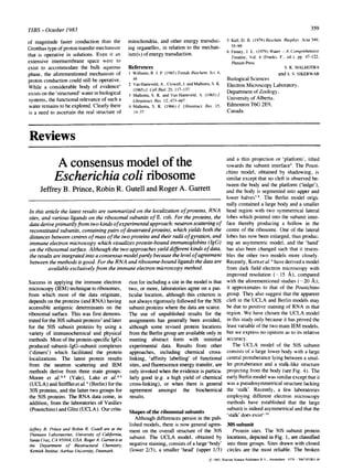
EXTERI 0 R I NTER FACE
Fig. 1. Consensus model of 30S subunit. (a) exterior surface; (b) subunit interface. Circlesdesignate posi-
tions of protein and RNA sites on two-dimensional projections of each subunit surface. Closed or broken
circles aresite locations with greater or lessercertainty, respectively. Brackets indicate region ofsite location
of proteins in margin (surface unspecified), mRNA - entry/exit site of mRNA; Pm= puromycin. Subunit
shapeafterLakeandco-workers'.
circles indicate less certain sites; for exam-
ple, they may lie intermediate between two
close sites assigned by different groups.
Finally, proteins that have been localized
within a specific section of the subunit, but
at different positions, are listed adjacent to
that section of the model. While the protein
sites in Fig. 1depict surface locations (with
the possible exceptions of $4 and $8), the
neutron scattering data yield the relative
positions of the centres of mass of the pro-
teins. To emphasize this distinction, the
12-protein model derived by Moore and
co-workers2.3 is also shown (Fig. 2); the
view is apparently equivalent to that of the
exterior surface presented in Fig. 1a. Loca-
tions are specified for thirteen proteins in
our map: $3, $4, $5, $6, $7, $8, $9, SI0,
SI1, S12, S13, S14 and S15. In addition,
proteins SI, S18, and S19 are assigned to
general regions. Although $6, Sll, and
S13 are exposed on both the exterior sur-
face and the interface side of the subunit,
these locations probably correspond to one
site on the subunit. Protein S19 is the only
protein currently assigned to two widely
separated sites (-100 /~ apart) by the
UCLA group, and although the Berlin
group agrees that it lies in the upper part of
the head", their positioning does not coin-
cide with either of the UCLA sites.
In general, the shapes of the proteins,
within the ribosomal subunits, approximate
to globular structures; of twelve proteins
that have been examined by neutron scatter-
ing only two (S 1and $4) have yielded gyra-
tion radii that are incompatible with
globular structures 2'3.
Our confidence in the consensus model
is reinforced by other structural and func-
tional evidence, and in particular the chem-
ical cross-linking data. In addition to S13
and S19, three pairs of adjacent proteins,
$5--$8, $6-S18 and $7-$9 have been
obtained in high yields in several
laboratories using different chemical reag-
ents; the model is clearly consistent with
these results. Of these six proteins all but
S18 and S19 are located. The Berlin group
locates S18 close to $6'~and the Yale group
reports preliminary evidence for an unde-
fined position neighbouring $62; we have
placed it, in brackets, in the upper body reg-
ion of the subunit. In addition, although the
UCLA and Berlin groups disagree on the
precise location of S19, they nonetheless
both place this protein close to S13~'".
Further structural evidence in support of the
protein sites derives from chemical and
photoaffinity labelling studies; proteins
labelled by analogs of tRNA and mRNA
which were pre-bound to the ribosome tend
to cluster in the head and upper body,
respectively (discussed below). The func-
tional and assembly evidence is less com-
pelling because proteins related by function
or during assembly need not be physically
close. Nevertheless, the three proteins $4,
$5 and S12 that can incur mutations which
alter the ribosome's response to strep-
tomycin and, therefore, the accuracy of
translation, are all clustered in the upper
body.
Recently, the UCLA and Berlin groups
have reported that proteins $45 and $86,
respectively, are not available for antibody
binding on the ribosomal surface. How-
ever, the former group has localized $84
and the latter, $4 (on both E. coli and
B. stearothermophilus subunits)6. This par-
TIBS - October 1983
ticular disagreement may, therefore, reflect
differences in either the antigenic
specificities of their immunoglobulins or
the structural state of their isolated ribo-
somal subunits.
16S RNA sites. Parts of the 16S RNA
structure have been mapped on the 30S
subunit by using antibodies raised against
either haptens covalently attached to one of
the termini of the RNA chain or naturally
occurring modified nucleotides. Using the
former approach, three groups have local-
ized the 3'-end of the 16S RNA at approxi-
mately the same position on the upper plat-
form7'11,12(see Fig. lb); the Berlin group
also demonstrated that subunits, reconsti-
tuted with the derivatized RNA, are active
in the formation of an initiation complex
with R17 mRNATM. A dinitrophenyl hap-
ten, attached to the 5'-terminus of the
RNA, has also been located by the Poust-
chino group in the lower bodyaalthough, in
the absence of any supporting evidence,
this result is considered tentative. The large
distance between the 3'- and 5'-ends of the
16S RNA (> 100/~) suggests that a major
conformational rearrangement occurs after
processing the 17S RNA precursor when
the two ends are presumably adjacent.
Two N",N'Ldimethyladenosine residues
(rn~A) which occur about 25 nucleotides
from the 3'-terminus of 16S RNA have
been mapped with antibodies raised against
the modified nucleoside13. The specificity
of the antibody reaction was established by
showing that no IgG would bind to subunits
isolated from a kasugamycin-resistant
strain ofE. coli that lacks m~A. Consistent
with the location of the 3'-end, the m~A
residues have been placed on the lower plat-
form (Fig. 1b). Another minor nucleoside,
7-methylguanosine (m7G), which occurs at
position 526 in E. coil 16S RNA, lies at the
junction of the upper body and head (Ref.
14 and Gutell, R. R., Politz, S. M.,
Meredith, R. D., Erlanger, B. F. and
Noller, H. F., unpublished results). (The
12
®
Fig. 2. Neutron scattering model of 30S subunit
proteins. Proteins are shown as spheres of the
appropriate volumes; S12 is behind $5. AfterMoore
andco-workers2.
3. TIBS - October 1983
same result was also obtained for the mTGat
position 474 in a chloroplast 16S RNA14.)
In addition to the sites directly visualized
by antibody labelling, the position of some
RNA regions can be inferred from the loca-
tions of the proteins which associate with
them. One of the secondary structural mod-
els (Fig. 3) which has been proposed for E.
coli 16S RNA by Noller and Woese~5 is
based upon chemical, enzymatic and
phylogenetic evidence. Numbers indicate
the approximate RNA binding site of the
corresponding protein as determined by
ribonuclease protection or photochemical
cross-linking studies. From the known pro-
tein-RNA associations, it is possible to
place domain II of the RNA on the left side
of the upper body (in the exterior view of
the UCLA model) and domain III in the
head of the subunit. In addition, the lower
surface of the 30S subunit, and the subunit
interface region, appear to be particularly
rich in RNA2 (see also Fig. 1). Of course,
our understanding of the RNA organization
is still very limited and will only improve as
more tertiary interactions are defined.
Ligand binding sites. The binding of a
few ligands, including tRNA and mRNA,
have been ascertained on the 30S subunit
either directly or indirectly. AfFinity label-
led puromycin, an inhibitor of peptidyl-
transferase, can be cross-linked to both
ribosomal subunits by ultraviolet light, with
Sl4 the predominant reactant on the 30S
subunit. Using antibodies raised against the
m~A moiety of puromycin, and 30S sub-
units which lack this modification at the
3'-end of their 16S RNA, the cross-linked
puromycin was visualized in the upper head
and close to the site determined for S14'' ~';
(Fig. lb). Since puromycin is an analog of
the 3'-terminus of aminoacyl-tRNA, the
data suggest that the aminoacyl moiety
binds proximal to the head of the 30S sub-
unit. Additional support for this view stems
from the labelling of proteins $3, $7, S13
and S 14 by affinity analogs of tRNA mod-
ified at the aminoacyl end. In contrast,
several lines of evidence place the decoding
site, i.e. the region which binds mRNA and
the tRNA anticodon, on the platform and
upper body. First, the polypyrimidine
sequence believed to base-pair with a
preinitiation sequence in mRNA
(Shine-Dalgarno interaction) lies very near
the 3'-end of the 16S RNA. In addition, the
wobble base of the tRNA anticodon can be
cross-linked to a cytidine residue at position
1400 ofE. coli 16S RNA~7,which may lie
close to the 3'-end by virtue of the RNA
secondary structure (Fig. 3). Finally, the
Poustchino groupTM have attached haptens
to the 5'- and 3'-ends of a 40 to 70-
nucleotide fragment of polyuridylic acid
(ribosomes protect about 30 nucleotides of
361
(6
11
ii~iiii~i~iI
mT(
( 7,9 ,13,19 )
ITI
5' El
rnRNA
3'END
Fig. 3. Schematic diagram orE. coli 16S RNA. Black dots are placed every 2O0nucleotides from the 5' end.
Roman numerals indicate RNA domains; numbers represent 30S proteins which protect regions (outlined)
from nuclease digestion or cross-link to sites (indicated by arrows) upon UV irradiation, mRNA -
Shine-Dalgarno preinitiation sequence; tRNA - site which cross-links to tRNA anticodon. Secondarystruc-
ture proposed by Noller and Woese (Ref. 15 and personal communication).
mRNA against ribonuclease digestion) and
have located a coincident entry and exit site
on the exterior surface of the 30S subunit,
adjacent to the decoding site, which sug-
gests that the mRNA forms a loop structure
during translation. The Berlin group also
report preliminary evidence for haptenated
poly-4-thiouridylic acid lying in the same
general region~.
50 S subunit
Less progress has been made with the
50S subunit proteins. No distances between
centres of mass have been determined by
the neutron scattering method and few pro-
tein sites appear on the latest modelsG.19.
Our consensus assignments are, therefore,
inevitably more subjective than those for
the 30S subunit. Account has been taken of
the amount and quality of the published
IEM data and whether the placement of two
or more proteins as neighbours [for exam-
ple LI8 and L25, or L10, Lll and
(L7/12h] is supported by strong biochemi-
cal evidence. By far the best characterized
of the proteins is L7/I 2, which exists on the
subunit as two dimers. Boublik et al. and
Strycharzetal. demonstrated that they lie in
a 'stalk-like' projection (Fig. 4 and see Ref.
20). More recent work, by M611erand col-
leagues, has demonstrated that the 'stalk'
can be generated by one protein dimer per
ribosome, and they provide evidence that
the other dimer, which has a different bind-
ing affinity for the ribosome, may fold into
the body of the subunit on the interface
side2°. Protein L10 which interacts with
L7/12 has been placed at the base of the
stalk (Fig. 4). LI 1, which is related to L10
by both structural and functional criteria, is
located adjacent to LIO in the revised Berlin
modelG.This group of proteins is known to
4. 362
be involved in EF-G-dependent GTP hyd-
rolysis and Vasiliev and colleagues have
localized antibodies against EF-G in this
neighbourhood21 (see Fig. 4b). There is
also preliminary evidence that immuno-
globulins raised against thiostrepton, which
inhibits EF-G binding to the 50S subunit,
also attach in this region22. The validity of
these results is strengthened by the observa-
tion that LI1, EF-G and thiostrepton all
attach to the same small RNA region.
The 3'-end of the 5S RNA was mapped
using the same immunochemical technique
as was developed for localizing the 3'-end
of the 16S RNA. The Poustchino group2s
showed that it occurred on the central pro-
tuberance (confLrmed by the Berlin
group24) and they predicted that protein
L25, which binds close to the 3'-end of 5S
RNA, would occur in the same region.
Recently, both of the strong 5S RNA bind-
ing proteins L25 and L18 have, indeed,
been placed in this locality by the Berlin
group° (Fig. 4).
Adjacent to the central protuberance,
several sites have been localized which lie
in, or close to, the peptidyl transferase. Pro-
tein L27, which has been chemically cross-
linked to the modified 3'-end of aminoacyl
tRNA, bound in either the peptidyl or the
aminoacyl site, lies on one side of the cen-
tral protuberance, and puromycin, which
binds to the peptidyl transferase centre, has
been chemically cross-linked primarily to
protein L232s and is localized adjacent to
the central protuberance25'2e. Protein L1,
the first protein to be localized, albeit on a
pseudosymmetrical model, lies on the
small protuberance as depicted in Fig.
4t',2~, adjacent to the peptidyl transferase
centre.
Bernabeu and Lake28, using a mutant of
E. coli that overproduces fl-galactosidase,
established that the nascent polypeptide
(the C-terminal 30--40 amino acids) leaves
CENTRAL
a. p/PROTUBERANCE
STALK ~
the 50S subunit on its exterior side, away
from the subunit interface. They proposed
that there may be a tunnel through the body
of the 50S subunit through which all nas-
cent proteins are threaded in an extended
form. The site is located some 150 (-+30) A
from the peptidyl transferase centre (Fig.
4).
The 3'-end of the 23S RNA was located
on the back of the 50S subunit by the Pous-
tchino group2gusing the same technique as
for the 3'-ends of the 16S and 5S RNAs,
and this location was confirmed by the Ber-
lin group=4; since the 3'- and 5'-terminal
sequences are base-paired3°, the latter must
also share this location. The protein-RNA
relationships are less well defined for the
50S subunit. The 23S RNA contains six
large structural domains; (L7/12h-L10,
L11 and EF-G have attachment sites within
domain II (nucleotides 579-1261 ) whereas
the 5S RNA-protein complex and L1 are
associated with domain V (2043-2625)3°.
70S ribosome
Using both single and double antibody
labelling of 70S ribosomes, Lake has local-
ized antigenic sites on the intact particle and
determined the relative orientation of the
two subunits al. With similar double-
labelling experiments, the Berlin group has
recently achieved similar results°. The two
models are depicted schematically in Fig.
5. An earlier model from the Berlin group,
in which the 30S subunit lies with its long
axis on a line between the stalk and small
protuberance of the 50S subunit, has thus
been superseded. Although the current
models are consistent with certain aspects
of ribosome function in that, for example,
they bring together the puromycin sites on
the 50S and 30S subunits into a single pep-
tidyl transferase centre, there are still major
discrepancies between these models and
other data. An alternative alignment of the
b.
I ;5o,,
EXTERIOR INTERFACE
Fig. 4. Consensus model of 50S subunit. (a) exterior view; (b) interface view. E = exit site of nascent poly-
peptide; for explanation of other symbols see legend to Fig. 1. Subunit shape after Lake and co-workers1".
TIBS - October 1983
subunits has been proposed by Boublik et
aL 32 Moreover, while chemical cross-
linking of protein pairs located at the sub-
unit interface, with short reagents33, has
provided some support for the models in
Fig. 5 (e.g. Sll-L1 and S13-L18 are
cross-linked), the majority of the detected
pairings are inconsistent with these models
(e.g. S4-L1, S8-L1 and S10-L1 contain
proteins located on the exterior surface of
the 30S subunit and distant from protein
L1). If the cross-linking data are valid,
these discrepancies may reflect either errors
in the subunit alignment, or that some pro-
teins extend through the subunit but are
inaccessible to antibodies on the subunit
interface.
Evolution of the IEM model
A striking aspect of the early work was
the finding that many proteins had widely
separated antigenic determinants on the
ribosomal surface; the most extreme exam-
ples, from the Berlin group, were protein
S15 (tool. wt 10 001) and S18 (tool. wt
8 896) with multiple sites about 250/~ and
200 A apart, respectively. It was proposed
that these, and other proteins, exhibited
highly extended conformations within the
ribosome. This conclusion received some
support from solution studies on proteins,
isolated in 6 M urea, which yielded high
estimates for the gyration radii; however,
many of the same proteins, when subjected
to limited proteolysis, also produced large
resistant fragments. More recently, pro-
teins prepared under mildly denaturing
conditions have yielded lower gyration
radii estimates (with the possible exception
of protein $4), but similar protease frag-
ments. It seems probable, therefore, that a
fraction (and possibly a large one) of the
proteins used in the earlier solution studies
was denatured. The antigenic sites that
were detected in the earlier Berlin and
UCLA models have been concisely com-
pared by Gaffney and Craven34 who
emphasized the extensive differences be-
tween the two models (this article also
covers the early literature).
As the maps have evolved the multiple
determinants for single proteins have been
eliminated leaving one and often no sites.
To some degree, the multiple sites can be
attributed to cross-contamination of the
antibody preparations, a problem which
underscores the difficulty in obtaining
highly purified ribosomal proteins by con-
ventional methods. This view is supported
by the higher levelof agreement obtained in
localizingRNA determinants where there is
a unique site. However, the frequency of
multiple sites was highest in the Berlin
model and, here, interpretive problems
may also have contributed, owing to their
5. TIBS - October 1983
O. b.
Fig. 5. Relative orientation of subunits in the 70S ribosome. Site locations offour proteins are shown for
comparison. (a) after LakeS'; (b) after St6ffler and co-workers6.
earlier use of pseudosymmetrical models
for both subunits; their double sites for $3
and S10, for example, were mirror-image
duplications of the single sites for each pro-
tein in the UCLA model.
A few control experiments have been
introduced in order to establish the specific-
ity of the localized IgG attachment sites. An
assessment of the total yield of IgG-linked
subunit 'dimers' is corroborative when the
figure is high, although low yields might
have more to do with steric factors imposed
by the requisite orientation of the subunits.
To measure the specificity more directly,
Lake and co-workers4 have performed two
types of reconstitution experiments. In one
method they omit a single protein from the
30S reconstitution mixture and demonstrate
a concomitant loss of'dimer' yield with lgG
raised against that protein. In a comparable
control experiment the Berlin group have
employed ribosomes isolated from mutants
which are deficient in a single protein 27.
The second method used by the UCLA
group is analogous, except that the omitted
protein is replaced with the equivalent pro-
tein from B. stearothermophilus. While
the reduction in dimer yield is generally
dramatic, these control experiments are
often difficult to interpret in view of the
decreased functional activity and poten-
tially altered conformation of the chimeric
30S subunits.
Conclusion
The purpose of this review is to produce a
minimal structural model of the ribosome
that can be used with some confidence in
future research. It can be added to (and
revised) as more data become available.
Our main criterion for reliability, namely
that at least two groups should agree, is
obviously not foolproof, especially when
one considers the large number of changes
that have occurred in the IEM data over the
past few years. However, the current
awareness of the technical difficulties, par-
ticularly in applying the immune electron
microscopy method to ribosomes, has
instilled considerable caution in purifying
immunoglobulins and in designing experi-
ments such that future results are likely to
be more accurate.
Acknowledgements
We thank all those colleagues who sent
manuscripts prior to publication or who crit-
ically read this review. The review was
made possible by a NATO travel grant
shared by R. A. Garter and Prof. H.
Noller, J. B. Prince is supported by the US
National Institutes of Health postdoctoral
fellowship GM-08504. R. R. Gutell is sup-
ported by the US National Institutes of
Health grant GM- 17129 (awarded to H. F.
Noller). R. A. Garrett received grants from
the Danish Science Research Council.
References
1 st6ffler, G., Hasenbank, R., Liitgehaus, M.,
Maschler, R, Morrison, C. A., Zeichhardt, H.
and Garrett, R. A. (1973)Mol. Gen. Genet. 127,
89-110
2 Ramakfislman, V. R., Yabaki, S., Sillers, I.-Y.,
Schindler, D. G., Engelman, D. M. and Moore,
P. B. (1981)J. Mol. BioL 153,739-760
3 Sillers, I.-Y. and Moore, P. B. (1981) J. Mol.
Biol. 153,761-780
4 K~an, L., Winkelmann, D. A. and Lake, J. A.
(1981)J. Mol. Biol. 145, 193-214
5 Winkelmann, D. A., Kahan, L. and Lake, J. A.
(1982) Proc. Natl Acad. Sci. USA 79,
5184-5188
6 Kastner, B., Noah, M., St6ffler-Meilicke, M. and
St/~ffler,G. (1983)Proc. 10th Int. Congr. Elect.
Micros. 3, 99-106
7 Shatsky, I. N., Mochalova, L. V., Kojouharova,
H. S., Bogdanov, A. A. and Vasiliev, V. D.
(1979)J. Mol. Biol. 133,501-515
363
8 Mochalova, L. V., Shatsky, I. N., Bogdanov,
A. A. and Vasiliev, V. D. (1982)J. Mol. Biol.
159, 637-650
9 Korn, A. P., Spitnik-Elson, P. and Elson, D.
(1982)J. BioL Chem. 257, 7155-7160
10 Spiess, E. (1979)Eur. J. Cell. Biol. 19, 120-130
11 Olson, H. M. and Glitz, D. G. (1979) Proc. Natl
Acad. Sci. USA 76, 3769-3773
12 LUlmrman, R., StSffler-Meilicke, M. and StSffler,
G. (1981)Mol. Gen. Genet. 182, 369-376
13 Politz, S. M. and Glitz, D. G. (1977) Proc. Natl
Acad. Sei. USA 74, 1468-1472
14 Trempe, M. R., Ohgi, K. and Glitz, D. G. (1982)
J. Biol. Chem. 257, 9822-9829
15 Noller, H. F. and Woese, C. R. (1981)Science
212, 403-411
16 Olson, H. M., Grant, P. G., Glitz, D. G. and
Cooperman, B. S. (1980)Proc. Natl Acad. Sci.
USA 77, 890--894
17 Prince, J. B., Taylor, B. H., Thurlow, D. L.,
Ofengand, J. and Zimmermann, R. A. (1982)
Proc. Natl Acad. Sci. USA 79, 5450-5454
18 Evstafieva, A. G., Shatsky, I. N., Bogdanov,
A. A., Semenkov, Y. P. and Vasiliev, V. D.
(1983) EMBO J. 2,799--804
19 Lake, J. A. and Strycharz, W. A. (1981)J. Mol.
BIOL 153,979-992
20 M611er, W., Schrier, P. I., Maassen, J. A.,
Zantema, A., Sehop, E., Reinalda, H., Cren~rs,
A. F. M. andMellema, J. E. (1983)J. Mol. Biol.
163, 553-573
21 Girshovich, A. S. J., Kurtskhalia, T. V.,
Ovehinnikov,T. U. A. and Vasiliev, V. D. (1981 )
FEBS Lett. 130, 54-59
22 St6ffler, G., Bald, R., Liihnnann, R.,
Tischendorf, G. and StSffler-Meilicke, M. (1980)
7th Europ. Congr. in Elect. Micros., Leiden,
Vol. 2, p. 566-567
23 Shatsky, I. N., Evstafieva, A. G., Bystrova,T. F.,
Bogdanov, A. A. and Vasiliev, V. D. (1980)
FEBS Lett. 121,97-100
24 St/Sffler-Meilicke, M., StOffler,G., Odom, O. W.,
Zinn, A., Kramer, G. and Hardesty, B. (1981)
Proc. Natl Acad. Sci. USA 78, 5538-5542
25 Olson, H. M., Grant, P. G., Cooperman, B. S. and
Glitz, D. G. (1982)J. Biol. Chem. 257,
2649-2656
26 Liihrmann, R., Bald, R., St6ffler-Meilicke, M.
and Stfffler, G. (1981) Proc. Natl Acad. Sci.
USA 78, 7276-7280
27 Dabbs, E. R., Ehrlich, R., Hasenbank, R.,
Schroeter, B.-H., St/~ffler-Meilicke, M. and
St6ffler, G. (1981)J. Mol. Biol. 149,553-578
28 Bernabeu, C. and Lake, J. A. (1982) Proc. Natl
Acad. Sci. USA 79, 3111-3115
29 Shatsky, I. N., Evstafieva, A. G., Bystrova,T. F.,
Bogdanov, A. A. and Vasiliev, V. D. (1980)
FEBS Lett. 122, 251-255
30 NoUer, H. F., Kop, J., Wbeaton, V., Brosius, J.,
Gutell, R. R., Kopylov, A. M., Dohme, F., Herr,
W., Staid, D. A., Gupta, R. and Woese, C. R.
(1981)Nuc/. Acids Res. 9, 6167-6189
31 Lake, J. A. (1982)J. Mol. Biol. 161, 89-106
32 Boublik, M., Hellmann, W. and Kleinschmidt,
A. K. (1977) Cytobiology 14, 293-300
33 Cover, J., Lambert, J. M., Norman, C. M. and
Traut, R. R. (1981) Biochem. 20, 2843-2852
34 Gaffney, P. T. and Craven, G. R. (1980) in
Ribosomes (Chambliss, G., et al., eds), pp.
237-253, University Park Press