SlideShare a Scribd company logo
1 of 11
Download to read offline
Nucleotides and Phosphorylation Bi-directionally Modulate
Ca2؉
/Calmodulin-dependent Protein Kinase II (CaMKII)
Binding to the N-Methyl-D-aspartate (NMDA) Receptor
Subunit GluN2B*□S
Received for publication,February 22, 2011, and in revised form, July 7, 2011 Published, JBC Papers in Press,July 18, 2011, DOI 10.1074/jbc.M111.233668
Heather O’Leary1
, Wallace H. Liu1
, Jacki M. Rorabaugh, Steven J. Coultrap, and K. Ulrich Bayer2
From the Department of Pharmacology, School of Medicine, University of Colorado Denver, Aurora, Colorado 80045
The Ca2؉
/calmodulin (CaM)-dependent protein kinase II
(CaMKII) and the NMDA-type glutamate receptor are key reg-
ulators of synaptic plasticity underlying learning and memory.
Direct binding of CaMKII to the NMDA receptor subunit
GluN2B (formerly known as NR2B) (i) is induced by Ca2؉
/CaM
but outlasts this initial Ca2؉
-stimulus, (ii) mediates CaMKII
translocation to synapses, and (iii) regulates synaptic strength.
CaMKII binds to GluN2B around S1303, the major CaMKII
phosphorylation site on GluN2B. We show here that a phospho-
mimetic S1303D mutation inhibited CaM-induced CaMKII
binding to GluN2B in vitro, presenting a conundrum how bind-
ing can occur within cells, where high ATP concentration
should promote S1303 phosphorylation. Surprisingly, addition
of ATP actually enhanced the binding. Mutational analysis
revealed that this positive net effect was caused by four modula-
tory effects of ATP, two positive (direct nucleotide binding and
CaMKII T286 autophosphorylation) and two negative (GluN2B
S1303 phosphorylation and CaMKII T305/6 autophosphoryla-
tion). Imaging showed positive regulation by nucleotide binding
also within transfected HEK cells and neurons. In fact, nucleo-
tide binding was a requirement for efficient CaMKII interaction
with GluN2B in cells, while T286 autophosphorylation was not.
Kinetic considerations support a model in which positive regu-
lation by nucleotide binding and T286 autophosphorylation
occurs faster than negative modulation by GluN2B S1303 and
CaMKII T305/6 phosphorylation, allowing efficient CaMKII
binding to GluN2B despite the inhibitory effects of the two
slower reactions.
The Ca2ϩ
/Calmodulin (CaM)3
-dependent protein kinase II
(CaMKII) is a major mediator of Ca2ϩ
signaling in a large vari-
ety of cell types; however, it is best known for its functions in
brain, where the CaMKII ␣ isoform is expressed in levels up to
1% of total protein (for review, see Refs. 1–5). Glutamate is the
major excitatory neurotransmitter in the mammalian brain,
and CaMKII mediates long term potentiation (LTP) of excit-
atory glutamatergic synapse strength (6, 7), a form of synaptic
plasticity thought to be crucial for learning and memory (for
review see Ref. 1, 8). Specifically, CaMKII is activated by Ca2ϩ
influx through the NMDA-type glutamate receptor, allowing
the kinase to potentiate AMPA-type glutamate receptor cur-
rents, likely both by increasing the number or receptors at the
synapse (9, 10) and by increasing their single channel conduc-
tance (11, 12). LTP-inducing NMDA-receptor stimulation also
leads to CaMKII translocation to excitatory synapses (13–17),
which is thought to be mediated by a Ca2ϩ
/CaM-induced direct
binding of CaMKII to the NMDA-receptor subunit GluN2B
(for review see Refs. 4, 5). Induction of LTP requires CaMKII
T286 autophophosphorylation (18, 19), a reaction that occurs
between two Ca2ϩ
/CaM-stimulated kinase subunits within the
12meric CaMKII holoenzyme (20, 21) and generates “autono-
mous” activity, i.e. partial kinase activity (ϳ20%) toward most
substrates even after dissociation of Ca2ϩ
/CaM (22–25). T286
autophosphorylation also induces CaMKII binding to GluN2B
in vitro (26, 27); however, T286 phosphorylation is not
required, as Ca2ϩ
/CaM stimulation alone is sufficient to induce
CaMKII binding to GluN2B (15, 17). This interaction of
CaMKII with GluN2B is indeed important in regulation of syn-
aptic strength (28–30). Binding to GluN2B keeps CaMKII in an
active conformation, which allows phosphorylation of GluN2B
even after the initial Ca2ϩ
stimulus has subsided, and even
when T286 is no longer phosphorylated (15, 17, 31, 32). In turn,
CaMKII activity is thought to regulate NMDA-receptor cur-
rents (33–35). Remarkably, the major CaMKII phosphorylation
site on GluN2B, S1303 (36), is located within the major CaMKII
binding site on the receptor (15, 27, for review see 4, 5), and
S1303 phosphorylation has been shown to interfere with
CaMKII binding (27, 37). Conditions that would induce CaM-
KII binding to GluN2B in cells (CaMKII activation by Ca2ϩ
/
CaM and/or by T286 autophosphorylation) should also trigger
GluN2B S1303 phosphorylation by CaMKII, which in turn
should prevent the binding (27). How, then, can CaMKII actu-
ally bind to GluN2B within cells, where ATP concentration is
high? To address this apparent conundrum, we added ATP to
our in vitro binding reactions. Remarkably, ATP actually
enhanced Ca2ϩ
/CaM-induced binding of CaMKII to GluN2B.
Further studies revealed that this positive net effect was the
result of four modulatory effects of ATP, two positive (directly
* The research was supported, in whole or in part, by National Institutes of
Health Grants T32GM007635 (to H. O’L., W. H. L., and J. M. R.), P30NS048154
(UCD Center Grant), and R01NS052644 (to K. U. B.).
□S
The on-line version of this article (available at http://www.jbc.org) contains
supplemental Figs. S1–S3.
1
Both authors contributed equally to this work.
2
To whom correspondence should be addressed. E-mail: ulli.bayer@
ucdenver.edu.
3
The abbreviations used are: CaM, Ca2ϩ
/calmodulin; NMDA, N-methyl-D-as-
partate; AMP-PNP, adenosine 5Ј-(␤,␥-imino)triphosphate; LTP, long term
potentiation.
THE JOURNAL OF BIOLOGICAL CHEMISTRY VOL. 286, NO. 36, pp. 31272–31281, September 9, 2011
© 2011 by The American Society for Biochemistry and Molecular Biology, Inc. Printed in the U.S.A.
31272 JOURNAL OF BIOLOGICAL CHEMISTRY VOLUME 286•NUMBER 36•SEPTEMBER 9, 2011
atUnivColorado-DenisonMemorialLibraryonJuly27,2015http://www.jbc.org/Downloadedfrom
by nucleotide binding and indirectly after CaMKII autophos-
phorylation at T286) and two negative (GluN2B phosphoryla-
tion at S1303 and CaMKII autophosphorylation at T305/6).
Previous localization studies in cells using corresponding
CaMKII or GluN2B mutants indicate biological relevance of
several of the regulatory effects of ATP shown here (17, 37, 38).
The importance of nucleotide binding for CaMKII transloca-
tion to GluN2B both in transfected HEK cells and in primary
hippocampal neurons was confirmed by comparing the nucle-
otide binding-impaired CaMKII mutant K42M with both
CaMKII wild type and T286A.
EXPERIMENTAL PROCEDURES
Protein Purification—CaM was purified after bacterial
expression, and CaMKII␣ was purified from a baculovirus/Sf9
cell expression system (15, 39). GFP-CaMKII wild type and
mutants were expressed in HEK 293 cells, and either used in
raw extracts (17) (in Fig. 6 and supplemental Fig. S2) or after the
same purification method used for unlabeled CaMKII. The
GFP protein used for CaMKII fusion was an A207K mutant of
EGFP, to eliminate residual GFP dimerization (17, 40), which is
of special importance when studying homo-multimeric pro-
teins such as CaMKII. GST-N2B-c, a fusion protein of GST
with the cytoplasmic C terminus of GluN2B (amino acids
1,120–1,482), was expressed in bacteria (15) and batch purified
using glutathione-Sepharose 4B (GE Healthcare) according to
manufacturer’s instructions. GST-N2B-c S1303 mutants were
generated by mutagenesis using QuikChange (Agilent), and
purified in the same manner.
Western Blot Analysis—Protein separation, transfer onto
PVDF membrane, and immunodetection was done as described
(41), using antibodies selective for CaMKII␣ (CB␣2), for phos-
pho-T305/6 CaMKII (PhosphoSolution), or for GST (Milli-
pore; for detection of the GST-N2B fusion protein). Chemolu-
minescence detection by Western Lightning (Perkin Elmer)
was visualized in a ChemiImager (Alpha Innotech), and the
“immuno detection values” (IDV) were quantified using Image
J software (after background subtraction). For comparing blots
from multiple experiments, the IDVs were normalized, gener-
ally to the amount of CaMKII wild type bound in absence of
nucleotide.
CaMKII Binding to GluN2B in Vitro—CaMKII/GluN2B
binding assays were done as described (15, 17, 41). GST-
GluN2B fusion proteins (GST-N2B) were immobilized on anti-
GST-antibody-coated microtiter plates (Thermo Scientific),
blocked for 30 min with 5% BSA, and then overlaid with 40 nM
CaMKII (subunit concentration) in PIPES-buffered saline (pH
7.2) for 15 min at room temperature. After extensive washes in
buffer containing 1 mM EGTA, GST-N2B and bound CaMKII
was eluted for 12 min in SDS-loading buffer at 95 °C. Addition
of Ca2ϩ
/CaM (1 mM/1 ␮M) induces persistent CaMKII binding
under these conditions (17). Other conditions tested for bind-
ing were: pre-T286-autophosphorylated CaMKII (25) in pres-
ence of 1 mM EGTA, and addition of 100 ␮M (or 2 mM) ATP,
ADP, or AMP-PNP. In assays testing the effect of these nucle-
otides, all parallel conditions additionally contained 10 mM
MgCl.
Live Imaging of HEK Cells—HEK cells were grown and trans-
fected with expression vectors for GFP-CaMKII mutants
and/or GluN2B (15, 17). For co-expression, the ratio of vector
amounts was 10:1 GluN2B to kinase, to assure that all trans-
fected cells identified by GFP-CaMKII fluorescence contain
GluN2B. GFP-CaMKII translocation in response to a Ca2ϩ
stimulus induced by 10 ␮M ionomycin was monitored for 12
min at 32 °C in imaging buffer (0.87ϫ Hanks Balanced Salt
Solution, 25 mM Hepes pH 7.4, 2 mM glucose, 2 mM CaCl2, 1 mM
MgCl2) by fluorescence microscopy. Images were aquired on
a Zeiss Axiovert 200 M equipped with a climate control
chamber (41), using SlideBook software (Intelligent Imaging
Innovations).
Immunocytochemistry—HEK cells co-expressing GFP-
CaMKII and GluN2B were treated as for live imaging, but were
fixed and stained for GluN2B as described (15) after 5 min of
ionomycin stimulation. Projection images were created from
deconvoluted z-stacks and analyzed for mean fluorescence
intensity (to compare relative levels of GluN2B and
GFP-CaMKII expression between the CaMKII mutants) and
for correlation of co-localization (using SlideBook software as
described; 42).
Live Imaging of Hippocampal Neurons—Medium density
cultures of hippocampal neurons were prepared from newborn
rat pups, and transfected on day in vitro (DIV) 12 using Lipo-
fectamine 2000, as described (17, 43). Live imaging was done as
for HEK cells on DIV 13, but neurons were stimulated instead
by glutamate/glycine (100 ␮M/10 ␮M). Z-stacks were acquired
prior to glutamate stimulation then again at 4 and 10 min after
stimulation. Single plane images were acquired at 30-s intervals
for all other time points.
RESULTS
The Phospho-mimetic GluN2B S1303D Mutation Inhibits
CaMKII Binding—S1303 phosphorylation has been demon-
strated to inhibit phospho-T286 CaMKII binding to GluN2B in
absence of Ca2ϩ
/CaM (27), however, it has recently been sug-
gested that it may not affect Ca2ϩ
/CaM-induced CaMKII bind-
ing (37). Thus, we compared phospho-T286- and Ca2ϩ
/CaM-
induced binding of purified CaMKII to purified GST-fusion
proteins of the GluN2B cytoplasmic C terminus (GST-N2B-c;
amino acids 1,120–1,482) and its mutants S1303A (cannot be
phosphorylated) and S1303D (phospho-mimetic) (Fig. 1). For
the in vitro binding assays, GST-N2B-c was immobilized on
anti-GST-antibody coated microplates and then overlaid with
CaMKII for 15 min at room temperature (which induces max-
imal persistent binding; 17). After extensive washes in presence
of EGTA, the complexes were eluted and analyzed by Western
blot (Fig. 1A). Quantification of such experiments showed that
the phospho-mimetic GluN2B S1303D mutation significantly
inhibited CaMKII binding, both when binding was induced by
T286-autophosphorylation only (i.e. after subsequent chelation
of Ca2ϩ
) or by Ca2ϩ
/CaM only (i.e. in absence of any ATP) (Fig.
1B). By contrast, mutation of GluN2B S1303 to A did not sig-
nificantly reduce CaMKII binding (Fig. 1B). Thus, taken
together, the phospho-mimetic S1303D mutation of GluN2B
reduces any persistent CaMKII binding independent of the
Nucleotides Enable Efficient CaMKII Binding to GluN2B
SEPTEMBER 9, 2011•VOLUME 286•NUMBER 36 JOURNAL OF BIOLOGICAL CHEMISTRY 31273
atUnivColorado-DenisonMemorialLibraryonJuly27,2015http://www.jbc.org/Downloadedfrom
inducing stimulus, which is consistent with the proposed model
of the CaMKII/GluN2B interaction (15, 17; see also Fig. 9).
Both ATP and ADP Enhance CaMKII Binding to GluN2B—If
phosphorylation of S1303, the major CaMKII phosphorylation
site on GluN2B, inhibits CaMKII binding to GluN2B, can this
binding actually occur in presence of ATP? Surprisingly, addi-
tion of ATP (100 ␮M; Ͼ10-fold kDa for CaMKII) actually
enhanced Ca2ϩ
/CaM-induced CaMKII binding to immobilized
GST-N2B-c in vitro (Fig. 2A). A similar effect has been reported
previously for ADP and non-hydrolyzable ATP homologues,
i.e. for nucleotides that do not support phosphorylation reac-
tions (31). Direct comparison between nucleotides showed that
Ca2ϩ
/CaM-induced CaMKII binding was even more strongly
enhanced by ADP compared with ATP (ϳ2.5-fold further
increase in detected bound CaMKII in presence of ADP com-
pared with ATP, ϳ10-fold compared with no nucleotide)(Fig.
2, B and C). The same effect as with ADP was also seen with the
non-hydrolyzable ATP analog AMP-PNP (Fig. 2D). The nucle-
otides reached their maximal effect at a concentration of 100
␮M, as increasing their concentrations to 2 mM (which is within
the range of the high nucleotide concentration in cells) did not
appear to further enhance CaMKII binding to GluN2B (Fig.
2D). By contrast, AMP did not increase CaMKII binding to
GluN2B (supplemental Fig. S1), consistent with the Ͼ500-fold
lower affinity of AMP compared with ADP or ATP described
for other kinases (44–46). Thus, taken together, nucleotide
binding to CaMKII enhances Ca2ϩ
/CaM-induced CaMKII
binding to GluN2B, without requirement of a phosphorylation
reaction. The results also indicate an additional, negative-reg-
ulatory effect of ATP (likely mediated by a phosphorylation
reaction), which reduces the enhancement by nucleotide.
GluN2B S1303 Phosphorylation Negatively Regulates
CaMKII Binding—If GluN2B phosphorylation at S1303 is
responsible for the observation that CaMKII binding to
GluN2B is enhanced less by ATP than by ADP (see Fig. 2, B and
C), mutation of GluN2B S1303 to A should alleviate the differ-
ential effect of the two nucleotides. Indeed, the increase of
Ca2ϩ
/CaM-stimulated CaMKII binding to GluN2B induced by
ATP was even more pronounced for the GluN2B S1303A
mutant compared with GluN2B wild type (Fig. 3, A and B). In
fact, for the GluN2B S1303A mutant, ATP enhanced CaMKII
binding even more strongly than ADP did (Fig. 3, A and B),
opposite to the observations for GluN2B wild type (see Fig. 2, B
and C). Thus, GluN2B S1303 phosphorylation indeed nega-
tively regulates CaMKII binding and is responsible for the lesser
enhancement of CaMKII binding by ATP compared with ADP.
Additionally, the results indicated that there may be an addi-
tional phosphorylation reaction that positively regulates CaM-
KII binding.
FIGURE 1. The phospho-mimetic GluN2B S1303D mutation impairs
CaMKII binding to GluN2B. A, binding of purified CaMKII␣ to immobilized
GST-N2B-c (a GST-fusion with the cytoplasmic C terminus of GluN2B) was
induced by Ca2ϩ
/CaM (Ca2ϩ
) or by pre-autophosphorylation at T286 (P), both
for N2B-c wild type and S1303A, but to a much lesser extent for the phospho-
mimetic S1303D mutant. Bound CaMKII (and immobilized GST-N2B-c) was
eluted and detected by Western blot analysis. B, quantification of binding
experiments as shown in panel A (n ϭ 3). CaMKII bound similarly to N2B-c wild
typeandS1303A(n.s.)butsignificantlylesstoS1303D(**,pϽ0.01;*,pϽ0.05;
in Neuman-Keuls multiple comparison test, after one-way ANOVA), both for
CaM-induced and phospho-T286-induced binding (each normalized for
bindingtoN2Bwildtype;IDVofphospho-T286-inducedbindingwas1.3–4.0-
fold higher in the individual experiment series). Error bars show S.E.
FIGURE 2. ATP and other nucleotides enhance Ca2؉
/CaM-induced CaMKII
binding to GluN2B. A, addition of ATP (100 ␮M) enhanced Ca2ϩ
/CaM-in-
duced CaMKII binding to GluN2B in vitro, as determined by Western blot anal-
ysis of the protein complex. B, addition of ADP (100 ␮M) enhanced Ca2ϩ
/CaM-
induced CaMKII binding to GluN2B even more than ATP (without the slight
band shift caused by autophosphorylation in presence of ATP). C, quantifica-
tion of experiments as shown in panels A and B (n ϭ 6; for ADP n ϭ 3)). The
significantly increased CaMKII binding in presence of ATP (*, p Ͻ 0.05) was
significantly further increased when ADP was present instead (**, p Ͻ 0.01; in
Neuman-Keuls multiple comparison test, after one-way ANOVA). Error bars
show S.E. D, increasing the amount of nucleotide (ATP, ADP, or AMP-PNP)
from 100 ␮M to 2 mM did not increase CaMKII binding to GluN2B any further,
as determined by Western blot analysis.
Nucleotides Enable Efficient CaMKII Binding to GluN2B
31274 JOURNAL OF BIOLOGICAL CHEMISTRY VOLUME 286•NUMBER 36•SEPTEMBER 9, 2011
atUnivColorado-DenisonMemorialLibraryonJuly27,2015http://www.jbc.org/Downloadedfrom
CaMKII T286 Phosphorylation Positively Regulates Binding
to GluN2B—A good candidate for the positive-regulatory
phosphorylation site in the CaMKII binding to GluN2B (as
indicated by the studies using the GluN2B S1303A mutant; see
Fig. 3) was T286 on CaMKII, an autophosphorylation site that
enhances CaM binding (47) and generates autonomous activity
(22–24). Thus, we compared CaMKII wild type with a T286A
mutant (Fig. 4A). If T286 is indeed a positive-regulatory site,
then ATP should enhance Ca2ϩ
/CaM-induced CaMKII bind-
ing to GluN2B less for a CaMKII T286A mutant than for
CaMKII wild type. Indeed, for the T286A mutant, ATP showed
not only less enhancement of the binding, but actually reduced
it significantly (Fig. 4B). Thus, lack of positive-regulatory phos-
phorylationatT286unmaskedanegative-regulatoryphosphor-
ylation that becomes dominant for the CaMKII T286A mutant.
GluN2B S1303 was already identified as a logical candidate for
this negative-regulatory phosphorylation (see above). Indeed, a
S1303A mutation prevented the negative effect of ATP on
CaMKII T286A binding to GluN2B (Fig. 4C); the S1303A muta-
tion even partially restored the positive effect of ATP also for
the CaMKII T286A mutant (Fig. 4C), consistent with the direct
positive effect of nucleotide binding indicated by our initial
experiments (see Fig. 2). Statistical significance of the effects
was demonstrated after quantification (Fig. 4D). Taken
together, these results demonstrate that T286 autophosphory-
lation has a strong positive-regulatory effect on Ca2ϩ
/CaM-
induced CaMKII binding to GluN2B in presence of ATP, and
provide further evidence for positive regulation also by direct
nucleotide binding.
GFP-CaMKII fusion protein purified after overexpression in
HEK cells were used for the experiments with CaMKII mutants
in Fig. 4. Unpurified GFP-CaMKII from raw HEK cell extracts
also showed Ca2ϩ
/CaM-induced binding to GluN2B in the
same in vitro assay (supplemental Fig. S2). However, addition of
exogenous ATP failed to further enhance binding of CaMKII
from these extracts (supplemental Fig. S2), likely because of
significant nucleotide presence in the extracts (ϳ40–80 ␮M in
the binding reaction, based on extract dilution and assumption
of a ϳ4 mM cytosol concentration).
CaMKII T305/306 Autophosphorylation Negatively Regu-
lates Binding to GluN2B—CaMKII autophoshorylation at
T305/306 inhibits Ca2ϩ
/CaM binding to CaMKII (48–50),
which in turn would be expected to interfere with Ca2ϩ
/CaM-
induced CaMKII binding to GluN2B. Indeed, the T305/306D
mutation, which mimics autophosphorylation at these resi-
dues, failed to bind to GluN2B in vitro (Fig. 5A), as expected.
Thus, the reduced binding of CaMKII T286A to GluN2B after
additionofATPcouldbeexplainedinpartbyT305/6phosphor-
ylation (in addition to the effect caused by GluN2B S1303 phos-
phorylation demonstrated in Fig. 4), if this mutant is more eas-
ily phosphorylated at T305/6 than wild type. However, this was
FIGURE 3. GluN2B S1303A mutation reverses the relative effect of ATP
and ADP on enhancing CaMKII binding. A, CaMKII binding to GluN2B
S1303A mutant was also further enhance by nucleotide, but more strongly by
ATP compared with ADP, as determined by Western blot analysis of the pro-
tein complex. B, quantification of experiments as shown in panel A (n ϭ 4). For
the GluN2B 1303A mutant, the increased CaMKII binding by nucleotide was
significantly greater of ATP (***, p Ͻ 0.001) compared with ADP (**, p Ͻ 0.01;
in Neuman-Keuls multiple comparison test, after one-way ANOVA). Error bars
show S.E.
FIGURE4.ATPenhancesCaMKIIbindingtoGluN2B,directlyandbyinduc-
ing T286 autophosphorylation. GFP-CaMKII purified after over-expression
in HEK 293 cells was used for these binding experiments. A, increase in CaMKII
binding to GluN2B wild type caused by ATP was not only abolished but even
reversed for the CaMKII T286A mutant, as determined by Western blot anal-
ysis of the protein complex. B, an increase in CaMKII binding to GluN2B S1303
caused by ATP was also seen for the CaMKII T286A mutant, but to a lesser
extent than for CaMKII wild type. C, similar amounts of GFP-CaMKII wild type
and T286A mutant were used in the binding experiments, as determined by
Western blot analysis of the input mix of the binding reaction. D, quantifica-
tion of experiments as shown in panels A (n ϭ 4) and B (n ϭ 3). Binding to
GluN2B wild type was significantly enhanced by ATP for CaMKII wild type, but
significantly reduced for CaMKII T286A (**, p Ͻ 0.005; t test). By contrast,
binding to GluN2B S1303A was enhanced by ATP for both CaMKII wild type
and the T286A mutant (*, p Ͻ 0.05; t test). Error bars show S.E.
Nucleotides Enable Efficient CaMKII Binding to GluN2B
SEPTEMBER 9, 2011•VOLUME 286•NUMBER 36 JOURNAL OF BIOLOGICAL CHEMISTRY 31275
atUnivColorado-DenisonMemorialLibraryonJuly27,2015http://www.jbc.org/Downloadedfrom
not the case: If any, the T286A mutant autophosphorylates
T305/6 to a lesser extend than wild type CaMKII (Fig. 5B).
During the GluN2B binding reactions in presence of ATP, both
CaMKII wild type and T286A became at least partially auto-
phosphorylated T305/6, as detected in the un-bound superna-
tant of the binding reaction (Fig. 5C). However, no T305/6
phosphorylation was detected in the bound kinase fraction (Fig.
5C), further indicating that this inhibitory phosphorylation
indeed interferes with CaMKII binding to GluN2B.
CaMKII K42M Mutation Affects Nucleotide-enhanced but
Not Basal GluN2B Binding—Presumably, nucleotide-enhance-
ment of CaMKII binding to GluN2B is mediated by nucleotide
binding to the nucleotide binding pocket of the kinase. This was
directly tested using a CaMKII K42M mutation that impairs
nucleotide interaction with this binding pocket. Indeed, in bio-
chemical GluN2B binding assay in presence of ADP (100 ␮M
added), GFP-CaMKII K42M showed much less Ca2ϩ
/CaM-in-
duced binding than wild type (Fig. 6A). The difference in bind-
ing observed between K42M and wild type (in presence of ADP)
was essentially the same as the difference observed between
wild type in absence and presence of ADP (Fig. 6A; compared
with Fig. 2C). Indeed, in absence of nucleotide, GFP-CaMKII wt
and K42M showed the same level of Ca2ϩ
/CaM-induced bind-
ing to GluN2B (supplemental Fig. S3). Thus, the effect of the
FIGURE 5. T305/6 autophosphorylation inhibits CaMKII binding to
GluN2B. A, Ca2ϩ
/CaM stimulated in vitro GluN2B binding of GFP-CaMKII wild
type but not of its T305/6D phospho-mimetic mutant, as determined by
Western blot analysis of the protein complex. B, Ca2ϩ
/CaM stimulation
induced more T305 autophosphorylation in GFP-CaMKII wild type compared
with its T286A mutant (*, p Ͻ 0.05, t test), as determined by quantification
(right; n ϭ 3) of Western blot analysis with phospho-T305 selective antibody
(left). C, in a GFP-CaMKII binding reaction to immobilized N2B, CaMKII auto-
phosphorylation at T305 was detected only in the unbound fraction, not in
the N2B-bound CaMKII, as detected by Western blot analysis of the protein
complex and the unbound supernatant.
FIGURE 6. Nucleotide binding is required for stimulus-induced transloca-
tion of GFP-CaMKII in heterologous cells co-expressing GluN2B. A, K42M
mutation(whichimpairsnucleotidebinding)significantlyreducedCa2ϩ
/CaM
stimulated binding to GluN2B in vitro (100 ␮M ADP added), compared with
both GFP-CaMKII wild type and T286A, as determined by Western blot anal-
ysis of the protein complex (**, p Ͻ 0.001; Neuman-Keuls multiple compari-
son test, after one-way ANOVA). B, GFP-CaMKII localization was monitored in
live HEK cells (with or without co-expressed GluN2B) before (0 min) and dur-
ing an ionomycin induced Ca2ϩ
stimulus (1–12 min). Translocation was seen
only after co-expression with GluNRB, and was almost completely abolished
by the K42M mutation. Number of cells that show translocation and total
number of cells monitored are indicated. C, quantification of the GFP-CaMKII
translocation experiment shown in panel B. The mean percentage (Ϯ S.E.) of
cells showing translocation during each independent stimulation (n ϭ 5; for
wild type n ϭ 4) is plotted. The T286A mutant translocated significantly less
than wild type after 2 and 3 min of stimulation (p Ͻ 0.05) but reached the
same level after 12 min. The K42M mutant showed almost no translocation at
all, even after 12 min. Statistical difference to T286A (and wild type) translo-
cation is indicated (*, p Ͻ 0.05; **, p Ͻ 0.001; in Bonferroni post hoc analysis
after two-way ANOVA).
Nucleotides Enable Efficient CaMKII Binding to GluN2B
31276 JOURNAL OF BIOLOGICAL CHEMISTRY VOLUME 286•NUMBER 36•SEPTEMBER 9, 2011
atUnivColorado-DenisonMemorialLibraryonJuly27,2015http://www.jbc.org/Downloadedfrom
K42M mutation on GluN2B binding is mediated by its lack of
nucleotide binding and not by other structural impairments.
Nucleotide Dependence of CaMKII Interaction with GluN2B
in HEK Cells—To examine the role of nucleotide binding to
CaMKII in cells, we utilized the nucleotide binding-impaired
K42M mutant. In transfected HEK cells, the K42M mutation
almost completely abolished Ca2ϩ
-induced GFP-CaMKII
translocation in cells co-expressing GluN2B (Fig. 6B). For these
experiments, GFP-CaMKII wild type and mutants were co-ex-
pressed with full-length GluN2B. Cells were monitored live by
GFP fluorescence microscopy before and during a Ca2ϩ
stimu-
lus induced by ionomycin. Without GluN2B, no CaMKII trans-
location was observed under the conditions used, even after 12
min of continuous stimulation (Fig. 6B). CaMKII K42M trans-
located only in 1 out of 158 cells even after 12 min of stimula-
tion, even in presence of GluN2B (compared with 49 of 96 and
81 of 183 for CaMKII wild type and T286A, respectively; Fig.
6B). The time course of translocation was faster for CaMKII
wild type compared with T286A, as a two-way ANOVA with
Bonferoni post-hoc analysis showed significantly more translo-
cation of wild type after 2 and 3 min of stimulation (Fig. 6C).
However, translocation of CaMKII wild type and T286A even-
tually reached the same level after 12 min of stimulation (Fig.
6C). Significantly stronger translocation for both CaMKII wild
type and T286A compared with K42M was immediately obvi-
ous, as K42M showed almost no translocation at all (Fig. 6C).
To test if CaMKII translocated indeed to the localization of
GluN2B, HEK cells were fixed after 5 min of ionomycin stimu-
lation and immunostained for GluN2B (Fig. 7A). Indeed, co-lo-
calization of GFP-CaMKII wild type and T286A, but not
K42M, was apparent visually (Fig. 7A) and was confirmed
quantitatively by Pearson’s correlation of co-localization
(Fig. 7B). Both GFP-CaMKII and GluN2B expression levels
were undistinguishable between CaMKII wild type and
K42M, as quantified based on fluorescence intensity (Fig.
7C), demonstrating that the difference in translocation was
not caused by differences in expression levels. The expres-
sion levels in T286A-transfected cells also fell within the
same range, even though the mean showed statistically sig-
nificant differences (Fig. 7C).
Nucleotide Dependence of Synaptic CaMKII Translocation in
Hippocampal Neurons—The hippocampus is a brain region
important in spatial learning and memory. Glutamate stimula-
tion causes GFP-CaMKII to translocate to post-synaptic sites in
hippocampal neurons in dissociated culture (13–15, 17), likely
mediated by regulated CaMKII binding to GluN2B (15, 17). To
test the effect of nucleotide binding to CaMKII on this translo-
cation, we compared translocation of GFP-CaMKII K42M
(nucleotide binding-impaired) to T286A (autophosphorylation
incompetent) and wild type in hippocampal neurons in primary
culture, during stimulation with glutamate/glycine (100 ␮M/10
␮M) (Fig. 8). As the K42M mutant fails to autophosphorylate,
and because T286 phosphorylation enhanced CaMKII binding
to GluN2B (see Fig. 4) and increases persistent synaptic trans-
location (17), comparison to CaMKII T286A in addition to wild
type was important. However, no significant difference
between translocation of CaMKII wild type and T286A was
observed during continuous glutamate stimulation (Fig. 8).
This is consistent with previous reports that found rather subtle
effects of the T286A mutation, which did not manifest in acute
translocation during stimulation (13, 15, 17) but mainly in per-
sistence of localization after removal of the stimulus (17). By
contrast, the nucleotide binding deficient K42M mutant
showed significantly less stimulation-induced translocation
than both GFP-CaMKII wild type and the T286A mutant, even
after 5–10 min of continuous stimulation (Fig. 8). Thus, nucle-
otide binding is required for efficient synaptic translocation of
CaMKII in neurons, consistent with its effects on Ca2ϩ
/CaM-
induced binding of CaMKII to GluN2B in vitro and in
heterologous.cells.
FIGURE 7. Expression and co-localization of GFP-CaMKII mutants and
GluN2B in heterologous cells after stimulation. A, in HEK cells stimulated
with ionomycin (10 ␮M, 5 min), GFP-CaMKII wild type, and T286A, but not
K42M, co-localized with co-expressed GluN2B, detected by immunocyto-
chemistry after fixation. B, quantification of co-localization experiments as
shown in panel A (n ϭ 22–23). GFP-CaMKII wild type and T28A showed similar
co-localizationwithGluN2B,whileK42Mco-localizedsignificantlyless(**,pϽ
0.001 in Neuman-Keuls multiple comparison test, after one-way ANOVA).
C, quantification of GFP-CaMKII and GluN2B-immunodetection fluorescence
intensity showed that both GFP-CaMKII wild type and T286A expressed to a
similarlevelastheK42Mmutant;thelevelofco-expressedGluN2Bwassimilar
for CaMKII wild type and K42M, but slightly higher for T286A (*, p Ͻ 0.01; all in
Neuman-Keuls multiple comparison test, after one-way ANOVA). Error bars
show S.E. in all panels.
Nucleotides Enable Efficient CaMKII Binding to GluN2B
SEPTEMBER 9, 2011•VOLUME 286•NUMBER 36 JOURNAL OF BIOLOGICAL CHEMISTRY 31277
atUnivColorado-DenisonMemorialLibraryonJuly27,2015http://www.jbc.org/Downloadedfrom
DISCUSSION
CaMKII binding to the NMDA receptor subunit GluN2B is
thought to be important for CaMKII translocation to synapses
in response to LTP-inducing stimuli and, in turn, for regulating
synaptic plasticity (15, 26, 28; for review see Ref. 4, 5). This study
showed that ATP significantly increases Ca2ϩ
/CaM-induced
CaMKII binding to GluN2B in vitro (despite also inducing neg-
ative-regulatory phosphorylation). Nucleotide binding was also
required for Ca2ϩ
-induced CaMKII translocation to GluN2B
co-expressed in heterologous cells, and for efficient glutamate-
induced synaptic CaMKII translocation in hippocampal neu-
rons. In vitro, the overall enhancement of CaMKII binding to
GluN2B was based on four modulatory effects of ATP, two
positive (directly by nucleotide binding and indirectly after
CaMKII autophosphorylation at T286) and two negative
(GluN2BphosphorylationatS1303andCaMKIIautophosphor-
ylation at T305/6).
Compelling evidence has been collected that indicate direct
binding to GluN2B as the mechanism underlying stimulus-in-
duced CaMKII translocation to synapses (for review see Refs. 4,
5). This study clarified the remaining conundrum of how
CaMKII could bind to GluN2B under high ATP conditions (as
found within cells), which should lead to the GluN2B S1303
phosphorylation (36) shown to prevent CaMKII binding (at
least the binding induced by T286 autophosphorylation) (27).
Another alternative has recently been suggested, i.e. that phos-
pho-S1303 GluN2B prevents only CaMKII binding induced by
T286 autophosphorylation, but not binding induced by Ca2ϩ
/
CaM (37). However, our results indicated that phospho-S1303
interferes with both phospho-T286- and Ca2ϩ
/CaM-induced
CaMKII binding, consistent with the binding model (see Fig. 9)
and with cellular studies using GluN2B mutated in the S1303
region (27, 28, 37). Another negative-regulatory phosphoryla-
tion for CaMKII binding to GluN2B has been suggested previ-
ously (51), but was directly demonstrated for the first time in
this study: the autophosphorylation at T305/6. This “inhibi-
tory” autophosphorylation prevents Ca2ϩ
/CaM binding to
CaMKII (48, 50) and thereby also autophosphorylation at T286.
Thus, T305/6 phosphorylation was predicted to interfere with
CaMKII binding to GluN2B by inhibiting the stimulation
required to induce the interaction. Indeed, T305/6D phospho-
mimic mutation also impairs synaptic CaMKII translocation in
hippocampal neurons (38, 52), while T305/6A mutation
enhances synaptic localization (14, 52). Thus, inhibition of
GluN2B binding in vitro by T305/6D mutation lends further
support for GluN2B binding as underlying mechanism for syn-
aptic CaMKII targeting. Interestingly, T305/6 phosphorylation
plays a role in plasticity and flexibility of learning, as T305/6A
mutant mice have a lower LTP threshold and impairments in
“un-learning” of no longer beneficial tasks (52). Autophospho-
rylation of T286, which generates autonomous CaMKII activ-
ity, is long known to induce CaMKII binding to GluN2B even in
absence of Ca2ϩ
/CaM-stimulation (15, 26, 27). It has been spec-
ulated that it may also further enhance Ca2ϩ
/CaM-stimulated
binding (for review see Ref. 4), consistent with our previous
findings that T286 phosphorylation further increased persis-
tent synaptic targeting after brief stimuli (17). The results of this
FIGURE 8. Nucleotide binding is required for efficient glutamate-induced
synaptic translocation of GFP-CaMKII in hippocampal neurons. A, GFP-
CaMKII localization was monitored in live rat hippocampal neurons (DIV 13)
before and during a 10 min glutamate/glycine (100 ␮M/10 ␮M) stimulus.
Translocation to synaptic sites was seen for both wild type and T286A CaMKII
within 4 min of stimulation, whereas K42M did not display this pattern of
localization even after 10 min of stimulation. Shown are sample projection
images created from deconvolved z-stacks of the same neuron at different
time points. B, translocation in time lapse images was quantified (for 20 neu-
rons in six independent experiments for each condition). CaMKII wild type
and the T286A mutant showed the same translocation, while K42M translo-
cated significantly less than T286A (and wild type) as indicated (*, p Ͻ 0.05; **,
p Ͻ 0.01; in Bonferroni post-hoc analysis after two-way ANOVA).
FIGURE 9. Schematic model: CaMKII binding to GluN2B (left) and aggre-
gation of multiple CaMKII holoenzymes (right) are based on similar
molecular interaction mechanisms. Displacement of the auto-inhibitory
␣-helix (which includes the region around T286) by Ca2ϩ
/CaM binding acti-
vates the kinase (by allowing substrate access to the S-site; orange) and
exposes the T-site (yellow), which then can interact with other binding part-
ners: either the region around S1303 in GluN2B (red) or the T286 region of a
kinase subunit on another holoenzyme (brown). Both interactions are
enhanced by nucleotide binding to CaMKII. However, while T286 autophos-
phorylation further enhances binding to GluN2B, it inhibits aggregation of
holoenzymes.
Nucleotides Enable Efficient CaMKII Binding to GluN2B
31278 JOURNAL OF BIOLOGICAL CHEMISTRY VOLUME 286•NUMBER 36•SEPTEMBER 9, 2011
atUnivColorado-DenisonMemorialLibraryonJuly27,2015http://www.jbc.org/Downloadedfrom
study show that autophosphorylation at T286 indeed directly
enhanced or accelerated Ca2ϩ
-stimulated binding to GluN2B,
both in vitro and in heterologous cells. Thus, our results provide
important new insight about three phosphorylation reactions
previously shown or speculated to be involved in regulation of
CaMKII binding to GluN2B. All of these findings further sup-
port regulated binding of CaMKII to GluN2B as the underlying
mechanism for stimulation induced translocation of CaMKII to
synapses.
As also found in a previous study (31), ADP enhanced
CaMKII binding to GluN2B in vitro, indicating a direct effect of
nucleotide binding to CaMKII without requirement for any
phosphorylation reaction. Consistent with these findings,
CaMKII binding to GluN2B does, vice versa, enhance nucleo-
tide binding to the kinase (53). The present study shows that the
positive effect of nucleotide binding on CaMKII/GluN2B inter-
action is important in order to overcome the negative effect
caused by GluN2B S1303 phosphorylation. In fact, efficient
CaMKII binding to GluN2B was not only possible in presence
of ATP, but even enhanced by it. Importantly, in heterologous
cells and in neurons, nucleotide binding was required to enable
stimulus-induced CaMKII interaction with GluN2B and effi-
cient synaptic translocation, as both were either almost com-
pletely abolished or significantly reduced by the K42M muta-
tion that impairs nucleotide binding to CaMKII. But is the
regulation by nucleotide within cells indeed only mediated
directly by the binding, or could it involve a phosphorylation
reaction? The only known positive regulatory phosphorylation
is at T286, and the non-phosphorylatable CaMKII T286A
mutant behaved more similarly to wild type than to K42M. In
vitro, nucleotide enhanced CaMKII binding to GluN2B even
more when phosphorylation was prevented at the same time
(for instance by using ADP compared with ATP), due to lack of
inhibitory phosphorylation of GluN2B S1303. Thus, direct reg-
ulation by the binding of nucleotide is the most plausible expla-
nation also for the effects observed within cells. However, some
additional contribution by an unidentified positive regulatory
phosphorylation by CaMKII cannot be formally ruled out. In
principle, this possibility could be tested by inhibiting CaMKII
activity. However, CaMKII inhibitors directly interfere with
binding to GluN2B, independently from their effect on kinase
activity (i.e. in vitro even in the absence of ATP): KN93 is com-
petitive with activation by Ca2ϩ
/CaM, and thus also removes
the stimulus required to induce interaction with GluN2B;
tatCN21 competes with GluN2B for the same binding region
on CaMKII (41, 43). It should be noted that a previous study
reported synaptic translocation of another CaMKII mutant
with impaired nucleotide binding (13). However, this study
used K42R (a more subtle mutation of the ATP binding pocket)
and, maybe more importantly, did not quantify the degree of
synaptic translocation. While synaptic translocation was signif-
icantly reduced in our study by the K42M mutation, it was not
completely abolished and some residual translocation was still
observed.
Interestingly, direct enhancement by nucleotide binding
makes the regulation of CaMKII binding to GluN2B even more
similar to the regulation of aggregation of multiple CaMKII
holoenzymes into large clusters (the former induced in neurons
by physiological glutamate stimuli, the latter by pathological
excitotoxic glutamate stimuli). Indeed, both binding reactions
were proposed to involve similar molecular interactions with
the CaMKII T-site (Fig. 9)(15, 39, 54). In the basal state of
CaMKII, the T-site interacts with the region around T286 in the
regulatory ␣-helix (which also blocks access to the adjacent
substrate binding S-site; see Fig. 9) (15, 55). After Ca2ϩ
/CaM
binds to the regulatory helix and displaces it, the T-site
becomes accessible for other interactions: with the region
around S1303 on GluN2B (which is homologous to the regions
around T286 on CaMKII; 15), or with the T286 region on the
regulatory helix of a CaMKII subunit in another holoenzyme
(thus leading to formation of holoenzyme clusters) (see Fig. 9).
Both GluN2B binding and holoenzyme aggregation require
stimulation by Ca2ϩ
/CaM and involve interaction of the
CaMKII T-site with a binding partner that is sensitive to phos-
phorylation by CaMKII. While aggregation is strictly depen-
dent on presence of nucleotide (39, 56), GluN2B binding was
ϳ10-fold enhanced by it. The underlying mechanism is
unclear, and the kinase domain structure of CaMKII has been
solved so far only in absence of nucleotides (55). However,
nucleotide can enhance Ca2ϩ
/CaM binding independent of
T286 autophosphorylation (57), possibly by facilitating dis-
placement of the regulatory helix, which would make the T-site
more accessible. Additionally, nucleotide may induce a confor-
mation in or around the T-site that is more favorable for exter-
nal interaction. In contrast to GluN2B binding, holoenzyme
aggregation additionally requires a pH below 6.8 (39, 56), a con-
dition found after the excitotoxic insults that trigger this aggre-
gation within neurons. The most plausible explanation is
requirement for protonation of the His-282, which is located at
the hinge that connects the regulatory helix with the core kinase
domain (55, 58), and may thus enable more dramatic displace-
ment of the regulatory helix. This may not be required for T-site
access, but for access to the T286 region, which is necessary for
binding between two kinase subunits, but not for GluN2B bind-
ing. Thus, both the regulation by pH and the more stringent
nucleotide requirement for CaMKII aggregation could be
explained by the necessity for more dramatic displacement of
the regulatory helix, to allow external interactions of its T286
region.
This study clearly showed that Ca2ϩ
/CaM-induced CaMKII
binding to GluN2B is possible in presence of ATP, even at phys-
iological mM concentrations. In fact, ATP significantly
enhanced this binding, as a positive net result caused by four
modulatory effects of ATP, two positive (nucleotide binding;
T286 autophosphorylation) and two negative (GluN2B S1303
phosphorylation; T305/6 autophosphorylation). But why was
the negative effect of GluN2B S1303 phosphorylation not dom-
inant? Indeed, GluN2B S1303 pre-phosphorylation or phos-
pho-mimetic S1303D mutation dramatically inhibited binding
(27) (and this study). Thus, the explanation must be based on
the kinetics of the reactions involved. A pre-requisite for any
phosphorylation reaction is ATP binding to the kinase, which
was shown to already directly enhance binding to GluN2B. The
fastest known phosphorylation reaction of CaMKII is auto-
phosphorylation at T286 (with a rate of ϳ20 sϪ1
) (59), again
promoting binding to GluN2B. This leaves CaMKII bound to
Nucleotides Enable Efficient CaMKII Binding to GluN2B
SEPTEMBER 9, 2011•VOLUME 286•NUMBER 36 JOURNAL OF BIOLOGICAL CHEMISTRY 31279
atUnivColorado-DenisonMemorialLibraryonJuly27,2015http://www.jbc.org/Downloadedfrom
ADP, which also directly stimulates binding to GluN2B but
without allowing any further phosphorylation until the ADP is
exchanged for ATP, with the relatively slow dissociation of
ADP (ϳ20 sϪ1
compared with ϳ500 sϪ1
for the actual phos-
pho-transfer) generally considered as a rate-limiting step for
kinase reactions (44, 60, 61). Only then can CaMKII mediate
the inhibitory phosphorylation of GluN2B at S1303. At this
point, significant CaMKII binding may have already occurred.
Once bound, CaMKII can still phosphorylate GluN2B, but
without causing any significant dissociation (17, 27), and it is
unclear if this phosphorylation can still happen at S1303 and/or
on the same GluN2B subunit. The other inhibitory phosphory-
lation, at CaMKII T305/6, is inhibited during CaMKII stimula-
tion by Ca2ϩ
/CaM binding to an overlapping site on the
regulatory helix (48, 50). Thus, fast kinetics of T305/6 auto-
phosphorylation are achieved only by T286 phosphorylated
“autonomous” CaMKII and only after dissociation of Ca2ϩ
/
CaM. These kinetic considerations are also consistent with the
observation that aggregation of CaMKII holoenzymes, while
dependent on nucleotide binding, is actually inhibited by high
physiological concentrations of ATP (39, 56)(which can be sig-
nificantly reduced under pathological ischemic conditions). In
this case, the fast T286 phosphorylation may still enhance
T-site access but would at the same time prevent its binding to
the now phosphorylated T286 region of another kinase subunit.
GluN2B is not the only binding partner for CaMKII at the
synapse (for review see 4, 5). While additional interactions may
contribute, they do not appear sufficient to mediate the stimu-
lus induced but persistent synaptic translocation of CaMKII.
Similar to T286 autophosphorylation, CaMKII binding to
GluN2B also directly generates Ca2ϩ
-independent “autono-
mous” activity (15), which can provide a “molecular memory” of
past Ca2ϩ
stimuli (for review see Ref. 62). Recent results have
shown that autonomy generated by T286 phosphorylation is
required for inducing but not for maintaining LTP at excitatory
synapses, and for learning rather than memory (18, 19). In fact,
T286 phosphorylation induced by LTP stimuli may only be very
short lived (63). However, a recent study indicated that storage
of synaptic memory previously proposed to be mediated by
T286 phosphorylation can instead be mediated by the NMDA
receptor-bound form of CaMKII (30), and it will be interesting
to examine the functional contribution to memory in behav-
ioral studies.
Acknowledgments—We thank Jacqueline R. Kulbe for technical
assistance. We thank Dr. Rebekah Vest for help with the schematic
illustration.
REFERENCES
1. Lisman, J., Schulman, H., and Cline, H. (2002) Nat. Rev. Neurosci. 3,
175–190
2. Hudmon, A., and Schulman, H. (2002) Annu. Rev. Biochem. 71, 473–510
3. Sheng, M., and Kim, M. J. (2002) Science 298, 776–780
4. Colbran, R. J. (2004) Biochem. J. 378, 1–16
5. Merrill, M. A., Chen, Y., Strack, S., and Hell, J. W. (2005) Trends Pharma-
col. Sci. 26, 645–653
6. Malinow, R., Schulman, H., and Tsien, R. W. (1989) Science 245, 862–866
7. Silva, A. J., Stevens, C. F., Tonegawa, S., and Wang, Y. (1992) Science 257,
201–206
8. Lee, Y. S., and Silva, A. J. (2009) Nat. Rev. Neurosci. 10, 126–140
9. Hayashi, Y., Shi, S. H., Esteban, J. A., Piccini, A., Poncer, J. C., and Malinow,
R. (2000) Science 287, 2262–2267
10. Opazo, P., Labrecque, S., Tigaret, C. M., Frouin, A., Wiseman, P. W., De
Koninck, P., and Choquet, D. (2010) Neuron 67, 239–252
11. Benke, T. A., Lu¨thi, A., Isaac, J. T., and Collingridge, G. L. (1998) Nature
393, 793–797
12. Derkach, V., Barria, A., and Soderling, T. R. (1999) Proc. Natl. Acad. Sci.
U.S.A. 96, 3269–3274
13. Shen, K., and Meyer, T. (1999) Science 284, 162–166
14. Shen, K., Teruel, M. N., Connor, J. H., Shenolikar, S., and Meyer, T. (2000)
Nat. Neurosci. 3, 881–886
15. Bayer, K. U., De Koninck, P., Leonard, A. S., Hell, J. W., and Schulman, H.
(2001) Nature 411, 801–805
16. Otmakhov, N., Tao-Cheng, J. H., Carpenter, S., Asrican, B., Dosemeci, A.,
Reese, T. S., and Lisman, J. (2004) J. Neurosci. 24, 9324–9331
17. Bayer, K. U., LeBel, E., McDonald, G. L., O’Leary, H., Schulman, H., and De
Koninck, P. (2006) J. Neurosci. 26, 1164–1174
18. Giese, K. P., Fedorov, N. B., Filipkowski, R. K., and Silva, A. J. (1998) Science
279, 870–873
19. Buard, I., Coultrap, S. J., Freund, R. K., Lee, Y. S., Dell’Acqua, M. L., Silva,
A. J., and Bayer, K. U. (2010) J. Neurosci. 30, 8214–8220
20. Hanson, P. I., Meyer, T., Stryer, L., and Schulman, H. (1994) Neuron 12,
943–956
21. Rich, R. C., and Schulman, H. (1998) J. Biol. Chem. 273, 28424–28429
22. Miller, S. G., and Kennedy, M. B. (1986) Cell 44, 861–870
23. Lou, L. L., Lloyd, S. J., and Schulman, H. (1986) Proc. Natl. Acad. Sci. U.S.A.
83, 9497–9501
24. Schworer, C. M., Colbran, R. J., and Soderling, T. R. (1986) J. Biol. Chem.
261, 8581–8584
25. Coultrap, S. J., Buard, I., Kulbe, J. R., Dell’Acqua, M. L., and Bayer, K. U.
(2010) J. Biol. Chem. 285, 17930–17937
26. Strack, S., and Colbran, R. J. (1998) J. Biol. Chem. 273, 20689–20692
27. Strack, S., McNeill, R. B., and Colbran, R. J. (2000) J. Biol. Chem. 275,
23798–23806
28. Barria, A., and Malinow, R. (2005) Neuron 48, 289–301
29. Zhou, Y., Takahashi, E., Li, W., Halt, A., Wiltgen, B., Ehninger, D., Li, G. D.,
Hell, J. W., Kennedy, M. B., and Silva, A. J. (2007) J. Neurosci. 27,
13843–13853
30. Sanhueza, M., Fernandez-Villalobos, G., Stein, I. S., Kasumova, G., Zhang,
P., Bayer, K. U., Otmakhov, N., Hell, J. W., and Lisman, J. (2011) J. Neurosci.
31, 9170–9178
31. Robison, A. J., Bartlett, R. K., Bass, M. A., and Colbran, R. J. (2005) J. Biol.
Chem. 280, 39316–39323
32. Pradeep, K. K., Cheriyan, J., Suma Priya, S. D., Rajeevkumar, R., Mayadevi,
M., Praseeda, M., and Omkumar, R. V. (2009) Biochem. J. 419, 123–132
33. Kitamura, Y., Miyazaki, A., Yamanaka, Y., and Nomura, Y. (1993) J. Neu-
rochem. 61, 100–109
34. Kolaj, M., Cerne, R., Cheng, G., Brickey, D. A., and Randic´, M. (1994)
J. Neurophysiol. 72, 2525–2531
35. Sessoms-Sikes, S., Honse, Y., Lovinger, D. M., and Colbran, R. J. (2005)
Mol. Cell Neurosci. 29, 139–147
36. Omkumar, R. V., Kiely, M. J., Rosenstein, A. J., Min, K. T., and Kennedy,
M. B. (1996) J. Biol. Chem. 271, 31670–31678
37. Raveendran, R., Devi Suma Priya, S., Mayadevi, M., Steephan, M., San-
thoshkumar, T. R., Cheriyan, J., Sanalkumar, R., Pradeep, K. K., James, J.,
and Omkumar, R. V. (2009) J. Neurochem. 110, 92–105
38. Hudmon, A., Lebel, E., Roy, H., Sik, A., Schulman, H., Waxham, M. N., and
De Koninck, P. (2005) J. Neurosci. 25, 6971–6983
39. Vest, R. S., O’Leary, H., and Bayer, K. U. (2009) FEBS Lett. 583, 3577–3581
40. Zacharias, D. A., Violin, J. D., Newton, A. C., and Tsien, R. Y. (2002)
Science 296, 913–916
41. Vest, R. S., Davies, K. D., O’Leary, H., Port, J. D., and Bayer, K. U. (2007)
Mol. Biol. Cell 18, 5024–5033
42. O’Leary, H., Lasda, E., and Bayer, K. U. (2006) Mol. Biol. Cell 17, 4656–4665
43. Vest, R. S., O’Leary, H., Coultrap, S. J., Kindy, M. S., and Bayer, K. U. (2010)
J. Biol. Chem. 285, 20675–20682
Nucleotides Enable Efficient CaMKII Binding to GluN2B
31280 JOURNAL OF BIOLOGICAL CHEMISTRY VOLUME 286•NUMBER 36•SEPTEMBER 9, 2011
atUnivColorado-DenisonMemorialLibraryonJuly27,2015http://www.jbc.org/Downloadedfrom
44. Cook, P. F., Neville, M. E., Jr., Vrana, K. E., Hartl, F. T., and Roskoski, R., Jr.
(1982) Biochemistry 21, 5794–5799
45. Bhatnagar, D., Roskoski, R. Jr., Rosendahl, M. S., and Leonard, N. J. (1983)
Biochemistry 22, 6310–6317
46. Aubol, B. E., Nolen, B., Shaffer, J., Ghosh, G., and Adams, J. A. (2003)
Biochemistry 42, 12813–12820
47. Meyer, T., Hanson, P. I., Stryer, L., and Schulman, H. (1992) Science 256,
1199–1202
48. Colbran, R. J., and Soderling, T. R. (1990) J. Biol. Chem. 265, 11213–11219
49. Lu, C. S., Hodge, J. J., Mehren, J., Sun, X. X., and Griffith, L. C. (2003)
Neuron 40, 1185–1197
50. Hanson, P. I., and Schulman, H. (1992) J. Biol. Chem. 267, 17216–17224
51. Leonard, A. S., Bayer, K. U., Merrill, M. A., Lim, I. A., Shea, M. A., Schul-
man, H., and Hell, J. W. (2002) J. Biol. Chem. 277, 48441–48448
52. Elgersma, Y., Fedorov, N. B., Ikonen, S., Choi, E. S., Elgersma, M., Car-
valho, O. M., Giese, K. P., and Silva, A. J. (2002) Neuron 36, 493–505
53. Cheriyan, J., Kumar, P., Mayadevi, M., Surolia, A., and Omkumar, R. V.
(2011) PLoS ONE 6, e16495
54. Hudmon, A., Kim, S. A., Kolb, S. J., Stoops, J. K., and Waxham, M. N.
(2001) J. Neurochem. 76, 1364–1375
55. Rosenberg, O. S., Deindl, S., Sung, R. J., Nairn, A. C., and Kuriyan, J. (2005)
Cell 123, 849–860
56. Hudmon, A., Aronowski, J., Kolb, S. J., and Waxham, M. N. (1996) J. Biol.
Chem. 271, 8800–8808
57. Tzortzopoulos, A., and To¨ro¨k, K. (2004) Biochemistry 43, 6404–6414
58. Smith, M. K., Colbran, R. J., Brickey, D. A., and Soderling, T. R. (1992)
J. Biol. Chem. 267, 1761–1768
59. Bradshaw, J. M., Hudmon, A., and Schulman, H. (2002) J. Biol. Chem. 277,
20991–20998
60. Zhou, J., and Adams, J. A. (1997) Biochemistry 36, 15733–15738
61. Lew, J., Taylor, S. S., and Adams, J. A. (1997) Biochemistry 36, 6717–6724
62. Lisman, J. E., and McIntyre, C. C. (2001) Curr. Biol. 11, R788–791
63. Lee, S. J., Escobedo-Lozoya, Y., Szatmari, E. M., and Yasuda, R. (2009)
Nature 458, 299–304
Nucleotides Enable Efficient CaMKII Binding to GluN2B
SEPTEMBER 9, 2011•VOLUME 286•NUMBER 36 JOURNAL OF BIOLOGICAL CHEMISTRY 31281
atUnivColorado-DenisonMemorialLibraryonJuly27,2015http://www.jbc.org/Downloadedfrom
Bayer
Rorabaugh, Steven J. Coultrap and K. Ulrich
Heather O'Leary, Wallace H. Liu, Jacki M.
Subunit GluN2B
-Methyl-d-aspartate (NMDA) Receptor
N(CaMKII) Binding to the
/Calmodulin-dependent Protein Kinase II
2+Bi-directionally Modulate Ca
Nucleotides and Phosphorylation
Neurobiology:
doi: 10.1074/jbc.M111.233668 originally published online July 18, 2011
2011, 286:31272-31281.J. Biol. Chem.
10.1074/jbc.M111.233668Access the most updated version of this article at doi:
.JBC Affinity SitesFind articles, minireviews, Reflections and Classics on similar topics on the
Alerts:
When a correction for this article is posted•
When this article is cited•
to choose from all of JBC's e-mail alertsClick here
Supplemental material:
http://www.jbc.org/content/suppl/2011/07/18/M111.233668.DC1.html
http://www.jbc.org/content/286/36/31272.full.html#ref-list-1
This article cites 63 references, 34 of which can be accessed free at
atUnivColorado-DenisonMemorialLibraryonJuly27,2015http://www.jbc.org/Downloadedfrom

More Related Content

What's hot

Biochemistry quiz 2- Rapid fire
Biochemistry quiz 2- Rapid fireBiochemistry quiz 2- Rapid fire
Biochemistry quiz 2- Rapid fireNamrata Chhabra
 
Complement System
Complement SystemComplement System
Complement SystemJESSE OWAKI
 
Aubrey Perrine Research Poster 2016
Aubrey Perrine Research Poster 2016Aubrey Perrine Research Poster 2016
Aubrey Perrine Research Poster 2016Aubrey Perrine
 
Ablooglu et al 2001 Nat Str Biol
Ablooglu et al 2001 Nat Str BiolAblooglu et al 2001 Nat Str Biol
Ablooglu et al 2001 Nat Str BiolArarat Ablooglu
 
The complement system
The complement systemThe complement system
The complement systemAnushi Jain
 
PERTURBAÇÕES INTUITIVAS DE 1901 CONFISCO (Baseados em conclusões do - b b...
PERTURBAÇÕES INTUITIVAS  DE  1901   CONFISCO (Baseados em conclusões do - b b...PERTURBAÇÕES INTUITIVAS  DE  1901   CONFISCO (Baseados em conclusões do - b b...
PERTURBAÇÕES INTUITIVAS DE 1901 CONFISCO (Baseados em conclusões do - b b...1901 Atos das Policias Internacionais
 
Discovery Day 2014 Final Poster - JP
Discovery Day 2014 Final Poster - JPDiscovery Day 2014 Final Poster - JP
Discovery Day 2014 Final Poster - JPJustin Pezick
 
Alcohol induced metabolic alterations - A Case based discussion
Alcohol induced metabolic alterations - A Case based discussionAlcohol induced metabolic alterations - A Case based discussion
Alcohol induced metabolic alterations - A Case based discussionNamrata Chhabra
 
Effect of DX and Phosphorylation of Gal3-Binding Partner Interactions Draft 08
Effect of DX and Phosphorylation of Gal3-Binding Partner Interactions Draft 08Effect of DX and Phosphorylation of Gal3-Binding Partner Interactions Draft 08
Effect of DX and Phosphorylation of Gal3-Binding Partner Interactions Draft 08Matthew Rotondi
 
ATP- The universal energy currency of cell
ATP- The universal energy currency of cellATP- The universal energy currency of cell
ATP- The universal energy currency of cellNamrata Chhabra
 
Midterm2 w10
Midterm2 w10Midterm2 w10
Midterm2 w10dumral831
 

What's hot (20)

Biochemistry quiz 2- Rapid fire
Biochemistry quiz 2- Rapid fireBiochemistry quiz 2- Rapid fire
Biochemistry quiz 2- Rapid fire
 
Final GSK John A L Short Report
Final GSK John A L Short ReportFinal GSK John A L Short Report
Final GSK John A L Short Report
 
Complement System
Complement SystemComplement System
Complement System
 
Complement System
Complement SystemComplement System
Complement System
 
Aubrey Perrine Research Poster 2016
Aubrey Perrine Research Poster 2016Aubrey Perrine Research Poster 2016
Aubrey Perrine Research Poster 2016
 
Complement system
Complement systemComplement system
Complement system
 
Complement06
Complement06Complement06
Complement06
 
Ablooglu et al 2001 Nat Str Biol
Ablooglu et al 2001 Nat Str BiolAblooglu et al 2001 Nat Str Biol
Ablooglu et al 2001 Nat Str Biol
 
ACS Poster TV4 TM2 JE
ACS Poster TV4 TM2 JEACS Poster TV4 TM2 JE
ACS Poster TV4 TM2 JE
 
The complement system
The complement systemThe complement system
The complement system
 
finalposterapril13
finalposterapril13finalposterapril13
finalposterapril13
 
Oncotarget
OncotargetOncotarget
Oncotarget
 
PERTURBAÇÕES INTUITIVAS DE 1901 CONFISCO (Baseados em conclusões do - b b...
PERTURBAÇÕES INTUITIVAS  DE  1901   CONFISCO (Baseados em conclusões do - b b...PERTURBAÇÕES INTUITIVAS  DE  1901   CONFISCO (Baseados em conclusões do - b b...
PERTURBAÇÕES INTUITIVAS DE 1901 CONFISCO (Baseados em conclusões do - b b...
 
Discovery Day 2014 Final Poster - JP
Discovery Day 2014 Final Poster - JPDiscovery Day 2014 Final Poster - JP
Discovery Day 2014 Final Poster - JP
 
Alcohol induced metabolic alterations - A Case based discussion
Alcohol induced metabolic alterations - A Case based discussionAlcohol induced metabolic alterations - A Case based discussion
Alcohol induced metabolic alterations - A Case based discussion
 
Effect of DX and Phosphorylation of Gal3-Binding Partner Interactions Draft 08
Effect of DX and Phosphorylation of Gal3-Binding Partner Interactions Draft 08Effect of DX and Phosphorylation of Gal3-Binding Partner Interactions Draft 08
Effect of DX and Phosphorylation of Gal3-Binding Partner Interactions Draft 08
 
Complement final
Complement finalComplement final
Complement final
 
ATP- The universal energy currency of cell
ATP- The universal energy currency of cellATP- The universal energy currency of cell
ATP- The universal energy currency of cell
 
Midterm2 w10
Midterm2 w10Midterm2 w10
Midterm2 w10
 
nihms653583
nihms653583nihms653583
nihms653583
 

Viewers also liked (20)

2006 O'Leary et al MBC
2006 O'Leary et al  MBC2006 O'Leary et al  MBC
2006 O'Leary et al MBC
 
3M 2011 IFRS Results
3M 2011 IFRS Results3M 2011 IFRS Results
3M 2011 IFRS Results
 
9M 2012 IFRS Results
9M 2012 IFRS Results9M 2012 IFRS Results
9M 2012 IFRS Results
 
Annual Report 2009 ENG
Annual Report 2009 ENGAnnual Report 2009 ENG
Annual Report 2009 ENG
 
Q1 2010 IFRS Report
Q1 2010 IFRS ReportQ1 2010 IFRS Report
Q1 2010 IFRS Report
 
2016 O'Leary et al NBD
2016 O'Leary et al NBD2016 O'Leary et al NBD
2016 O'Leary et al NBD
 
Annual Report 1999 RUS
Annual Report 1999 RUSAnnual Report 1999 RUS
Annual Report 1999 RUS
 
9M 2009 IFRS results
9M 2009 IFRS results9M 2009 IFRS results
9M 2009 IFRS results
 
H1 2010 IFRS results
H1 2010 IFRS resultsH1 2010 IFRS results
H1 2010 IFRS results
 
Luxembourg food waste conference
Luxembourg food waste conferenceLuxembourg food waste conference
Luxembourg food waste conference
 
ShipSticks Pres
ShipSticks PresShipSticks Pres
ShipSticks Pres
 
Annual Report 2001 ENG
Annual Report 2001 ENGAnnual Report 2001 ENG
Annual Report 2001 ENG
 
FY 2009 IFRS results
FY 2009 IFRS resultsFY 2009 IFRS results
FY 2009 IFRS results
 
Sans contenu vous êtes nus
Sans contenu vous êtes nusSans contenu vous êtes nus
Sans contenu vous êtes nus
 
áRea de conocimiento rodrigo perdomo
áRea de conocimiento rodrigo perdomoáRea de conocimiento rodrigo perdomo
áRea de conocimiento rodrigo perdomo
 
Food waste campaign brochure
Food waste campaign brochureFood waste campaign brochure
Food waste campaign brochure
 
Unit 1 a
Unit 1 aUnit 1 a
Unit 1 a
 
YOUTUBE COMO HERRAMIENTA DE MARKETING
YOUTUBE COMO HERRAMIENTA DE MARKETINGYOUTUBE COMO HERRAMIENTA DE MARKETING
YOUTUBE COMO HERRAMIENTA DE MARKETING
 
cyber crime midterm paper--nosal
cyber crime midterm paper--nosalcyber crime midterm paper--nosal
cyber crime midterm paper--nosal
 
Task 2 campaigning film
Task 2   campaigning filmTask 2   campaigning film
Task 2 campaigning film
 

Similar to 2011 O'LearyandLiu et al JBC

Brenna O-GlcNAc Final Poster
Brenna O-GlcNAc Final Poster Brenna O-GlcNAc Final Poster
Brenna O-GlcNAc Final Poster Brenna Seawalt
 
ASMS Conference Poster 2015_Andrey
ASMS Conference Poster 2015_AndreyASMS Conference Poster 2015_Andrey
ASMS Conference Poster 2015_AndreyAndrey Oliveira
 
cell inhibitor.docx
cell inhibitor.docxcell inhibitor.docx
cell inhibitor.docxShinaKhan4
 
A reading report for <Phosphoproteomics Identifies CK2 as a Negative Regula...
  A reading report for <Phosphoproteomics Identifies CK2 as a Negative Regula...  A reading report for <Phosphoproteomics Identifies CK2 as a Negative Regula...
A reading report for <Phosphoproteomics Identifies CK2 as a Negative Regula...星云 王
 
Receptors and signal transduction
Receptors and signal transductionReceptors and signal transduction
Receptors and signal transductionaljeirou
 
Site-Directed Mutagenesis of β-2 Microglobulin Research Symposium Poster
Site-Directed Mutagenesis of β-2 Microglobulin Research Symposium PosterSite-Directed Mutagenesis of β-2 Microglobulin Research Symposium Poster
Site-Directed Mutagenesis of β-2 Microglobulin Research Symposium PosterTyler Liang
 
Discovery and Mechanistic Study of Mycobacterium tuberculosis PafA Inhibitors...
Discovery and Mechanistic Study of Mycobacterium tuberculosis PafA Inhibitors...Discovery and Mechanistic Study of Mycobacterium tuberculosis PafA Inhibitors...
Discovery and Mechanistic Study of Mycobacterium tuberculosis PafA Inhibitors...Dr.Shuaib Ahmad
 
Chinese physiology
Chinese physiologyChinese physiology
Chinese physiologydoc30845
 
Cell cycle regulation and checkpoints
Cell cycle regulation and checkpointsCell cycle regulation and checkpoints
Cell cycle regulation and checkpointsDhaval Bhatt
 
GABA-AT_1
GABA-AT_1GABA-AT_1
GABA-AT_1Rui Wu
 
Cell_Acknowledgement_Glucose
Cell_Acknowledgement_GlucoseCell_Acknowledgement_Glucose
Cell_Acknowledgement_GlucoseYikun Guo
 
Temp-Sensitive Inhibition of Development in Dictyostelium - Dev Bio 251 18-26...
Temp-Sensitive Inhibition of Development in Dictyostelium - Dev Bio 251 18-26...Temp-Sensitive Inhibition of Development in Dictyostelium - Dev Bio 251 18-26...
Temp-Sensitive Inhibition of Development in Dictyostelium - Dev Bio 251 18-26...James Silverman
 
Poster Filippa Pettersson ICDT 2010
Poster Filippa Pettersson ICDT 2010Poster Filippa Pettersson ICDT 2010
Poster Filippa Pettersson ICDT 2010FilippaP
 
Signal transduction processes connected to the changes in cytosolic calcium c...
Signal transduction processes connected to the changes in cytosolic calcium c...Signal transduction processes connected to the changes in cytosolic calcium c...
Signal transduction processes connected to the changes in cytosolic calcium c...Pradeep Singh Narwat
 
Multiplex energy metabolism
Multiplex energy metabolismMultiplex energy metabolism
Multiplex energy metabolismMatt Sanderson
 
Dynamic modification of PMMA chips using PVA for GAG disaccharide isomer sepa...
Dynamic modification of PMMA chips using PVA for GAG disaccharide isomer sepa...Dynamic modification of PMMA chips using PVA for GAG disaccharide isomer sepa...
Dynamic modification of PMMA chips using PVA for GAG disaccharide isomer sepa...Yong Zhang
 
A reading report for <A Secreted Slit2 Fragment Regulates Adipose Tissue Ther...
A reading report for <A Secreted Slit2 Fragment Regulates Adipose Tissue Ther...A reading report for <A Secreted Slit2 Fragment Regulates Adipose Tissue Ther...
A reading report for <A Secreted Slit2 Fragment Regulates Adipose Tissue Ther...星云 王
 

Similar to 2011 O'LearyandLiu et al JBC (20)

Brenna O-GlcNAc Final Poster
Brenna O-GlcNAc Final Poster Brenna O-GlcNAc Final Poster
Brenna O-GlcNAc Final Poster
 
ASMS Conference Poster 2015_Andrey
ASMS Conference Poster 2015_AndreyASMS Conference Poster 2015_Andrey
ASMS Conference Poster 2015_Andrey
 
cell inhibitor.docx
cell inhibitor.docxcell inhibitor.docx
cell inhibitor.docx
 
SAM
SAMSAM
SAM
 
A reading report for <Phosphoproteomics Identifies CK2 as a Negative Regula...
  A reading report for <Phosphoproteomics Identifies CK2 as a Negative Regula...  A reading report for <Phosphoproteomics Identifies CK2 as a Negative Regula...
A reading report for <Phosphoproteomics Identifies CK2 as a Negative Regula...
 
meinkoth1993.pdf
meinkoth1993.pdfmeinkoth1993.pdf
meinkoth1993.pdf
 
emm201548a
emm201548aemm201548a
emm201548a
 
Receptors and signal transduction
Receptors and signal transductionReceptors and signal transduction
Receptors and signal transduction
 
Site-Directed Mutagenesis of β-2 Microglobulin Research Symposium Poster
Site-Directed Mutagenesis of β-2 Microglobulin Research Symposium PosterSite-Directed Mutagenesis of β-2 Microglobulin Research Symposium Poster
Site-Directed Mutagenesis of β-2 Microglobulin Research Symposium Poster
 
Discovery and Mechanistic Study of Mycobacterium tuberculosis PafA Inhibitors...
Discovery and Mechanistic Study of Mycobacterium tuberculosis PafA Inhibitors...Discovery and Mechanistic Study of Mycobacterium tuberculosis PafA Inhibitors...
Discovery and Mechanistic Study of Mycobacterium tuberculosis PafA Inhibitors...
 
Chinese physiology
Chinese physiologyChinese physiology
Chinese physiology
 
Cell cycle regulation and checkpoints
Cell cycle regulation and checkpointsCell cycle regulation and checkpoints
Cell cycle regulation and checkpoints
 
GABA-AT_1
GABA-AT_1GABA-AT_1
GABA-AT_1
 
Cell_Acknowledgement_Glucose
Cell_Acknowledgement_GlucoseCell_Acknowledgement_Glucose
Cell_Acknowledgement_Glucose
 
Temp-Sensitive Inhibition of Development in Dictyostelium - Dev Bio 251 18-26...
Temp-Sensitive Inhibition of Development in Dictyostelium - Dev Bio 251 18-26...Temp-Sensitive Inhibition of Development in Dictyostelium - Dev Bio 251 18-26...
Temp-Sensitive Inhibition of Development in Dictyostelium - Dev Bio 251 18-26...
 
Poster Filippa Pettersson ICDT 2010
Poster Filippa Pettersson ICDT 2010Poster Filippa Pettersson ICDT 2010
Poster Filippa Pettersson ICDT 2010
 
Signal transduction processes connected to the changes in cytosolic calcium c...
Signal transduction processes connected to the changes in cytosolic calcium c...Signal transduction processes connected to the changes in cytosolic calcium c...
Signal transduction processes connected to the changes in cytosolic calcium c...
 
Multiplex energy metabolism
Multiplex energy metabolismMultiplex energy metabolism
Multiplex energy metabolism
 
Dynamic modification of PMMA chips using PVA for GAG disaccharide isomer sepa...
Dynamic modification of PMMA chips using PVA for GAG disaccharide isomer sepa...Dynamic modification of PMMA chips using PVA for GAG disaccharide isomer sepa...
Dynamic modification of PMMA chips using PVA for GAG disaccharide isomer sepa...
 
A reading report for <A Secreted Slit2 Fragment Regulates Adipose Tissue Ther...
A reading report for <A Secreted Slit2 Fragment Regulates Adipose Tissue Ther...A reading report for <A Secreted Slit2 Fragment Regulates Adipose Tissue Ther...
A reading report for <A Secreted Slit2 Fragment Regulates Adipose Tissue Ther...
 

2011 O'LearyandLiu et al JBC

  • 1. Nucleotides and Phosphorylation Bi-directionally Modulate Ca2؉ /Calmodulin-dependent Protein Kinase II (CaMKII) Binding to the N-Methyl-D-aspartate (NMDA) Receptor Subunit GluN2B*□S Received for publication,February 22, 2011, and in revised form, July 7, 2011 Published, JBC Papers in Press,July 18, 2011, DOI 10.1074/jbc.M111.233668 Heather O’Leary1 , Wallace H. Liu1 , Jacki M. Rorabaugh, Steven J. Coultrap, and K. Ulrich Bayer2 From the Department of Pharmacology, School of Medicine, University of Colorado Denver, Aurora, Colorado 80045 The Ca2؉ /calmodulin (CaM)-dependent protein kinase II (CaMKII) and the NMDA-type glutamate receptor are key reg- ulators of synaptic plasticity underlying learning and memory. Direct binding of CaMKII to the NMDA receptor subunit GluN2B (formerly known as NR2B) (i) is induced by Ca2؉ /CaM but outlasts this initial Ca2؉ -stimulus, (ii) mediates CaMKII translocation to synapses, and (iii) regulates synaptic strength. CaMKII binds to GluN2B around S1303, the major CaMKII phosphorylation site on GluN2B. We show here that a phospho- mimetic S1303D mutation inhibited CaM-induced CaMKII binding to GluN2B in vitro, presenting a conundrum how bind- ing can occur within cells, where high ATP concentration should promote S1303 phosphorylation. Surprisingly, addition of ATP actually enhanced the binding. Mutational analysis revealed that this positive net effect was caused by four modula- tory effects of ATP, two positive (direct nucleotide binding and CaMKII T286 autophosphorylation) and two negative (GluN2B S1303 phosphorylation and CaMKII T305/6 autophosphoryla- tion). Imaging showed positive regulation by nucleotide binding also within transfected HEK cells and neurons. In fact, nucleo- tide binding was a requirement for efficient CaMKII interaction with GluN2B in cells, while T286 autophosphorylation was not. Kinetic considerations support a model in which positive regu- lation by nucleotide binding and T286 autophosphorylation occurs faster than negative modulation by GluN2B S1303 and CaMKII T305/6 phosphorylation, allowing efficient CaMKII binding to GluN2B despite the inhibitory effects of the two slower reactions. The Ca2ϩ /Calmodulin (CaM)3 -dependent protein kinase II (CaMKII) is a major mediator of Ca2ϩ signaling in a large vari- ety of cell types; however, it is best known for its functions in brain, where the CaMKII ␣ isoform is expressed in levels up to 1% of total protein (for review, see Refs. 1–5). Glutamate is the major excitatory neurotransmitter in the mammalian brain, and CaMKII mediates long term potentiation (LTP) of excit- atory glutamatergic synapse strength (6, 7), a form of synaptic plasticity thought to be crucial for learning and memory (for review see Ref. 1, 8). Specifically, CaMKII is activated by Ca2ϩ influx through the NMDA-type glutamate receptor, allowing the kinase to potentiate AMPA-type glutamate receptor cur- rents, likely both by increasing the number or receptors at the synapse (9, 10) and by increasing their single channel conduc- tance (11, 12). LTP-inducing NMDA-receptor stimulation also leads to CaMKII translocation to excitatory synapses (13–17), which is thought to be mediated by a Ca2ϩ /CaM-induced direct binding of CaMKII to the NMDA-receptor subunit GluN2B (for review see Refs. 4, 5). Induction of LTP requires CaMKII T286 autophophosphorylation (18, 19), a reaction that occurs between two Ca2ϩ /CaM-stimulated kinase subunits within the 12meric CaMKII holoenzyme (20, 21) and generates “autono- mous” activity, i.e. partial kinase activity (ϳ20%) toward most substrates even after dissociation of Ca2ϩ /CaM (22–25). T286 autophosphorylation also induces CaMKII binding to GluN2B in vitro (26, 27); however, T286 phosphorylation is not required, as Ca2ϩ /CaM stimulation alone is sufficient to induce CaMKII binding to GluN2B (15, 17). This interaction of CaMKII with GluN2B is indeed important in regulation of syn- aptic strength (28–30). Binding to GluN2B keeps CaMKII in an active conformation, which allows phosphorylation of GluN2B even after the initial Ca2ϩ stimulus has subsided, and even when T286 is no longer phosphorylated (15, 17, 31, 32). In turn, CaMKII activity is thought to regulate NMDA-receptor cur- rents (33–35). Remarkably, the major CaMKII phosphorylation site on GluN2B, S1303 (36), is located within the major CaMKII binding site on the receptor (15, 27, for review see 4, 5), and S1303 phosphorylation has been shown to interfere with CaMKII binding (27, 37). Conditions that would induce CaM- KII binding to GluN2B in cells (CaMKII activation by Ca2ϩ / CaM and/or by T286 autophosphorylation) should also trigger GluN2B S1303 phosphorylation by CaMKII, which in turn should prevent the binding (27). How, then, can CaMKII actu- ally bind to GluN2B within cells, where ATP concentration is high? To address this apparent conundrum, we added ATP to our in vitro binding reactions. Remarkably, ATP actually enhanced Ca2ϩ /CaM-induced binding of CaMKII to GluN2B. Further studies revealed that this positive net effect was the result of four modulatory effects of ATP, two positive (directly * The research was supported, in whole or in part, by National Institutes of Health Grants T32GM007635 (to H. O’L., W. H. L., and J. M. R.), P30NS048154 (UCD Center Grant), and R01NS052644 (to K. U. B.). □S The on-line version of this article (available at http://www.jbc.org) contains supplemental Figs. S1–S3. 1 Both authors contributed equally to this work. 2 To whom correspondence should be addressed. E-mail: ulli.bayer@ ucdenver.edu. 3 The abbreviations used are: CaM, Ca2ϩ /calmodulin; NMDA, N-methyl-D-as- partate; AMP-PNP, adenosine 5Ј-(␤,␥-imino)triphosphate; LTP, long term potentiation. THE JOURNAL OF BIOLOGICAL CHEMISTRY VOL. 286, NO. 36, pp. 31272–31281, September 9, 2011 © 2011 by The American Society for Biochemistry and Molecular Biology, Inc. Printed in the U.S.A. 31272 JOURNAL OF BIOLOGICAL CHEMISTRY VOLUME 286•NUMBER 36•SEPTEMBER 9, 2011 atUnivColorado-DenisonMemorialLibraryonJuly27,2015http://www.jbc.org/Downloadedfrom
  • 2. by nucleotide binding and indirectly after CaMKII autophos- phorylation at T286) and two negative (GluN2B phosphoryla- tion at S1303 and CaMKII autophosphorylation at T305/6). Previous localization studies in cells using corresponding CaMKII or GluN2B mutants indicate biological relevance of several of the regulatory effects of ATP shown here (17, 37, 38). The importance of nucleotide binding for CaMKII transloca- tion to GluN2B both in transfected HEK cells and in primary hippocampal neurons was confirmed by comparing the nucle- otide binding-impaired CaMKII mutant K42M with both CaMKII wild type and T286A. EXPERIMENTAL PROCEDURES Protein Purification—CaM was purified after bacterial expression, and CaMKII␣ was purified from a baculovirus/Sf9 cell expression system (15, 39). GFP-CaMKII wild type and mutants were expressed in HEK 293 cells, and either used in raw extracts (17) (in Fig. 6 and supplemental Fig. S2) or after the same purification method used for unlabeled CaMKII. The GFP protein used for CaMKII fusion was an A207K mutant of EGFP, to eliminate residual GFP dimerization (17, 40), which is of special importance when studying homo-multimeric pro- teins such as CaMKII. GST-N2B-c, a fusion protein of GST with the cytoplasmic C terminus of GluN2B (amino acids 1,120–1,482), was expressed in bacteria (15) and batch purified using glutathione-Sepharose 4B (GE Healthcare) according to manufacturer’s instructions. GST-N2B-c S1303 mutants were generated by mutagenesis using QuikChange (Agilent), and purified in the same manner. Western Blot Analysis—Protein separation, transfer onto PVDF membrane, and immunodetection was done as described (41), using antibodies selective for CaMKII␣ (CB␣2), for phos- pho-T305/6 CaMKII (PhosphoSolution), or for GST (Milli- pore; for detection of the GST-N2B fusion protein). Chemolu- minescence detection by Western Lightning (Perkin Elmer) was visualized in a ChemiImager (Alpha Innotech), and the “immuno detection values” (IDV) were quantified using Image J software (after background subtraction). For comparing blots from multiple experiments, the IDVs were normalized, gener- ally to the amount of CaMKII wild type bound in absence of nucleotide. CaMKII Binding to GluN2B in Vitro—CaMKII/GluN2B binding assays were done as described (15, 17, 41). GST- GluN2B fusion proteins (GST-N2B) were immobilized on anti- GST-antibody-coated microtiter plates (Thermo Scientific), blocked for 30 min with 5% BSA, and then overlaid with 40 nM CaMKII (subunit concentration) in PIPES-buffered saline (pH 7.2) for 15 min at room temperature. After extensive washes in buffer containing 1 mM EGTA, GST-N2B and bound CaMKII was eluted for 12 min in SDS-loading buffer at 95 °C. Addition of Ca2ϩ /CaM (1 mM/1 ␮M) induces persistent CaMKII binding under these conditions (17). Other conditions tested for bind- ing were: pre-T286-autophosphorylated CaMKII (25) in pres- ence of 1 mM EGTA, and addition of 100 ␮M (or 2 mM) ATP, ADP, or AMP-PNP. In assays testing the effect of these nucle- otides, all parallel conditions additionally contained 10 mM MgCl. Live Imaging of HEK Cells—HEK cells were grown and trans- fected with expression vectors for GFP-CaMKII mutants and/or GluN2B (15, 17). For co-expression, the ratio of vector amounts was 10:1 GluN2B to kinase, to assure that all trans- fected cells identified by GFP-CaMKII fluorescence contain GluN2B. GFP-CaMKII translocation in response to a Ca2ϩ stimulus induced by 10 ␮M ionomycin was monitored for 12 min at 32 °C in imaging buffer (0.87ϫ Hanks Balanced Salt Solution, 25 mM Hepes pH 7.4, 2 mM glucose, 2 mM CaCl2, 1 mM MgCl2) by fluorescence microscopy. Images were aquired on a Zeiss Axiovert 200 M equipped with a climate control chamber (41), using SlideBook software (Intelligent Imaging Innovations). Immunocytochemistry—HEK cells co-expressing GFP- CaMKII and GluN2B were treated as for live imaging, but were fixed and stained for GluN2B as described (15) after 5 min of ionomycin stimulation. Projection images were created from deconvoluted z-stacks and analyzed for mean fluorescence intensity (to compare relative levels of GluN2B and GFP-CaMKII expression between the CaMKII mutants) and for correlation of co-localization (using SlideBook software as described; 42). Live Imaging of Hippocampal Neurons—Medium density cultures of hippocampal neurons were prepared from newborn rat pups, and transfected on day in vitro (DIV) 12 using Lipo- fectamine 2000, as described (17, 43). Live imaging was done as for HEK cells on DIV 13, but neurons were stimulated instead by glutamate/glycine (100 ␮M/10 ␮M). Z-stacks were acquired prior to glutamate stimulation then again at 4 and 10 min after stimulation. Single plane images were acquired at 30-s intervals for all other time points. RESULTS The Phospho-mimetic GluN2B S1303D Mutation Inhibits CaMKII Binding—S1303 phosphorylation has been demon- strated to inhibit phospho-T286 CaMKII binding to GluN2B in absence of Ca2ϩ /CaM (27), however, it has recently been sug- gested that it may not affect Ca2ϩ /CaM-induced CaMKII bind- ing (37). Thus, we compared phospho-T286- and Ca2ϩ /CaM- induced binding of purified CaMKII to purified GST-fusion proteins of the GluN2B cytoplasmic C terminus (GST-N2B-c; amino acids 1,120–1,482) and its mutants S1303A (cannot be phosphorylated) and S1303D (phospho-mimetic) (Fig. 1). For the in vitro binding assays, GST-N2B-c was immobilized on anti-GST-antibody coated microplates and then overlaid with CaMKII for 15 min at room temperature (which induces max- imal persistent binding; 17). After extensive washes in presence of EGTA, the complexes were eluted and analyzed by Western blot (Fig. 1A). Quantification of such experiments showed that the phospho-mimetic GluN2B S1303D mutation significantly inhibited CaMKII binding, both when binding was induced by T286-autophosphorylation only (i.e. after subsequent chelation of Ca2ϩ ) or by Ca2ϩ /CaM only (i.e. in absence of any ATP) (Fig. 1B). By contrast, mutation of GluN2B S1303 to A did not sig- nificantly reduce CaMKII binding (Fig. 1B). Thus, taken together, the phospho-mimetic S1303D mutation of GluN2B reduces any persistent CaMKII binding independent of the Nucleotides Enable Efficient CaMKII Binding to GluN2B SEPTEMBER 9, 2011•VOLUME 286•NUMBER 36 JOURNAL OF BIOLOGICAL CHEMISTRY 31273 atUnivColorado-DenisonMemorialLibraryonJuly27,2015http://www.jbc.org/Downloadedfrom
  • 3. inducing stimulus, which is consistent with the proposed model of the CaMKII/GluN2B interaction (15, 17; see also Fig. 9). Both ATP and ADP Enhance CaMKII Binding to GluN2B—If phosphorylation of S1303, the major CaMKII phosphorylation site on GluN2B, inhibits CaMKII binding to GluN2B, can this binding actually occur in presence of ATP? Surprisingly, addi- tion of ATP (100 ␮M; Ͼ10-fold kDa for CaMKII) actually enhanced Ca2ϩ /CaM-induced CaMKII binding to immobilized GST-N2B-c in vitro (Fig. 2A). A similar effect has been reported previously for ADP and non-hydrolyzable ATP homologues, i.e. for nucleotides that do not support phosphorylation reac- tions (31). Direct comparison between nucleotides showed that Ca2ϩ /CaM-induced CaMKII binding was even more strongly enhanced by ADP compared with ATP (ϳ2.5-fold further increase in detected bound CaMKII in presence of ADP com- pared with ATP, ϳ10-fold compared with no nucleotide)(Fig. 2, B and C). The same effect as with ADP was also seen with the non-hydrolyzable ATP analog AMP-PNP (Fig. 2D). The nucle- otides reached their maximal effect at a concentration of 100 ␮M, as increasing their concentrations to 2 mM (which is within the range of the high nucleotide concentration in cells) did not appear to further enhance CaMKII binding to GluN2B (Fig. 2D). By contrast, AMP did not increase CaMKII binding to GluN2B (supplemental Fig. S1), consistent with the Ͼ500-fold lower affinity of AMP compared with ADP or ATP described for other kinases (44–46). Thus, taken together, nucleotide binding to CaMKII enhances Ca2ϩ /CaM-induced CaMKII binding to GluN2B, without requirement of a phosphorylation reaction. The results also indicate an additional, negative-reg- ulatory effect of ATP (likely mediated by a phosphorylation reaction), which reduces the enhancement by nucleotide. GluN2B S1303 Phosphorylation Negatively Regulates CaMKII Binding—If GluN2B phosphorylation at S1303 is responsible for the observation that CaMKII binding to GluN2B is enhanced less by ATP than by ADP (see Fig. 2, B and C), mutation of GluN2B S1303 to A should alleviate the differ- ential effect of the two nucleotides. Indeed, the increase of Ca2ϩ /CaM-stimulated CaMKII binding to GluN2B induced by ATP was even more pronounced for the GluN2B S1303A mutant compared with GluN2B wild type (Fig. 3, A and B). In fact, for the GluN2B S1303A mutant, ATP enhanced CaMKII binding even more strongly than ADP did (Fig. 3, A and B), opposite to the observations for GluN2B wild type (see Fig. 2, B and C). Thus, GluN2B S1303 phosphorylation indeed nega- tively regulates CaMKII binding and is responsible for the lesser enhancement of CaMKII binding by ATP compared with ADP. Additionally, the results indicated that there may be an addi- tional phosphorylation reaction that positively regulates CaM- KII binding. FIGURE 1. The phospho-mimetic GluN2B S1303D mutation impairs CaMKII binding to GluN2B. A, binding of purified CaMKII␣ to immobilized GST-N2B-c (a GST-fusion with the cytoplasmic C terminus of GluN2B) was induced by Ca2ϩ /CaM (Ca2ϩ ) or by pre-autophosphorylation at T286 (P), both for N2B-c wild type and S1303A, but to a much lesser extent for the phospho- mimetic S1303D mutant. Bound CaMKII (and immobilized GST-N2B-c) was eluted and detected by Western blot analysis. B, quantification of binding experiments as shown in panel A (n ϭ 3). CaMKII bound similarly to N2B-c wild typeandS1303A(n.s.)butsignificantlylesstoS1303D(**,pϽ0.01;*,pϽ0.05; in Neuman-Keuls multiple comparison test, after one-way ANOVA), both for CaM-induced and phospho-T286-induced binding (each normalized for bindingtoN2Bwildtype;IDVofphospho-T286-inducedbindingwas1.3–4.0- fold higher in the individual experiment series). Error bars show S.E. FIGURE 2. ATP and other nucleotides enhance Ca2؉ /CaM-induced CaMKII binding to GluN2B. A, addition of ATP (100 ␮M) enhanced Ca2ϩ /CaM-in- duced CaMKII binding to GluN2B in vitro, as determined by Western blot anal- ysis of the protein complex. B, addition of ADP (100 ␮M) enhanced Ca2ϩ /CaM- induced CaMKII binding to GluN2B even more than ATP (without the slight band shift caused by autophosphorylation in presence of ATP). C, quantifica- tion of experiments as shown in panels A and B (n ϭ 6; for ADP n ϭ 3)). The significantly increased CaMKII binding in presence of ATP (*, p Ͻ 0.05) was significantly further increased when ADP was present instead (**, p Ͻ 0.01; in Neuman-Keuls multiple comparison test, after one-way ANOVA). Error bars show S.E. D, increasing the amount of nucleotide (ATP, ADP, or AMP-PNP) from 100 ␮M to 2 mM did not increase CaMKII binding to GluN2B any further, as determined by Western blot analysis. Nucleotides Enable Efficient CaMKII Binding to GluN2B 31274 JOURNAL OF BIOLOGICAL CHEMISTRY VOLUME 286•NUMBER 36•SEPTEMBER 9, 2011 atUnivColorado-DenisonMemorialLibraryonJuly27,2015http://www.jbc.org/Downloadedfrom
  • 4. CaMKII T286 Phosphorylation Positively Regulates Binding to GluN2B—A good candidate for the positive-regulatory phosphorylation site in the CaMKII binding to GluN2B (as indicated by the studies using the GluN2B S1303A mutant; see Fig. 3) was T286 on CaMKII, an autophosphorylation site that enhances CaM binding (47) and generates autonomous activity (22–24). Thus, we compared CaMKII wild type with a T286A mutant (Fig. 4A). If T286 is indeed a positive-regulatory site, then ATP should enhance Ca2ϩ /CaM-induced CaMKII bind- ing to GluN2B less for a CaMKII T286A mutant than for CaMKII wild type. Indeed, for the T286A mutant, ATP showed not only less enhancement of the binding, but actually reduced it significantly (Fig. 4B). Thus, lack of positive-regulatory phos- phorylationatT286unmaskedanegative-regulatoryphosphor- ylation that becomes dominant for the CaMKII T286A mutant. GluN2B S1303 was already identified as a logical candidate for this negative-regulatory phosphorylation (see above). Indeed, a S1303A mutation prevented the negative effect of ATP on CaMKII T286A binding to GluN2B (Fig. 4C); the S1303A muta- tion even partially restored the positive effect of ATP also for the CaMKII T286A mutant (Fig. 4C), consistent with the direct positive effect of nucleotide binding indicated by our initial experiments (see Fig. 2). Statistical significance of the effects was demonstrated after quantification (Fig. 4D). Taken together, these results demonstrate that T286 autophosphory- lation has a strong positive-regulatory effect on Ca2ϩ /CaM- induced CaMKII binding to GluN2B in presence of ATP, and provide further evidence for positive regulation also by direct nucleotide binding. GFP-CaMKII fusion protein purified after overexpression in HEK cells were used for the experiments with CaMKII mutants in Fig. 4. Unpurified GFP-CaMKII from raw HEK cell extracts also showed Ca2ϩ /CaM-induced binding to GluN2B in the same in vitro assay (supplemental Fig. S2). However, addition of exogenous ATP failed to further enhance binding of CaMKII from these extracts (supplemental Fig. S2), likely because of significant nucleotide presence in the extracts (ϳ40–80 ␮M in the binding reaction, based on extract dilution and assumption of a ϳ4 mM cytosol concentration). CaMKII T305/306 Autophosphorylation Negatively Regu- lates Binding to GluN2B—CaMKII autophoshorylation at T305/306 inhibits Ca2ϩ /CaM binding to CaMKII (48–50), which in turn would be expected to interfere with Ca2ϩ /CaM- induced CaMKII binding to GluN2B. Indeed, the T305/306D mutation, which mimics autophosphorylation at these resi- dues, failed to bind to GluN2B in vitro (Fig. 5A), as expected. Thus, the reduced binding of CaMKII T286A to GluN2B after additionofATPcouldbeexplainedinpartbyT305/6phosphor- ylation (in addition to the effect caused by GluN2B S1303 phos- phorylation demonstrated in Fig. 4), if this mutant is more eas- ily phosphorylated at T305/6 than wild type. However, this was FIGURE 3. GluN2B S1303A mutation reverses the relative effect of ATP and ADP on enhancing CaMKII binding. A, CaMKII binding to GluN2B S1303A mutant was also further enhance by nucleotide, but more strongly by ATP compared with ADP, as determined by Western blot analysis of the pro- tein complex. B, quantification of experiments as shown in panel A (n ϭ 4). For the GluN2B 1303A mutant, the increased CaMKII binding by nucleotide was significantly greater of ATP (***, p Ͻ 0.001) compared with ADP (**, p Ͻ 0.01; in Neuman-Keuls multiple comparison test, after one-way ANOVA). Error bars show S.E. FIGURE4.ATPenhancesCaMKIIbindingtoGluN2B,directlyandbyinduc- ing T286 autophosphorylation. GFP-CaMKII purified after over-expression in HEK 293 cells was used for these binding experiments. A, increase in CaMKII binding to GluN2B wild type caused by ATP was not only abolished but even reversed for the CaMKII T286A mutant, as determined by Western blot anal- ysis of the protein complex. B, an increase in CaMKII binding to GluN2B S1303 caused by ATP was also seen for the CaMKII T286A mutant, but to a lesser extent than for CaMKII wild type. C, similar amounts of GFP-CaMKII wild type and T286A mutant were used in the binding experiments, as determined by Western blot analysis of the input mix of the binding reaction. D, quantifica- tion of experiments as shown in panels A (n ϭ 4) and B (n ϭ 3). Binding to GluN2B wild type was significantly enhanced by ATP for CaMKII wild type, but significantly reduced for CaMKII T286A (**, p Ͻ 0.005; t test). By contrast, binding to GluN2B S1303A was enhanced by ATP for both CaMKII wild type and the T286A mutant (*, p Ͻ 0.05; t test). Error bars show S.E. Nucleotides Enable Efficient CaMKII Binding to GluN2B SEPTEMBER 9, 2011•VOLUME 286•NUMBER 36 JOURNAL OF BIOLOGICAL CHEMISTRY 31275 atUnivColorado-DenisonMemorialLibraryonJuly27,2015http://www.jbc.org/Downloadedfrom
  • 5. not the case: If any, the T286A mutant autophosphorylates T305/6 to a lesser extend than wild type CaMKII (Fig. 5B). During the GluN2B binding reactions in presence of ATP, both CaMKII wild type and T286A became at least partially auto- phosphorylated T305/6, as detected in the un-bound superna- tant of the binding reaction (Fig. 5C). However, no T305/6 phosphorylation was detected in the bound kinase fraction (Fig. 5C), further indicating that this inhibitory phosphorylation indeed interferes with CaMKII binding to GluN2B. CaMKII K42M Mutation Affects Nucleotide-enhanced but Not Basal GluN2B Binding—Presumably, nucleotide-enhance- ment of CaMKII binding to GluN2B is mediated by nucleotide binding to the nucleotide binding pocket of the kinase. This was directly tested using a CaMKII K42M mutation that impairs nucleotide interaction with this binding pocket. Indeed, in bio- chemical GluN2B binding assay in presence of ADP (100 ␮M added), GFP-CaMKII K42M showed much less Ca2ϩ /CaM-in- duced binding than wild type (Fig. 6A). The difference in bind- ing observed between K42M and wild type (in presence of ADP) was essentially the same as the difference observed between wild type in absence and presence of ADP (Fig. 6A; compared with Fig. 2C). Indeed, in absence of nucleotide, GFP-CaMKII wt and K42M showed the same level of Ca2ϩ /CaM-induced bind- ing to GluN2B (supplemental Fig. S3). Thus, the effect of the FIGURE 5. T305/6 autophosphorylation inhibits CaMKII binding to GluN2B. A, Ca2ϩ /CaM stimulated in vitro GluN2B binding of GFP-CaMKII wild type but not of its T305/6D phospho-mimetic mutant, as determined by Western blot analysis of the protein complex. B, Ca2ϩ /CaM stimulation induced more T305 autophosphorylation in GFP-CaMKII wild type compared with its T286A mutant (*, p Ͻ 0.05, t test), as determined by quantification (right; n ϭ 3) of Western blot analysis with phospho-T305 selective antibody (left). C, in a GFP-CaMKII binding reaction to immobilized N2B, CaMKII auto- phosphorylation at T305 was detected only in the unbound fraction, not in the N2B-bound CaMKII, as detected by Western blot analysis of the protein complex and the unbound supernatant. FIGURE 6. Nucleotide binding is required for stimulus-induced transloca- tion of GFP-CaMKII in heterologous cells co-expressing GluN2B. A, K42M mutation(whichimpairsnucleotidebinding)significantlyreducedCa2ϩ /CaM stimulated binding to GluN2B in vitro (100 ␮M ADP added), compared with both GFP-CaMKII wild type and T286A, as determined by Western blot anal- ysis of the protein complex (**, p Ͻ 0.001; Neuman-Keuls multiple compari- son test, after one-way ANOVA). B, GFP-CaMKII localization was monitored in live HEK cells (with or without co-expressed GluN2B) before (0 min) and dur- ing an ionomycin induced Ca2ϩ stimulus (1–12 min). Translocation was seen only after co-expression with GluNRB, and was almost completely abolished by the K42M mutation. Number of cells that show translocation and total number of cells monitored are indicated. C, quantification of the GFP-CaMKII translocation experiment shown in panel B. The mean percentage (Ϯ S.E.) of cells showing translocation during each independent stimulation (n ϭ 5; for wild type n ϭ 4) is plotted. The T286A mutant translocated significantly less than wild type after 2 and 3 min of stimulation (p Ͻ 0.05) but reached the same level after 12 min. The K42M mutant showed almost no translocation at all, even after 12 min. Statistical difference to T286A (and wild type) translo- cation is indicated (*, p Ͻ 0.05; **, p Ͻ 0.001; in Bonferroni post hoc analysis after two-way ANOVA). Nucleotides Enable Efficient CaMKII Binding to GluN2B 31276 JOURNAL OF BIOLOGICAL CHEMISTRY VOLUME 286•NUMBER 36•SEPTEMBER 9, 2011 atUnivColorado-DenisonMemorialLibraryonJuly27,2015http://www.jbc.org/Downloadedfrom
  • 6. K42M mutation on GluN2B binding is mediated by its lack of nucleotide binding and not by other structural impairments. Nucleotide Dependence of CaMKII Interaction with GluN2B in HEK Cells—To examine the role of nucleotide binding to CaMKII in cells, we utilized the nucleotide binding-impaired K42M mutant. In transfected HEK cells, the K42M mutation almost completely abolished Ca2ϩ -induced GFP-CaMKII translocation in cells co-expressing GluN2B (Fig. 6B). For these experiments, GFP-CaMKII wild type and mutants were co-ex- pressed with full-length GluN2B. Cells were monitored live by GFP fluorescence microscopy before and during a Ca2ϩ stimu- lus induced by ionomycin. Without GluN2B, no CaMKII trans- location was observed under the conditions used, even after 12 min of continuous stimulation (Fig. 6B). CaMKII K42M trans- located only in 1 out of 158 cells even after 12 min of stimula- tion, even in presence of GluN2B (compared with 49 of 96 and 81 of 183 for CaMKII wild type and T286A, respectively; Fig. 6B). The time course of translocation was faster for CaMKII wild type compared with T286A, as a two-way ANOVA with Bonferoni post-hoc analysis showed significantly more translo- cation of wild type after 2 and 3 min of stimulation (Fig. 6C). However, translocation of CaMKII wild type and T286A even- tually reached the same level after 12 min of stimulation (Fig. 6C). Significantly stronger translocation for both CaMKII wild type and T286A compared with K42M was immediately obvi- ous, as K42M showed almost no translocation at all (Fig. 6C). To test if CaMKII translocated indeed to the localization of GluN2B, HEK cells were fixed after 5 min of ionomycin stimu- lation and immunostained for GluN2B (Fig. 7A). Indeed, co-lo- calization of GFP-CaMKII wild type and T286A, but not K42M, was apparent visually (Fig. 7A) and was confirmed quantitatively by Pearson’s correlation of co-localization (Fig. 7B). Both GFP-CaMKII and GluN2B expression levels were undistinguishable between CaMKII wild type and K42M, as quantified based on fluorescence intensity (Fig. 7C), demonstrating that the difference in translocation was not caused by differences in expression levels. The expres- sion levels in T286A-transfected cells also fell within the same range, even though the mean showed statistically sig- nificant differences (Fig. 7C). Nucleotide Dependence of Synaptic CaMKII Translocation in Hippocampal Neurons—The hippocampus is a brain region important in spatial learning and memory. Glutamate stimula- tion causes GFP-CaMKII to translocate to post-synaptic sites in hippocampal neurons in dissociated culture (13–15, 17), likely mediated by regulated CaMKII binding to GluN2B (15, 17). To test the effect of nucleotide binding to CaMKII on this translo- cation, we compared translocation of GFP-CaMKII K42M (nucleotide binding-impaired) to T286A (autophosphorylation incompetent) and wild type in hippocampal neurons in primary culture, during stimulation with glutamate/glycine (100 ␮M/10 ␮M) (Fig. 8). As the K42M mutant fails to autophosphorylate, and because T286 phosphorylation enhanced CaMKII binding to GluN2B (see Fig. 4) and increases persistent synaptic trans- location (17), comparison to CaMKII T286A in addition to wild type was important. However, no significant difference between translocation of CaMKII wild type and T286A was observed during continuous glutamate stimulation (Fig. 8). This is consistent with previous reports that found rather subtle effects of the T286A mutation, which did not manifest in acute translocation during stimulation (13, 15, 17) but mainly in per- sistence of localization after removal of the stimulus (17). By contrast, the nucleotide binding deficient K42M mutant showed significantly less stimulation-induced translocation than both GFP-CaMKII wild type and the T286A mutant, even after 5–10 min of continuous stimulation (Fig. 8). Thus, nucle- otide binding is required for efficient synaptic translocation of CaMKII in neurons, consistent with its effects on Ca2ϩ /CaM- induced binding of CaMKII to GluN2B in vitro and in heterologous.cells. FIGURE 7. Expression and co-localization of GFP-CaMKII mutants and GluN2B in heterologous cells after stimulation. A, in HEK cells stimulated with ionomycin (10 ␮M, 5 min), GFP-CaMKII wild type, and T286A, but not K42M, co-localized with co-expressed GluN2B, detected by immunocyto- chemistry after fixation. B, quantification of co-localization experiments as shown in panel A (n ϭ 22–23). GFP-CaMKII wild type and T28A showed similar co-localizationwithGluN2B,whileK42Mco-localizedsignificantlyless(**,pϽ 0.001 in Neuman-Keuls multiple comparison test, after one-way ANOVA). C, quantification of GFP-CaMKII and GluN2B-immunodetection fluorescence intensity showed that both GFP-CaMKII wild type and T286A expressed to a similarlevelastheK42Mmutant;thelevelofco-expressedGluN2Bwassimilar for CaMKII wild type and K42M, but slightly higher for T286A (*, p Ͻ 0.01; all in Neuman-Keuls multiple comparison test, after one-way ANOVA). Error bars show S.E. in all panels. Nucleotides Enable Efficient CaMKII Binding to GluN2B SEPTEMBER 9, 2011•VOLUME 286•NUMBER 36 JOURNAL OF BIOLOGICAL CHEMISTRY 31277 atUnivColorado-DenisonMemorialLibraryonJuly27,2015http://www.jbc.org/Downloadedfrom
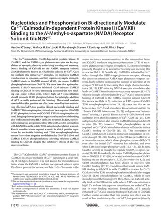
  • 7. DISCUSSION CaMKII binding to the NMDA receptor subunit GluN2B is thought to be important for CaMKII translocation to synapses in response to LTP-inducing stimuli and, in turn, for regulating synaptic plasticity (15, 26, 28; for review see Ref. 4, 5). This study showed that ATP significantly increases Ca2ϩ /CaM-induced CaMKII binding to GluN2B in vitro (despite also inducing neg- ative-regulatory phosphorylation). Nucleotide binding was also required for Ca2ϩ -induced CaMKII translocation to GluN2B co-expressed in heterologous cells, and for efficient glutamate- induced synaptic CaMKII translocation in hippocampal neu- rons. In vitro, the overall enhancement of CaMKII binding to GluN2B was based on four modulatory effects of ATP, two positive (directly by nucleotide binding and indirectly after CaMKII autophosphorylation at T286) and two negative (GluN2BphosphorylationatS1303andCaMKIIautophosphor- ylation at T305/6). Compelling evidence has been collected that indicate direct binding to GluN2B as the mechanism underlying stimulus-in- duced CaMKII translocation to synapses (for review see Refs. 4, 5). This study clarified the remaining conundrum of how CaMKII could bind to GluN2B under high ATP conditions (as found within cells), which should lead to the GluN2B S1303 phosphorylation (36) shown to prevent CaMKII binding (at least the binding induced by T286 autophosphorylation) (27). Another alternative has recently been suggested, i.e. that phos- pho-S1303 GluN2B prevents only CaMKII binding induced by T286 autophosphorylation, but not binding induced by Ca2ϩ / CaM (37). However, our results indicated that phospho-S1303 interferes with both phospho-T286- and Ca2ϩ /CaM-induced CaMKII binding, consistent with the binding model (see Fig. 9) and with cellular studies using GluN2B mutated in the S1303 region (27, 28, 37). Another negative-regulatory phosphoryla- tion for CaMKII binding to GluN2B has been suggested previ- ously (51), but was directly demonstrated for the first time in this study: the autophosphorylation at T305/6. This “inhibi- tory” autophosphorylation prevents Ca2ϩ /CaM binding to CaMKII (48, 50) and thereby also autophosphorylation at T286. Thus, T305/6 phosphorylation was predicted to interfere with CaMKII binding to GluN2B by inhibiting the stimulation required to induce the interaction. Indeed, T305/6D phospho- mimic mutation also impairs synaptic CaMKII translocation in hippocampal neurons (38, 52), while T305/6A mutation enhances synaptic localization (14, 52). Thus, inhibition of GluN2B binding in vitro by T305/6D mutation lends further support for GluN2B binding as underlying mechanism for syn- aptic CaMKII targeting. Interestingly, T305/6 phosphorylation plays a role in plasticity and flexibility of learning, as T305/6A mutant mice have a lower LTP threshold and impairments in “un-learning” of no longer beneficial tasks (52). Autophospho- rylation of T286, which generates autonomous CaMKII activ- ity, is long known to induce CaMKII binding to GluN2B even in absence of Ca2ϩ /CaM-stimulation (15, 26, 27). It has been spec- ulated that it may also further enhance Ca2ϩ /CaM-stimulated binding (for review see Ref. 4), consistent with our previous findings that T286 phosphorylation further increased persis- tent synaptic targeting after brief stimuli (17). The results of this FIGURE 8. Nucleotide binding is required for efficient glutamate-induced synaptic translocation of GFP-CaMKII in hippocampal neurons. A, GFP- CaMKII localization was monitored in live rat hippocampal neurons (DIV 13) before and during a 10 min glutamate/glycine (100 ␮M/10 ␮M) stimulus. Translocation to synaptic sites was seen for both wild type and T286A CaMKII within 4 min of stimulation, whereas K42M did not display this pattern of localization even after 10 min of stimulation. Shown are sample projection images created from deconvolved z-stacks of the same neuron at different time points. B, translocation in time lapse images was quantified (for 20 neu- rons in six independent experiments for each condition). CaMKII wild type and the T286A mutant showed the same translocation, while K42M translo- cated significantly less than T286A (and wild type) as indicated (*, p Ͻ 0.05; **, p Ͻ 0.01; in Bonferroni post-hoc analysis after two-way ANOVA). FIGURE 9. Schematic model: CaMKII binding to GluN2B (left) and aggre- gation of multiple CaMKII holoenzymes (right) are based on similar molecular interaction mechanisms. Displacement of the auto-inhibitory ␣-helix (which includes the region around T286) by Ca2ϩ /CaM binding acti- vates the kinase (by allowing substrate access to the S-site; orange) and exposes the T-site (yellow), which then can interact with other binding part- ners: either the region around S1303 in GluN2B (red) or the T286 region of a kinase subunit on another holoenzyme (brown). Both interactions are enhanced by nucleotide binding to CaMKII. However, while T286 autophos- phorylation further enhances binding to GluN2B, it inhibits aggregation of holoenzymes. Nucleotides Enable Efficient CaMKII Binding to GluN2B 31278 JOURNAL OF BIOLOGICAL CHEMISTRY VOLUME 286•NUMBER 36•SEPTEMBER 9, 2011 atUnivColorado-DenisonMemorialLibraryonJuly27,2015http://www.jbc.org/Downloadedfrom
  • 8. study show that autophosphorylation at T286 indeed directly enhanced or accelerated Ca2ϩ -stimulated binding to GluN2B, both in vitro and in heterologous cells. Thus, our results provide important new insight about three phosphorylation reactions previously shown or speculated to be involved in regulation of CaMKII binding to GluN2B. All of these findings further sup- port regulated binding of CaMKII to GluN2B as the underlying mechanism for stimulation induced translocation of CaMKII to synapses. As also found in a previous study (31), ADP enhanced CaMKII binding to GluN2B in vitro, indicating a direct effect of nucleotide binding to CaMKII without requirement for any phosphorylation reaction. Consistent with these findings, CaMKII binding to GluN2B does, vice versa, enhance nucleo- tide binding to the kinase (53). The present study shows that the positive effect of nucleotide binding on CaMKII/GluN2B inter- action is important in order to overcome the negative effect caused by GluN2B S1303 phosphorylation. In fact, efficient CaMKII binding to GluN2B was not only possible in presence of ATP, but even enhanced by it. Importantly, in heterologous cells and in neurons, nucleotide binding was required to enable stimulus-induced CaMKII interaction with GluN2B and effi- cient synaptic translocation, as both were either almost com- pletely abolished or significantly reduced by the K42M muta- tion that impairs nucleotide binding to CaMKII. But is the regulation by nucleotide within cells indeed only mediated directly by the binding, or could it involve a phosphorylation reaction? The only known positive regulatory phosphorylation is at T286, and the non-phosphorylatable CaMKII T286A mutant behaved more similarly to wild type than to K42M. In vitro, nucleotide enhanced CaMKII binding to GluN2B even more when phosphorylation was prevented at the same time (for instance by using ADP compared with ATP), due to lack of inhibitory phosphorylation of GluN2B S1303. Thus, direct reg- ulation by the binding of nucleotide is the most plausible expla- nation also for the effects observed within cells. However, some additional contribution by an unidentified positive regulatory phosphorylation by CaMKII cannot be formally ruled out. In principle, this possibility could be tested by inhibiting CaMKII activity. However, CaMKII inhibitors directly interfere with binding to GluN2B, independently from their effect on kinase activity (i.e. in vitro even in the absence of ATP): KN93 is com- petitive with activation by Ca2ϩ /CaM, and thus also removes the stimulus required to induce interaction with GluN2B; tatCN21 competes with GluN2B for the same binding region on CaMKII (41, 43). It should be noted that a previous study reported synaptic translocation of another CaMKII mutant with impaired nucleotide binding (13). However, this study used K42R (a more subtle mutation of the ATP binding pocket) and, maybe more importantly, did not quantify the degree of synaptic translocation. While synaptic translocation was signif- icantly reduced in our study by the K42M mutation, it was not completely abolished and some residual translocation was still observed. Interestingly, direct enhancement by nucleotide binding makes the regulation of CaMKII binding to GluN2B even more similar to the regulation of aggregation of multiple CaMKII holoenzymes into large clusters (the former induced in neurons by physiological glutamate stimuli, the latter by pathological excitotoxic glutamate stimuli). Indeed, both binding reactions were proposed to involve similar molecular interactions with the CaMKII T-site (Fig. 9)(15, 39, 54). In the basal state of CaMKII, the T-site interacts with the region around T286 in the regulatory ␣-helix (which also blocks access to the adjacent substrate binding S-site; see Fig. 9) (15, 55). After Ca2ϩ /CaM binds to the regulatory helix and displaces it, the T-site becomes accessible for other interactions: with the region around S1303 on GluN2B (which is homologous to the regions around T286 on CaMKII; 15), or with the T286 region on the regulatory helix of a CaMKII subunit in another holoenzyme (thus leading to formation of holoenzyme clusters) (see Fig. 9). Both GluN2B binding and holoenzyme aggregation require stimulation by Ca2ϩ /CaM and involve interaction of the CaMKII T-site with a binding partner that is sensitive to phos- phorylation by CaMKII. While aggregation is strictly depen- dent on presence of nucleotide (39, 56), GluN2B binding was ϳ10-fold enhanced by it. The underlying mechanism is unclear, and the kinase domain structure of CaMKII has been solved so far only in absence of nucleotides (55). However, nucleotide can enhance Ca2ϩ /CaM binding independent of T286 autophosphorylation (57), possibly by facilitating dis- placement of the regulatory helix, which would make the T-site more accessible. Additionally, nucleotide may induce a confor- mation in or around the T-site that is more favorable for exter- nal interaction. In contrast to GluN2B binding, holoenzyme aggregation additionally requires a pH below 6.8 (39, 56), a con- dition found after the excitotoxic insults that trigger this aggre- gation within neurons. The most plausible explanation is requirement for protonation of the His-282, which is located at the hinge that connects the regulatory helix with the core kinase domain (55, 58), and may thus enable more dramatic displace- ment of the regulatory helix. This may not be required for T-site access, but for access to the T286 region, which is necessary for binding between two kinase subunits, but not for GluN2B bind- ing. Thus, both the regulation by pH and the more stringent nucleotide requirement for CaMKII aggregation could be explained by the necessity for more dramatic displacement of the regulatory helix, to allow external interactions of its T286 region. This study clearly showed that Ca2ϩ /CaM-induced CaMKII binding to GluN2B is possible in presence of ATP, even at phys- iological mM concentrations. In fact, ATP significantly enhanced this binding, as a positive net result caused by four modulatory effects of ATP, two positive (nucleotide binding; T286 autophosphorylation) and two negative (GluN2B S1303 phosphorylation; T305/6 autophosphorylation). But why was the negative effect of GluN2B S1303 phosphorylation not dom- inant? Indeed, GluN2B S1303 pre-phosphorylation or phos- pho-mimetic S1303D mutation dramatically inhibited binding (27) (and this study). Thus, the explanation must be based on the kinetics of the reactions involved. A pre-requisite for any phosphorylation reaction is ATP binding to the kinase, which was shown to already directly enhance binding to GluN2B. The fastest known phosphorylation reaction of CaMKII is auto- phosphorylation at T286 (with a rate of ϳ20 sϪ1 ) (59), again promoting binding to GluN2B. This leaves CaMKII bound to Nucleotides Enable Efficient CaMKII Binding to GluN2B SEPTEMBER 9, 2011•VOLUME 286•NUMBER 36 JOURNAL OF BIOLOGICAL CHEMISTRY 31279 atUnivColorado-DenisonMemorialLibraryonJuly27,2015http://www.jbc.org/Downloadedfrom
  • 9. ADP, which also directly stimulates binding to GluN2B but without allowing any further phosphorylation until the ADP is exchanged for ATP, with the relatively slow dissociation of ADP (ϳ20 sϪ1 compared with ϳ500 sϪ1 for the actual phos- pho-transfer) generally considered as a rate-limiting step for kinase reactions (44, 60, 61). Only then can CaMKII mediate the inhibitory phosphorylation of GluN2B at S1303. At this point, significant CaMKII binding may have already occurred. Once bound, CaMKII can still phosphorylate GluN2B, but without causing any significant dissociation (17, 27), and it is unclear if this phosphorylation can still happen at S1303 and/or on the same GluN2B subunit. The other inhibitory phosphory- lation, at CaMKII T305/6, is inhibited during CaMKII stimula- tion by Ca2ϩ /CaM binding to an overlapping site on the regulatory helix (48, 50). Thus, fast kinetics of T305/6 auto- phosphorylation are achieved only by T286 phosphorylated “autonomous” CaMKII and only after dissociation of Ca2ϩ / CaM. These kinetic considerations are also consistent with the observation that aggregation of CaMKII holoenzymes, while dependent on nucleotide binding, is actually inhibited by high physiological concentrations of ATP (39, 56)(which can be sig- nificantly reduced under pathological ischemic conditions). In this case, the fast T286 phosphorylation may still enhance T-site access but would at the same time prevent its binding to the now phosphorylated T286 region of another kinase subunit. GluN2B is not the only binding partner for CaMKII at the synapse (for review see 4, 5). While additional interactions may contribute, they do not appear sufficient to mediate the stimu- lus induced but persistent synaptic translocation of CaMKII. Similar to T286 autophosphorylation, CaMKII binding to GluN2B also directly generates Ca2ϩ -independent “autono- mous” activity (15), which can provide a “molecular memory” of past Ca2ϩ stimuli (for review see Ref. 62). Recent results have shown that autonomy generated by T286 phosphorylation is required for inducing but not for maintaining LTP at excitatory synapses, and for learning rather than memory (18, 19). In fact, T286 phosphorylation induced by LTP stimuli may only be very short lived (63). However, a recent study indicated that storage of synaptic memory previously proposed to be mediated by T286 phosphorylation can instead be mediated by the NMDA receptor-bound form of CaMKII (30), and it will be interesting to examine the functional contribution to memory in behav- ioral studies. Acknowledgments—We thank Jacqueline R. Kulbe for technical assistance. We thank Dr. Rebekah Vest for help with the schematic illustration. REFERENCES 1. Lisman, J., Schulman, H., and Cline, H. (2002) Nat. Rev. Neurosci. 3, 175–190 2. Hudmon, A., and Schulman, H. (2002) Annu. Rev. Biochem. 71, 473–510 3. Sheng, M., and Kim, M. J. (2002) Science 298, 776–780 4. Colbran, R. J. (2004) Biochem. J. 378, 1–16 5. Merrill, M. A., Chen, Y., Strack, S., and Hell, J. W. (2005) Trends Pharma- col. Sci. 26, 645–653 6. Malinow, R., Schulman, H., and Tsien, R. W. (1989) Science 245, 862–866 7. Silva, A. J., Stevens, C. F., Tonegawa, S., and Wang, Y. (1992) Science 257, 201–206 8. Lee, Y. S., and Silva, A. J. (2009) Nat. Rev. Neurosci. 10, 126–140 9. Hayashi, Y., Shi, S. H., Esteban, J. A., Piccini, A., Poncer, J. C., and Malinow, R. (2000) Science 287, 2262–2267 10. Opazo, P., Labrecque, S., Tigaret, C. M., Frouin, A., Wiseman, P. W., De Koninck, P., and Choquet, D. (2010) Neuron 67, 239–252 11. Benke, T. A., Lu¨thi, A., Isaac, J. T., and Collingridge, G. L. (1998) Nature 393, 793–797 12. Derkach, V., Barria, A., and Soderling, T. R. (1999) Proc. Natl. Acad. Sci. U.S.A. 96, 3269–3274 13. Shen, K., and Meyer, T. (1999) Science 284, 162–166 14. Shen, K., Teruel, M. N., Connor, J. H., Shenolikar, S., and Meyer, T. (2000) Nat. Neurosci. 3, 881–886 15. Bayer, K. U., De Koninck, P., Leonard, A. S., Hell, J. W., and Schulman, H. (2001) Nature 411, 801–805 16. Otmakhov, N., Tao-Cheng, J. H., Carpenter, S., Asrican, B., Dosemeci, A., Reese, T. S., and Lisman, J. (2004) J. Neurosci. 24, 9324–9331 17. Bayer, K. U., LeBel, E., McDonald, G. L., O’Leary, H., Schulman, H., and De Koninck, P. (2006) J. Neurosci. 26, 1164–1174 18. Giese, K. P., Fedorov, N. B., Filipkowski, R. K., and Silva, A. J. (1998) Science 279, 870–873 19. Buard, I., Coultrap, S. J., Freund, R. K., Lee, Y. S., Dell’Acqua, M. L., Silva, A. J., and Bayer, K. U. (2010) J. Neurosci. 30, 8214–8220 20. Hanson, P. I., Meyer, T., Stryer, L., and Schulman, H. (1994) Neuron 12, 943–956 21. Rich, R. C., and Schulman, H. (1998) J. Biol. Chem. 273, 28424–28429 22. Miller, S. G., and Kennedy, M. B. (1986) Cell 44, 861–870 23. Lou, L. L., Lloyd, S. J., and Schulman, H. (1986) Proc. Natl. Acad. Sci. U.S.A. 83, 9497–9501 24. Schworer, C. M., Colbran, R. J., and Soderling, T. R. (1986) J. Biol. Chem. 261, 8581–8584 25. Coultrap, S. J., Buard, I., Kulbe, J. R., Dell’Acqua, M. L., and Bayer, K. U. (2010) J. Biol. Chem. 285, 17930–17937 26. Strack, S., and Colbran, R. J. (1998) J. Biol. Chem. 273, 20689–20692 27. Strack, S., McNeill, R. B., and Colbran, R. J. (2000) J. Biol. Chem. 275, 23798–23806 28. Barria, A., and Malinow, R. (2005) Neuron 48, 289–301 29. Zhou, Y., Takahashi, E., Li, W., Halt, A., Wiltgen, B., Ehninger, D., Li, G. D., Hell, J. W., Kennedy, M. B., and Silva, A. J. (2007) J. Neurosci. 27, 13843–13853 30. Sanhueza, M., Fernandez-Villalobos, G., Stein, I. S., Kasumova, G., Zhang, P., Bayer, K. U., Otmakhov, N., Hell, J. W., and Lisman, J. (2011) J. Neurosci. 31, 9170–9178 31. Robison, A. J., Bartlett, R. K., Bass, M. A., and Colbran, R. J. (2005) J. Biol. Chem. 280, 39316–39323 32. Pradeep, K. K., Cheriyan, J., Suma Priya, S. D., Rajeevkumar, R., Mayadevi, M., Praseeda, M., and Omkumar, R. V. (2009) Biochem. J. 419, 123–132 33. Kitamura, Y., Miyazaki, A., Yamanaka, Y., and Nomura, Y. (1993) J. Neu- rochem. 61, 100–109 34. Kolaj, M., Cerne, R., Cheng, G., Brickey, D. A., and Randic´, M. (1994) J. Neurophysiol. 72, 2525–2531 35. Sessoms-Sikes, S., Honse, Y., Lovinger, D. M., and Colbran, R. J. (2005) Mol. Cell Neurosci. 29, 139–147 36. Omkumar, R. V., Kiely, M. J., Rosenstein, A. J., Min, K. T., and Kennedy, M. B. (1996) J. Biol. Chem. 271, 31670–31678 37. Raveendran, R., Devi Suma Priya, S., Mayadevi, M., Steephan, M., San- thoshkumar, T. R., Cheriyan, J., Sanalkumar, R., Pradeep, K. K., James, J., and Omkumar, R. V. (2009) J. Neurochem. 110, 92–105 38. Hudmon, A., Lebel, E., Roy, H., Sik, A., Schulman, H., Waxham, M. N., and De Koninck, P. (2005) J. Neurosci. 25, 6971–6983 39. Vest, R. S., O’Leary, H., and Bayer, K. U. (2009) FEBS Lett. 583, 3577–3581 40. Zacharias, D. A., Violin, J. D., Newton, A. C., and Tsien, R. Y. (2002) Science 296, 913–916 41. Vest, R. S., Davies, K. D., O’Leary, H., Port, J. D., and Bayer, K. U. (2007) Mol. Biol. Cell 18, 5024–5033 42. O’Leary, H., Lasda, E., and Bayer, K. U. (2006) Mol. Biol. Cell 17, 4656–4665 43. Vest, R. S., O’Leary, H., Coultrap, S. J., Kindy, M. S., and Bayer, K. U. (2010) J. Biol. Chem. 285, 20675–20682 Nucleotides Enable Efficient CaMKII Binding to GluN2B 31280 JOURNAL OF BIOLOGICAL CHEMISTRY VOLUME 286•NUMBER 36•SEPTEMBER 9, 2011 atUnivColorado-DenisonMemorialLibraryonJuly27,2015http://www.jbc.org/Downloadedfrom
  • 10. 44. Cook, P. F., Neville, M. E., Jr., Vrana, K. E., Hartl, F. T., and Roskoski, R., Jr. (1982) Biochemistry 21, 5794–5799 45. Bhatnagar, D., Roskoski, R. Jr., Rosendahl, M. S., and Leonard, N. J. (1983) Biochemistry 22, 6310–6317 46. Aubol, B. E., Nolen, B., Shaffer, J., Ghosh, G., and Adams, J. A. (2003) Biochemistry 42, 12813–12820 47. Meyer, T., Hanson, P. I., Stryer, L., and Schulman, H. (1992) Science 256, 1199–1202 48. Colbran, R. J., and Soderling, T. R. (1990) J. Biol. Chem. 265, 11213–11219 49. Lu, C. S., Hodge, J. J., Mehren, J., Sun, X. X., and Griffith, L. C. (2003) Neuron 40, 1185–1197 50. Hanson, P. I., and Schulman, H. (1992) J. Biol. Chem. 267, 17216–17224 51. Leonard, A. S., Bayer, K. U., Merrill, M. A., Lim, I. A., Shea, M. A., Schul- man, H., and Hell, J. W. (2002) J. Biol. Chem. 277, 48441–48448 52. Elgersma, Y., Fedorov, N. B., Ikonen, S., Choi, E. S., Elgersma, M., Car- valho, O. M., Giese, K. P., and Silva, A. J. (2002) Neuron 36, 493–505 53. Cheriyan, J., Kumar, P., Mayadevi, M., Surolia, A., and Omkumar, R. V. (2011) PLoS ONE 6, e16495 54. Hudmon, A., Kim, S. A., Kolb, S. J., Stoops, J. K., and Waxham, M. N. (2001) J. Neurochem. 76, 1364–1375 55. Rosenberg, O. S., Deindl, S., Sung, R. J., Nairn, A. C., and Kuriyan, J. (2005) Cell 123, 849–860 56. Hudmon, A., Aronowski, J., Kolb, S. J., and Waxham, M. N. (1996) J. Biol. Chem. 271, 8800–8808 57. Tzortzopoulos, A., and To¨ro¨k, K. (2004) Biochemistry 43, 6404–6414 58. Smith, M. K., Colbran, R. J., Brickey, D. A., and Soderling, T. R. (1992) J. Biol. Chem. 267, 1761–1768 59. Bradshaw, J. M., Hudmon, A., and Schulman, H. (2002) J. Biol. Chem. 277, 20991–20998 60. Zhou, J., and Adams, J. A. (1997) Biochemistry 36, 15733–15738 61. Lew, J., Taylor, S. S., and Adams, J. A. (1997) Biochemistry 36, 6717–6724 62. Lisman, J. E., and McIntyre, C. C. (2001) Curr. Biol. 11, R788–791 63. Lee, S. J., Escobedo-Lozoya, Y., Szatmari, E. M., and Yasuda, R. (2009) Nature 458, 299–304 Nucleotides Enable Efficient CaMKII Binding to GluN2B SEPTEMBER 9, 2011•VOLUME 286•NUMBER 36 JOURNAL OF BIOLOGICAL CHEMISTRY 31281 atUnivColorado-DenisonMemorialLibraryonJuly27,2015http://www.jbc.org/Downloadedfrom
  • 11. Bayer Rorabaugh, Steven J. Coultrap and K. Ulrich Heather O'Leary, Wallace H. Liu, Jacki M. Subunit GluN2B -Methyl-d-aspartate (NMDA) Receptor N(CaMKII) Binding to the /Calmodulin-dependent Protein Kinase II 2+Bi-directionally Modulate Ca Nucleotides and Phosphorylation Neurobiology: doi: 10.1074/jbc.M111.233668 originally published online July 18, 2011 2011, 286:31272-31281.J. Biol. Chem. 10.1074/jbc.M111.233668Access the most updated version of this article at doi: .JBC Affinity SitesFind articles, minireviews, Reflections and Classics on similar topics on the Alerts: When a correction for this article is posted• When this article is cited• to choose from all of JBC's e-mail alertsClick here Supplemental material: http://www.jbc.org/content/suppl/2011/07/18/M111.233668.DC1.html http://www.jbc.org/content/286/36/31272.full.html#ref-list-1 This article cites 63 references, 34 of which can be accessed free at atUnivColorado-DenisonMemorialLibraryonJuly27,2015http://www.jbc.org/Downloadedfrom