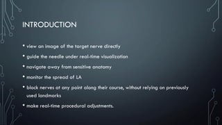
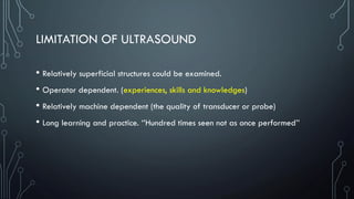
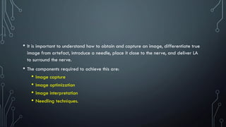
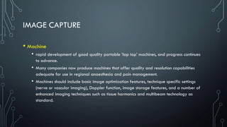
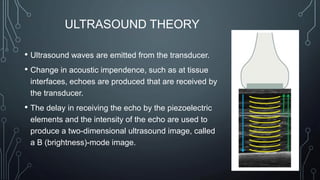
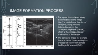
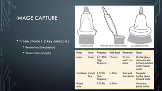
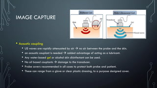
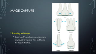
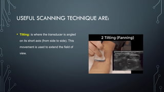
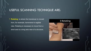
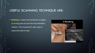
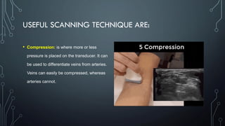
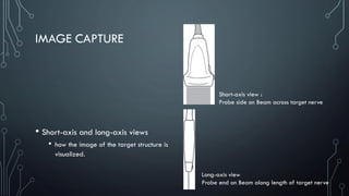
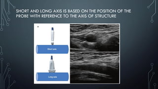
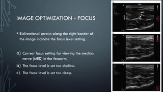
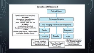
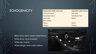
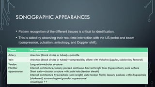
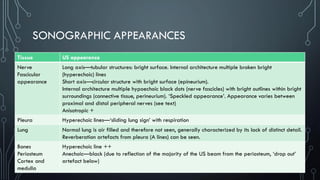
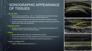
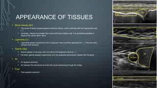
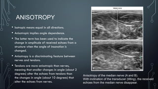
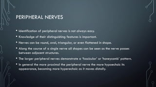
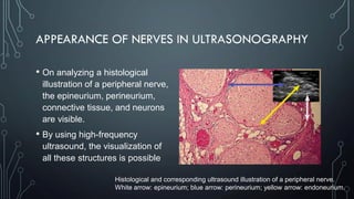
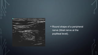
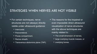
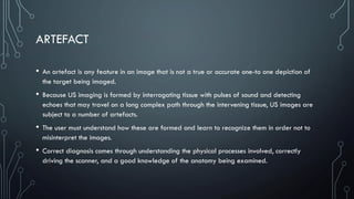
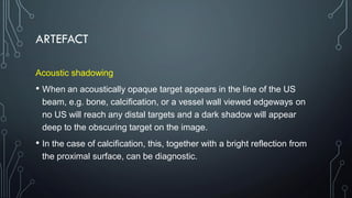
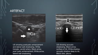
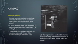
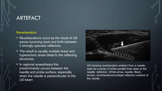
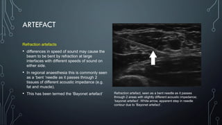
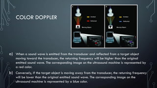
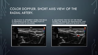
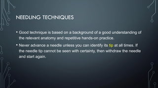
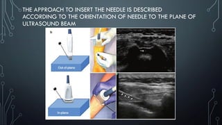
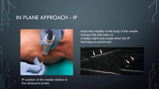
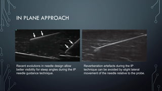
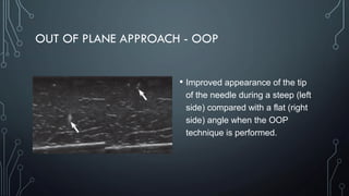
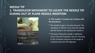
Ultrasonography is useful for regional anesthesia procedures. It allows visualization of key anatomical structures like nerves, blood vessels, muscles and bones. There are different ultrasound imaging modes used including B-mode, M-mode and Doppler. Factors like frequency, depth and gain affect image resolution. The needle can be inserted in-plane or out-of-plane relative to the ultrasound beam. Proper scanning technique and following guidelines help ensure safe and effective ultrasound-guided regional anesthesia.
Introduction to ultrasound & regional anesthesiaSaad Al-Shamma
Â
Ultrasound is used to guide regional anesthesia by visualizing anatomical structures and needle placement. The document discusses ultrasound image generation, modes, tissue appearance, artifacts, and techniques for brachial plexus blocks. Safety guidelines are outlined, including recommendations to inject local anesthetic under direct ultrasound visualization and consider nerve stimulation to confirm needle position. Complications like nerve injury, local anesthetic toxicity, and pneumothorax are addressed.
This document provides an overview of ultrasonography principles:
- Ultrasonography uses high-frequency sound waves to generate images and is a useful, noninvasive diagnostic tool.
- Sound waves have properties like frequency, wavelength, and velocity that affect image quality. Higher frequencies produce better surface details but poorer penetration.
- Images are produced when sound waves emitted from a transducer's piezoelectric crystals enter the body, encounter tissues, and return echoes that are converted into a visual display.
- Different transducer types and ultrasound modes like B-mode produce various image types used for diagnostic purposes. Artifacts like shadows and reverberations can occur and should be recognized to avoid diagnostic errors.
The document discusses the basic components and functioning of an ultrasound machine. It describes the transmitter/pulser, transducer, receiver and processor, display, and recording components. The transducer is made of piezoelectric crystals and converts electrical energy to ultrasound energy and vice versa. Different controls like gain, zoom, and Doppler are used by the radiographer to optimize the ultrasound image.
This document discusses various topics related to ultrasound imaging including goals, early pioneers, transducer types, Doppler instrumentation and physics, harmonic imaging, spatial compounding, extended field of view, fusion imaging, 3D and 4D ultrasound, and contrast enhanced ultrasound. It provides details on transducer selection, control settings, tissue harmonic imaging principles, spatial compounding benefits, fusion imaging steps, and contrast agent interactions.
Ultrasound technology works by using piezoelectric crystals that produce sound waves when electric current is applied. These waves travel through the body and reflect off tissues, returning echoes that are converted back to electric signals to form an image. Higher frequency waves have shorter wavelengths and provide better resolution, while lower frequencies penetrate deeper. Ultrasound modes like B-mode produce 2D images from multiple scan lines, while Doppler mode detects motion and flow. Tissue properties like density and elasticity affect how waves propagate and are displayed on ultrasound images.
This document discusses various types of image artifacts that can occur in CT scans, their causes, and methods for reducing or correcting artifacts. It describes streaking, shading, ring, and band artifacts and attributes them to issues like inconsistent measurements, detector errors, and metal objects. Artifacts can be caused by the scanner design, x-ray tube, detectors, patients, or operators. Methods to address artifacts involve adjusting scanning parameters, using specialized techniques, software corrections, improving patient positioning, and operator skill. Artifacts degrade image quality and must be minimized for accurate diagnosis.
Lecture 3 & 4 anam sanam chick ldkfdlsfldfjdlsjfdlks .pptxfaiz3334
Â
Computed tomography (CT) scans create cross-sectional images of the body by using X-rays and computer processing. An X-ray tube rotates around the body and produces multiple images from different angles, which are used to reconstruct cross-sectional slices using back projection. These slices can be combined to create 3D images. CT scans provide more detailed images than basic X-rays due to their ability to distinguish between different tissue densities and visualize structures throughout the body.
The document provides an introduction to CT scans, describing their components and how they work. It explains that a CT scan uses X-rays that rotate around the body, with some radiation absorbed and some passed through to detectors. Detectors convert the data into images with varying shades of white, black and grey. Key components are the gantry, X-ray tube, detectors, and operating console. The document also describes how bones appear white due to absorbing more radiation, while soft tissues vary in shades of grey depending on density.
BScan and Ascan in ophthalmology and eye fieldAsif469093
Â
This document provides an overview of B-scan ultrasound. It defines a B-scan as a brightness intensity-modulated display that is two-dimensional, with echoes displayed as dots where brightness indicates echo strength. It then discusses basic physics concepts for ultrasound such as velocity, reflectivity, absorption and artifacts. It also covers instrumentation, examination techniques including patient positioning and probe use, and types of scans based on probe position.
Ultrasound artifacts are structures in an image that do not represent the actual tissue being scanned. Common artifacts include reverberation caused by repeated reflections, acoustic shadowing behind strongly attenuating tissue, acoustic enhancement behind fluid-filled structures due to time-gain compensation, and edge shadowing at the edges of rounded structures due to refraction and reflection of the beam. Careful use and adjustment of ultrasound machine controls and transducers can help reduce artifacts.
Optical coherence tomography (OCT) provides high-resolution cross-sectional images of the retina using infrared light. It has advanced from time domain OCT to spectral domain OCT, improving resolution and scan speed. OCT is used to qualitatively and quantitatively analyze retinal thickness, layers, and structures. It is useful for diagnosing and monitoring many retinal conditions like macular holes, edema, age-related macular degeneration, and more. Artifacts can occur but OCT provides crucial information with advantages of being non-invasive and having micron-level resolution.
Fundamentals of musculoskeletal ultrasoundSamar Tharwat
Â
Ultrasonography is a useful tool for musculoskeletal imaging that is safe, low-cost, and provides real-time dynamic studies without radiation. Higher ultrasound frequencies provide better resolution but poorer penetration depth. The transducer probe and machine settings like gain, depth, and focal zone must be optimized to provide clear images. Various artifacts can occur from issues like anisotropy, shadowing, or reverberation that require understanding to avoid pitfalls in interpretation. Proper technique and knowledge of ultrasound physics and machine settings are needed to obtain high quality images for accurate musculoskeletal assessment.
Ultrasound uses high frequency sound waves to visualize internal structures. It works by transmitting sound waves into the body using a transducer probe, which detects the echoes as they bounce off tissues and organs. The echoes are processed to form images on the ultrasound machine screen in real-time. Common applications include obstetrics, cardiology, and urology. The Philips HD11 is an ultrasound system with curvilinear, linear, and phased array probes for different exams. It provides grey scale, Doppler, and color imaging modes. Ultrasound has benefits of being non-invasive, portable, and having no radiation, but has limitations of being operator dependent and unable to penetrate bone.
Ultrasonography uses high frequency sound waves to non-invasively image soft tissues. It is used for both diagnostic purposes like organ imaging and measurement as well as therapeutic purposes like HIFU and lithotripsy. Ultrasound has a range above human hearing and most diagnostic instruments use 1-10 MHz. It provides greyscale images based on tissue density and is useful for visualizing soft tissue structures and blood flow.
Ultrasound artifacts and contrast enhanced ultrasoundArjun Reddy
Â
This document discusses various ultrasound artifacts and the use of contrast-enhanced ultrasound (CEUS). It describes several types of artifacts caused by beam characteristics, multiple echoes, velocity errors, and attenuation errors. It also discusses artifacts associated with reverberation, comet tails, ring down, and mirror images. CEUS involves injecting microbubbles as contrast agents, which enhance lesion detection and characterization. CEUS can help diagnose conditions like hemangiomas, HCC, metastases and has various clinical applications for organs like the liver, kidneys and vasculature. It provides a safe and effective method to evaluate blood flow and tissue perfusion.
This document provides an overview of techniques for optimizing echo images. It discusses adjusting various ultrasound machine parameters like transducer type, echo modes, sector size, depth, frequency, dynamic range, gain, and focus to obtain high quality images. The objectives are to learn the manufacturer's techniques for achieving the best possible contrast in echo images by manipulating these different parameters.
This document provides an overview of B-scan ultrasonography. It begins with an introduction to B-scans and their use in providing qualitative and quantitative assessment of the eye and orbit. It then discusses the physics and principles behind ultrasound, including reflection, absorption, resolution and other key concepts. The document outlines the components and use of B-scan ultrasound machines, including different probe orientations and scanning techniques. It concludes with clinical applications and indications for B-scan ultrasonography in evaluating ocular pathology.
Ultra sonography indications in maxillofacial region /prosthodontic coursesIndian dental academy
Â
The Indian Dental Academy is the Leader in continuing dental education , training dentists in all aspects of dentistry and
offering a wide range of dental certified courses in different formats.
This document provides an overview of ultrasound uses in regional anesthesia. It discusses how ultrasound works by generating sound waves and visualizing tissue structures based on echo reflection. Key advantages of ultrasound include visualization of soft tissues and avoidance of radiation exposure compared to fluoroscopy. The document covers ultrasound machine controls, tissue appearance, probe positioning and handling, scanning techniques, and needle guidance using ultrasound.
Point of Care Ultrasound - Hyperechoic Future in Family Practice?cbyrne2014
Â
The document discusses the potential for point-of-care ultrasound (POCUS) in family practice. It begins with an overview of ultrasound fundamentals and image interpretation. It then argues that POCUS could benefit family practice by enhancing the physical exam, aiding diagnoses, and improving patient care. Examples are given of how POCUS can accurately identify or rule out pneumothorax, pleural effusion, and pericardial effusion. The document concludes by encouraging physicians to gain experience with POCUS and consider its applications.
The document discusses key concepts in ultrasound physics including:
1. Higher ultrasound frequency results in shorter wavelength and better resolution, while lower frequency has longer wavelength. The speed that ultrasound travels is determined by the medium and remains constant regardless of frequency.
2. Temporal resolution is improved with low line density images, as they use fewer pulses per frame, while spatial resolution is better with high line density images that use more pulses.
3. An ultrasound transducer transmits sound waves into the body and receives echoes to create images, with frequency inversely related to depth of penetration but directly related to resolution.
Learn from our Slideshare about the differences between ultrasound transducers. We also cover tips on how to treat your probes and how to select the right one.
ULTRASONOGRAPHY (USG) AND ULTRASOUND BIOMICROSCOPY(UBM)Dr. Gaurav Shukla
Â
Ultrasonography and ultrasound biomicroscopy are important tools for diagnosing ocular and orbital abnormalities. Ultrasonography uses high frequency sound waves transmitted into the eye via a probe to image intraocular structures. A-scans display returning echoes in one dimension while B-scans create a two-dimensional image by accumulating A-scan echoes. B-scans are useful for evaluating lesions' topography, reflectivity, internal structure, and mobility. Common applications include detecting retinal detachments, vitreous opacities, intraocular tumors, and foreign bodies. Ultrasonography is valuable for screening and characterizing many ocular pathologies.
Giloy in Ayurveda - Classical Categorization and SynonymsPlanet Ayurveda
Â
Giloy, also known as Guduchi or Amrita in classical Ayurvedic texts, is a revered herb renowned for its myriad health benefits. It is categorized as a Rasayana, meaning it has rejuvenating properties that enhance vitality and longevity. Giloy is celebrated for its ability to boost the immune system, detoxify the body, and promote overall wellness. Its anti-inflammatory, antipyretic, and antioxidant properties make it a staple in managing conditions like fever, diabetes, and stress. The versatility and efficacy of Giloy in supporting health naturally highlight its importance in Ayurveda. At Planet Ayurveda, we provide a comprehensive range of health services and 100% herbal supplements that harness the power of natural ingredients like Giloy. Our products are globally available and affordable, ensuring that everyone can benefit from the ancient wisdom of Ayurveda. If you or your loved ones are dealing with health issues, contact Planet Ayurveda at 01725214040 to book an online video consultation with our professional doctors. Let us help you achieve optimal health and wellness naturally.
More Related Content
Similar to 2. Basic US Guiding - Alamsyah Ambo Ala H.pdf
Lecture 3 & 4 anam sanam chick ldkfdlsfldfjdlsjfdlks .pptxfaiz3334
Â
Computed tomography (CT) scans create cross-sectional images of the body by using X-rays and computer processing. An X-ray tube rotates around the body and produces multiple images from different angles, which are used to reconstruct cross-sectional slices using back projection. These slices can be combined to create 3D images. CT scans provide more detailed images than basic X-rays due to their ability to distinguish between different tissue densities and visualize structures throughout the body.
The document provides an introduction to CT scans, describing their components and how they work. It explains that a CT scan uses X-rays that rotate around the body, with some radiation absorbed and some passed through to detectors. Detectors convert the data into images with varying shades of white, black and grey. Key components are the gantry, X-ray tube, detectors, and operating console. The document also describes how bones appear white due to absorbing more radiation, while soft tissues vary in shades of grey depending on density.
BScan and Ascan in ophthalmology and eye fieldAsif469093
Â
This document provides an overview of B-scan ultrasound. It defines a B-scan as a brightness intensity-modulated display that is two-dimensional, with echoes displayed as dots where brightness indicates echo strength. It then discusses basic physics concepts for ultrasound such as velocity, reflectivity, absorption and artifacts. It also covers instrumentation, examination techniques including patient positioning and probe use, and types of scans based on probe position.
Ultrasound artifacts are structures in an image that do not represent the actual tissue being scanned. Common artifacts include reverberation caused by repeated reflections, acoustic shadowing behind strongly attenuating tissue, acoustic enhancement behind fluid-filled structures due to time-gain compensation, and edge shadowing at the edges of rounded structures due to refraction and reflection of the beam. Careful use and adjustment of ultrasound machine controls and transducers can help reduce artifacts.
Optical coherence tomography (OCT) provides high-resolution cross-sectional images of the retina using infrared light. It has advanced from time domain OCT to spectral domain OCT, improving resolution and scan speed. OCT is used to qualitatively and quantitatively analyze retinal thickness, layers, and structures. It is useful for diagnosing and monitoring many retinal conditions like macular holes, edema, age-related macular degeneration, and more. Artifacts can occur but OCT provides crucial information with advantages of being non-invasive and having micron-level resolution.
Fundamentals of musculoskeletal ultrasoundSamar Tharwat
Â
Ultrasonography is a useful tool for musculoskeletal imaging that is safe, low-cost, and provides real-time dynamic studies without radiation. Higher ultrasound frequencies provide better resolution but poorer penetration depth. The transducer probe and machine settings like gain, depth, and focal zone must be optimized to provide clear images. Various artifacts can occur from issues like anisotropy, shadowing, or reverberation that require understanding to avoid pitfalls in interpretation. Proper technique and knowledge of ultrasound physics and machine settings are needed to obtain high quality images for accurate musculoskeletal assessment.
Ultrasound uses high frequency sound waves to visualize internal structures. It works by transmitting sound waves into the body using a transducer probe, which detects the echoes as they bounce off tissues and organs. The echoes are processed to form images on the ultrasound machine screen in real-time. Common applications include obstetrics, cardiology, and urology. The Philips HD11 is an ultrasound system with curvilinear, linear, and phased array probes for different exams. It provides grey scale, Doppler, and color imaging modes. Ultrasound has benefits of being non-invasive, portable, and having no radiation, but has limitations of being operator dependent and unable to penetrate bone.
Ultrasonography uses high frequency sound waves to non-invasively image soft tissues. It is used for both diagnostic purposes like organ imaging and measurement as well as therapeutic purposes like HIFU and lithotripsy. Ultrasound has a range above human hearing and most diagnostic instruments use 1-10 MHz. It provides greyscale images based on tissue density and is useful for visualizing soft tissue structures and blood flow.
Ultrasound artifacts and contrast enhanced ultrasoundArjun Reddy
Â
This document discusses various ultrasound artifacts and the use of contrast-enhanced ultrasound (CEUS). It describes several types of artifacts caused by beam characteristics, multiple echoes, velocity errors, and attenuation errors. It also discusses artifacts associated with reverberation, comet tails, ring down, and mirror images. CEUS involves injecting microbubbles as contrast agents, which enhance lesion detection and characterization. CEUS can help diagnose conditions like hemangiomas, HCC, metastases and has various clinical applications for organs like the liver, kidneys and vasculature. It provides a safe and effective method to evaluate blood flow and tissue perfusion.
This document provides an overview of techniques for optimizing echo images. It discusses adjusting various ultrasound machine parameters like transducer type, echo modes, sector size, depth, frequency, dynamic range, gain, and focus to obtain high quality images. The objectives are to learn the manufacturer's techniques for achieving the best possible contrast in echo images by manipulating these different parameters.
This document provides an overview of B-scan ultrasonography. It begins with an introduction to B-scans and their use in providing qualitative and quantitative assessment of the eye and orbit. It then discusses the physics and principles behind ultrasound, including reflection, absorption, resolution and other key concepts. The document outlines the components and use of B-scan ultrasound machines, including different probe orientations and scanning techniques. It concludes with clinical applications and indications for B-scan ultrasonography in evaluating ocular pathology.
Ultra sonography indications in maxillofacial region /prosthodontic coursesIndian dental academy
Â
The Indian Dental Academy is the Leader in continuing dental education , training dentists in all aspects of dentistry and
offering a wide range of dental certified courses in different formats.
This document provides an overview of ultrasound uses in regional anesthesia. It discusses how ultrasound works by generating sound waves and visualizing tissue structures based on echo reflection. Key advantages of ultrasound include visualization of soft tissues and avoidance of radiation exposure compared to fluoroscopy. The document covers ultrasound machine controls, tissue appearance, probe positioning and handling, scanning techniques, and needle guidance using ultrasound.
Point of Care Ultrasound - Hyperechoic Future in Family Practice?cbyrne2014
Â
The document discusses the potential for point-of-care ultrasound (POCUS) in family practice. It begins with an overview of ultrasound fundamentals and image interpretation. It then argues that POCUS could benefit family practice by enhancing the physical exam, aiding diagnoses, and improving patient care. Examples are given of how POCUS can accurately identify or rule out pneumothorax, pleural effusion, and pericardial effusion. The document concludes by encouraging physicians to gain experience with POCUS and consider its applications.
The document discusses key concepts in ultrasound physics including:
1. Higher ultrasound frequency results in shorter wavelength and better resolution, while lower frequency has longer wavelength. The speed that ultrasound travels is determined by the medium and remains constant regardless of frequency.
2. Temporal resolution is improved with low line density images, as they use fewer pulses per frame, while spatial resolution is better with high line density images that use more pulses.
3. An ultrasound transducer transmits sound waves into the body and receives echoes to create images, with frequency inversely related to depth of penetration but directly related to resolution.
Learn from our Slideshare about the differences between ultrasound transducers. We also cover tips on how to treat your probes and how to select the right one.
ULTRASONOGRAPHY (USG) AND ULTRASOUND BIOMICROSCOPY(UBM)Dr. Gaurav Shukla
Â
Ultrasonography and ultrasound biomicroscopy are important tools for diagnosing ocular and orbital abnormalities. Ultrasonography uses high frequency sound waves transmitted into the eye via a probe to image intraocular structures. A-scans display returning echoes in one dimension while B-scans create a two-dimensional image by accumulating A-scan echoes. B-scans are useful for evaluating lesions' topography, reflectivity, internal structure, and mobility. Common applications include detecting retinal detachments, vitreous opacities, intraocular tumors, and foreign bodies. Ultrasonography is valuable for screening and characterizing many ocular pathologies.
Similar to 2. Basic US Guiding - Alamsyah Ambo Ala H.pdf (20)
Giloy in Ayurveda - Classical Categorization and SynonymsPlanet Ayurveda
Â
Giloy, also known as Guduchi or Amrita in classical Ayurvedic texts, is a revered herb renowned for its myriad health benefits. It is categorized as a Rasayana, meaning it has rejuvenating properties that enhance vitality and longevity. Giloy is celebrated for its ability to boost the immune system, detoxify the body, and promote overall wellness. Its anti-inflammatory, antipyretic, and antioxidant properties make it a staple in managing conditions like fever, diabetes, and stress. The versatility and efficacy of Giloy in supporting health naturally highlight its importance in Ayurveda. At Planet Ayurveda, we provide a comprehensive range of health services and 100% herbal supplements that harness the power of natural ingredients like Giloy. Our products are globally available and affordable, ensuring that everyone can benefit from the ancient wisdom of Ayurveda. If you or your loved ones are dealing with health issues, contact Planet Ayurveda at 01725214040 to book an online video consultation with our professional doctors. Let us help you achieve optimal health and wellness naturally.
Dr. Tan's Balance Method.pdf (From Academy of Oriental Medicine at Austin)GeorgeKieling1
Â
Home
Organization
Academy of Oriental Medicine at Austin
About AOMA: The Academy of Oriental Medicine at Austin offers a masters-level graduate program in acupuncture and Oriental medicine, preparing its students for careers as skilled, professional practitioners. AOMA is known for its internationally recognized faculty, award-winning student clinical internship program, and herbal medicine program. Since its founding in 1993, AOMA has grown rapidly in size and reputation, drawing students from around the nation and faculty from around the world. AOMA also conducts more than 20,000 patient visits annually in its student and professional clinics. AOMA collaborates with Western healthcare institutions including the Seton Family of Hospitals, and gives back to the community through partnerships with nonprofit organizations and by providing free and reduced price treatments to people who cannot afford them. The Academy of Oriental Medicine at Austin is located at 2700 West Anderson Lane. AOMA also serves patients and retail customers at its south Austin location, 4701 West Gate Blvd. For more information see www.aoma.edu or call 512-492-303434.
Storyboard on Skin- Innovative Learning (M-pharm) 2nd sem. (Cosmetics)MuskanShingari
Â
Skin is the largest organ of the human body, serving crucial functions that include protection, sensation, regulation, and synthesis. Structurally, it consists of three main layers: the epidermis, dermis, and hypodermis (subcutaneous layer).
1. **Epidermis**: The outermost layer primarily composed of epithelial cells called keratinocytes. It provides a protective barrier against environmental factors, pathogens, and UV radiation.
2. **Dermis**: Located beneath the epidermis, the dermis contains connective tissue, blood vessels, hair follicles, and sweat glands. It plays a vital role in supporting and nourishing the epidermis, regulating body temperature, and housing sensory receptors for touch, pressure, temperature, and pain.
3. **Hypodermis**: Also known as the subcutaneous layer, it consists of fat and connective tissue that anchors the skin to underlying structures like muscles and bones. It provides insulation, cushioning, and energy storage.
Skin performs essential functions such as regulating body temperature through sweat production and blood flow control, synthesizing vitamin D when exposed to sunlight, and serving as a sensory interface with the external environment.
Maintaining skin health is crucial for overall well-being, involving proper hygiene, hydration, protection from sun exposure, and avoiding harmful substances. Skin conditions and diseases range from minor irritations to chronic disorders, emphasizing the importance of regular care and medical attention when needed.
Breast cancer: Post menopausal endocrine therapyDr. Sumit KUMAR
Â
Breast cancer in postmenopausal women with hormone receptor-positive (HR+) status is a common and complex condition that necessitates a multifaceted approach to management. HR+ breast cancer means that the cancer cells grow in response to hormones such as estrogen and progesterone. This subtype is prevalent among postmenopausal women and typically exhibits a more indolent course compared to other forms of breast cancer, which allows for a variety of treatment options.
Diagnosis and Staging
The diagnosis of HR+ breast cancer begins with clinical evaluation, imaging, and biopsy. Imaging modalities such as mammography, ultrasound, and MRI help in assessing the extent of the disease. Histopathological examination and immunohistochemical staining of the biopsy sample confirm the diagnosis and hormone receptor status by identifying the presence of estrogen receptors (ER) and progesterone receptors (PR) on the tumor cells.
Staging involves determining the size of the tumor (T), the involvement of regional lymph nodes (N), and the presence of distant metastasis (M). The American Joint Committee on Cancer (AJCC) staging system is commonly used. Accurate staging is critical as it guides treatment decisions.
Treatment Options
Endocrine Therapy
Endocrine therapy is the cornerstone of treatment for HR+ breast cancer in postmenopausal women. The primary goal is to reduce the levels of estrogen or block its effects on cancer cells. Commonly used agents include:
Selective Estrogen Receptor Modulators (SERMs): Tamoxifen is a SERM that binds to estrogen receptors, blocking estrogen from stimulating breast cancer cells. It is effective but may have side effects such as increased risk of endometrial cancer and thromboembolic events.
Aromatase Inhibitors (AIs): These drugs, including anastrozole, letrozole, and exemestane, lower estrogen levels by inhibiting the aromatase enzyme, which converts androgens to estrogen in peripheral tissues. AIs are generally preferred in postmenopausal women due to their efficacy and safety profile compared to tamoxifen.
Selective Estrogen Receptor Downregulators (SERDs): Fulvestrant is a SERD that degrades estrogen receptors and is used in cases where resistance to other endocrine therapies develops.
Combination Therapies
Combining endocrine therapy with other treatments enhances efficacy. Examples include:
Endocrine Therapy with CDK4/6 Inhibitors: Palbociclib, ribociclib, and abemaciclib are CDK4/6 inhibitors that, when combined with endocrine therapy, significantly improve progression-free survival in advanced HR+ breast cancer.
Endocrine Therapy with mTOR Inhibitors: Everolimus, an mTOR inhibitor, can be added to endocrine therapy for patients who have developed resistance to aromatase inhibitors.
Chemotherapy
Chemotherapy is generally reserved for patients with high-risk features, such as large tumor size, high-grade histology, or extensive lymph node involvement. Regimens often include anthracyclines and taxanes.
The Children are very vulnerable to get affected with respiratory disease.
In our country, the respiratory Disease conditions are consider as major cause for mortality and Morbidity in Child.
CLASSIFICATION OF H1 ANTIHISTAMINICS-
FIRST GENERATION ANTIHISTAMINICS-
1)HIGHLY SEDATIVE-DIPHENHYDRAMINE,DIMENHYDRINATE,PROMETHAZINE,HYDROXYZINE 2)MODERATELY SEDATIVE- PHENARIMINE,CYPROHEPTADINE, MECLIZINE,CINNARIZINE
3)MILD SEDATIVE-CHLORPHENIRAMINE,DEXCHLORPHENIRAMINE
TRIPROLIDINE,CLEMASTINE
SECOND GENERATION ANTIHISTAMINICS-FEXOFENADINE,
LORATADINE,DESLORATADINE,CETIRIZINE,LEVOCETIRIZINE,
AZELASTINE,MIZOLASTINE,EBASTINE,RUPATADINE. Mechanism of action of 2nd generation antihistaminics-
These drugs competitively antagonize actions of
histamine at the H1 receptors.
Pharmacological actions-
Antagonism of histamine-The H1 antagonists effectively block histamine induced bronchoconstriction, contraction of intestinal and other smooth muscle and triple response especially wheal, flare and itch. Constriction of larger blood vessel by histamine is also antagonized.
2) Antiallergic actions-Many manifestations of immediate hypersensitivity (type I reactions)are suppressed. Urticaria, itching and angioedema are well controlled.3) CNS action-The older antihistamines produce variable degree of CNS depression.But in case of 2nd gen antihistaminics there is less CNS depressant property as these cross BBB to significantly lesser extent.
4) Anticholinergic action- many H1 blockers
in addition antagonize muscarinic actions of ACh. BUT IN 2ND gen histaminics there is Higher H1 selectivitiy : no anticholinergic side effects
Computer in pharmaceutical research and development-Mpharm(Pharmaceutics)MuskanShingari
Â
Statistics- Statistics is the science of collecting, organizing, presenting, analyzing and interpreting numerical data to assist in making more effective decisions.
 A statistics is a measure which is used to estimate the population parameter
 Parameters-It is used to describe the properties of an entire population.
Examples-Measures of central tendency Dispersion, Variance, Standard Deviation (SD), Absolute Error, Mean Absolute Error (MAE), Eigen Value
Nano-gold for Cancer Therapy chemistry investigatory projectSIVAVINAYAKPK
Â
chemistry investigatory project
The development of nanogold-based cancer therapy could revolutionize oncology by providing a more targeted, less invasive treatment option. This project contributes to the growing body of research aimed at harnessing nanotechnology for medical applications, paving the way for future clinical trials and potential commercial applications.
Cancer remains one of the leading causes of death worldwide, prompting the need for innovative treatment methods. Nanotechnology offers promising new approaches, including the use of gold nanoparticles (nanogold) for targeted cancer therapy. Nanogold particles possess unique physical and chemical properties that make them suitable for drug delivery, imaging, and photothermal therapy.
Discover the benefits of homeopathic medicine for irregular periods with our guide on 5 common remedies. Learn how these natural treatments can help regulate menstrual cycles and improve overall menstrual health.
Visit Us:Â https://drdeepikashomeopathy.com/service/irregular-periods-treatment/
This presentation gives information on the pharmacology of Prostaglandins, Thromboxanes and Leukotrienes i.e. Eicosanoids. Eicosanoids are signaling molecules derived from polyunsaturated fatty acids like arachidonic acid. They are involved in complex control over inflammation, immunity, and the central nervous system. Eicosanoids are synthesized through the enzymatic oxidation of fatty acids by cyclooxygenase and lipoxygenase enzymes. They have short half-lives and act locally through autocrine and paracrine signaling.
2. INTRODUCTION
⢠view an image of the target nerve directly
⢠guide the needle under real-time visualization
⢠navigate away from sensitive anatomy
⢠monitor the spread of LA
⢠block nerves at any point along their course, without relying on previously
used landmarks
⢠make real-time procedural adjustments.
3. LIMITATION OF ULTRASOUND
⢠Relatively superficial structures could be examined.
⢠Operator dependent. (experiences, skills and knowledges)
⢠Relatively machine dependent (the quality of transducer or probe)
⢠Long learning and practice. ââHundred times seen not as once performedââ
4. ⢠It is important to understand how to obtain and capture an image, differentiate true
image from artefact, introduce a needle, place it close to the nerve, and deliver LA
to surround the nerve.
⢠The components required to achieve this are:
⢠Image capture
⢠Image optimization
⢠Image interpretation
⢠Needling techniques.
6. IMAGE CAPTURE
⢠Machine
⢠rapid development of good quality portable âlap topâ machines, and progress continues
to advance.
⢠Many companies now produce machines that offer quality and resolution capabilities
adequate for use in regional anaesthesia and pain management.
⢠Machines should include basic image optimization features, technique specific settings
(nerve or vascular imaging), Doppler function, image storage features, and a number of
enhanced imaging techniques such as tissue harmonics and multibeam technology as
standard.
7. ULTRASOUND THEORY
⢠Ultrasound waves are emitted from the transducer.
⢠Change in acoustic impendence, such as at tissue
interfaces, echoes are produced that are received by
the transducer.
⢠The delay in receiving the echo by the piezoelectric
elements and the intensity of the echo are used to
produce a two-dimensional ultrasound image, called
a B (brightness)-mode image.
8. IMAGE FORMATION PROCESS
⢠The signal from a beam along
the dotted line in the image
(right) is plotted as a function of
time (left), along with the
detected power vs the
corresponding depth (centre),
which is then mapped to grey
scale value in the image.
⢠The complete image for a single
frame is formed by repeating this
process for each beam across
the Regio Of Interest (ROI).
10. IMAGE CAPTURE
⢠Acoustic coupling
⢠US waves are rapidly attenuated by air â no air between the probe and the skin.
⢠an acoustic couplant is needed â added advantage of acting as a lubricant.
⢠Any water-based gel or alcohol skin disinfectant can be used.
⢠No oil based couplants â damage to the transducer.
⢠Probe covers recommended in all cases to protect both probe and patient.
⢠These can range from a glove or clear plastic dressing, to a purpose designed cover.
11. IMAGE CAPTURE
⢠Scanning technique
⢠basic hand/transducer movements, are
employed to improve view and locate
the target structure.
12. USEFUL SCANNING TECHNIQUE ARE:
⢠Sliding: is when the transducer is moved
over the surface of the skin. This is
essential for locating structures.
13. USEFUL SCANNING TECHNIQUE ARE:
⢠Tilting: is where the transducer is angled
on its short axis (from side to side). This
movement is used to extend the field of
view.
14. USEFUL SCANNING TECHNIQUE ARE:
⢠Rotating: is where the transducer is moved
from, for example, transverse to sagittal
view. Rotating is necessary to move from a
short axis to a long axis view of a structure.
15. USEFUL SCANNING TECHNIQUE ARE:
⢠Rocking: is where the transducer is angled
on its long axis (to and from the orientation
marker). This movement is also used to
extend the field of view.
16. USEFUL SCANNING TECHNIQUE ARE:
⢠Compression: is where more or less
pressure is placed on the transducer. It can
be used to differentiate veins from arteries.
Veins can easily be compressed, whereas
arteries cannot.
17. IMAGE CAPTURE
⢠Short-axis and long-axis views
⢠how the image of the target structure is
visualized.
Short-axis view :
Probe side on Beam across target nerve
Long-axis view
Probe end on Beam along length of target nerve
18. SHORT AND LONG AXIS IS BASED ON THE POSITION OF THE
PROBE WITH REFERENCE TO THE AXIS OF STRUCTURE
19. IMAGE OPTIMIZATION
Once the target structure has been identified it is important to optimize the image by making
adjustments to:
⢠Pre-sets
⢠designed to optimize image quality for individual probes and different tissue types.
⢠Standard pre-sets are: nerve, vascular, musculoskeletal, breast, and small parts.
⢠Depth
⢠adjusted to keep the target structure in the middle of the screen.
⢠allows visualization of all the structures around the target and is also frequently the best focused area of the image.
⢠Gain
⢠Gain, or contrast, should be set so that it is consistent throughout the screen. This is important as echoes from
similar structures should give rise to similar screen brightness, regardless of their depth.
⢠Focus
⢠On some machines the focus is fixed to the middle of the screen. If not, then it is important to adjust the focus to
the depth of the target structure in order to maximize resolution.
20. IMAGE OPTIMIZATION - PRE-SETS
Selecting the correct application preset is similar in that it will automatically select the ideal frequency,
depth, and gain for that application
22. IMAGE OPTIMIZATION - GAIN
⢠Ultrasound gain simply means how bright or dark you want your image to appear. It increases
or decreases the strength of the returning ultrasound signals that you visualize on the screen.
Undergained Overgained Optimal
23. IMAGE OPTIMIZATION - FOCUS
⢠Bidirectional arrows along the right border of
the image indicate the focus level setting.
a) Correct focus setting for viewing the median
nerve (MED) in the forearm.
b) The focus level is set too shallow.
c) The focus level is set too deep.
24.
25. IMAGE INTERPRETATION
⢠Image interpretation is a critical part of successful Ultrasound Guiding.
⢠Requires a good knowledge of anatomy, an understanding of the appearance of
different tissues, consideration of how artefact can affect images, and familiarity
with the methods available for interrogating a structure to aid its identification.
⢠Important factors include:
⢠Echogenicity
⢠Sonographic appearances
⢠Peripheral nerves
⢠Artefact
⢠Interrogation
26. ECHOGENICITY
⢠The degree to which the US beam reflects from a structure and
returns to the probe determines the returning signal intensity.
⢠This creates images that are black, white, or varying degrees of grey:
⢠Structures that strongly reflect the US beam generate large returning signal
intensities at the transducer and appear white or hyperechoic.
⢠Structures that only weakly reflect US generate lower signal intensities and
appear darker or hypoechoic.
⢠Structures that reflect none of the US beam appear black or anechoic.
27. ECHOGENICITY
⢠Black arrow, blood vessel (radial artery);
⢠White arrow, nerve (median);
⢠White star, muscle;
⢠White triangle, bone cortex (radius).
28. SONOGRAPHIC APPEARANCES
⢠Pattern recognition of the different tissues is critical to identification.
⢠This is aided by observing their real-time interaction with the US probe and beam
(compression, pulsation, anisotropy, and Doppler shift).
Tissue US appearance
Artery Anechoic (black circles or tubes)âpulsatile
Vein Anechoic (black circles or tubes)âcompressible, dilate with Valsalva (jugular, subclavian, femoral)
Tendon
Fibrillar
appearance
Long axisâtubular structure
Internal architecture, loosely packed continuous blurred bright lines (hyperechoic), pale surface
Short axisâcircular structure with pale halo (tendon sheath)
Internal architecture hyperechoic (semi-bright) dots (tendon fibrils) loosely packed, within hypoechoic
(darkened) surroundingsââgranular appearanceâ
Anisotropic ++
29. SONOGRAPHIC APPEARANCES
Tissue US appearance
Nerve
Fascicular
appearance
Long axisâtubular structures: bright surface. Internal architecture multiple broken bright
(hyperechoic) lines
Short axisâcircular structure with bright surface (epineurium).
Internal architecture multiple hypoechoic black dots (nerve fascicles) with bright outlines within bright
surroundings (connective tissue, perineurium). âSpeckled appearanceâ. Appearance varies between
proximal and distal peripheral nerves (see text)
Anisotropic +
Pleura Hyperechoic linesââsliding lung signâ with respiration
Lung Normal lung is air filled and therefore not seen, generally characterized by its lack of distinct detail.
Reverberation artefacts from pleura (A lines) can be seen.
Bones
Periosteum
Cortex and
medulla
Hyperechoic line ++
Anechoicâblack (due to reflection of the majority of the US beam from the periosteum, âdrop outâ
artefact below)
30. SONOGRAPHIC APPEARANCE
OF TISSUES
⢠Muscle (MU)
⢠Muscle appears hypoechoic (A and B). The perimysium, the connective tissue
surrounding individual muscle fascicles, appears hyperechoic.
⢠A muscle can be identified by moving joints that contract or relax the muscle.
⢠During a contraction, the muscle will thicken. Muscles can also be traced to their
attachments to help with identification.
⢠Myofascia (MY)
⢠Myofascia appears as hyperechoic layers (B). The hyperechoic appearance of
myofascia makes it easy to delineate muscles.
⢠Nerve
⢠Nerves appear medium gray with a heterogeneous texture.
⢠In long axis, they have a striated appearance due to their fascicular structure (C).
⢠In short axis, nerves have a characteristic honeycomb appearance (B).
⢠Hyaline cartilage (HC)
⢠Hyaline cartilage appears hypoechoic (A).
31. SONOGRAPHIC APPEARANCE
OF TISSUES
⢠Subcutaneous fat (Sf)
⢠Subcutaneous fat appears hypoechoic with characteristic interposed curved
hyperechoic lines that are formed by connective tissue septa (E). Fat
scatters ultrasonic waves, which can diminish the image quality of deeper
structures.
⢠Tendon (TE)
⢠Tendons appear hyperechoic (light) and in long axis, are striated (G).
⢠Fibrocartilage
⢠Fibrocartilage appears hyperechoic and has a homogeneous texture (D).
⢠Bone
⢠The surface of bone (the cortex) appears highly echogenic due to the large
difference in acoustic impedance between the overlying soft tissue and the
bone itself (D).
⢠Since most ultrasonic waves are reflected back to the surface, underlying
bone is devoid of signal.
32. APPEARANCE OF TISSUES
⢠Blood vessels (BV)
⢠The lumen of blood vessels appears anechoic (black), which contrasts with the hyperechoic wall
(F).
⢠Generally, arteries are smaller than veins and have a thicker wall. It is sometimes possible to
observe the valves within veins.
⢠Ligaments (Li)
⢠Ligaments appear hyperechoic and in long axis, have a laminar appearance (D). They are more
compact than tendons.
⢠Glands (Gla)
⢠Glands appear a mid-gray color and have a homogeneous texture (F).
⢠Fat within glands appears hyperechoic and can suppress transmission deeper into the gland.
⢠Air
⢠Air appears anechoic.
⢠Air between the transducer and skin will cause shadowing through the image.
⢠Fluid
⢠Fluid appears anechoic.
33. ANISOTROPY
⢠Isotropic means equal in all directions.
⢠Anisotropic implies angle dependence.
⢠The latter term has been used to indicate the
change in amplitude of received echoes from a
structure when the angle of insonation is
changed.
⢠Anisotropy is a discriminating feature between
nerves and tendons.
⢠Tendons are more anisotropic than nerves,
meaning that smaller changes in angle (about 2
degrees) alter the echoes from tendons than
the changes in angle (about 10 degrees) that
alter the echoes from nerves.
Anisotropy of the median nerve (A and B).
With inclination of the transducer (tilting), the received
echoes from the median nerve disappear.
34. PERIPHERAL NERVES
⢠Identification of peripheral nerves is not always easy.
⢠Knowledge of their distinguishing features is important.
⢠Nerves can be round, oval, triangular, or even flattened in shape.
⢠Along the course of a single nerve all shapes can be seen as the nerve passes
between adjacent structures.
⢠The larger peripheral nerves demonstrate a âfascicularâ or âhoneycombâ pattern.
⢠In general the more proximal the peripheral nerve the more hypoechoic its
appearance, becoming more hyperechoic as it moves distally.
35. APPEARANCE OF NERVES IN ULTRASONOGRAPHY
⢠On analyzing a histological
illustration of a peripheral nerve,
the epineurium, perineurium,
connective tissue, and neurons
are visible.
⢠By using high-frequency
ultrasound, the visualization of
all these structures is possible
Histological and corresponding ultrasound illustration of a peripheral nerve.
White arrow: epineurium; blue arrow: perineurium; yellow arrow: endoneurium.
36. ⢠The arrow indicates a hypoechoic
central nerve structure (C5 root in
the interscalene space) with a
hyperechoic border in the area of
the interscalene groove.
⢠Tracking of this nerve structure in a
proximal and distal direction is
possible and is an important
method of identification.
37. ⢠The arrow indicates a
hyperechoic peripheral nerve
structure (ulnar nerve at the level
of the lower third of the forearm).
⢠Tracking of this nerve structure
in a proximal and distal direction
is possible and is an important
method of identification.
39. Typical form of the ulnar nerve at three different anatomical positions.
⢠Above the elbow joint: oval;
⢠proximal third of the forearm: triangular;
⢠distal third of the forearm: round.
40. ⢠Round shape of a peripheral
nerve (tibial nerve at the
popliteal level).
41. STRATEGIES WHEN NERVES ARE NOT VISIBLE
⢠For certain techniques, nerve
structures are not (always) directly
visible under ultrasound guidance:
⢠Intercostal.
⢠Paravertebral.
⢠Psoas compartment.
⢠Rectus sheath.
⢠Transversus abdominis plane (TAP).
⢠The reasons for the impaired or
even impossible direct ultrasound
visualization of nerve structures
with the above techniques are
mainly related to :
⢠The small dimension of nerves,
⢠large overlying muscle masses, or
⢠overlying bones.
42. ARTEFACT
⢠An artefact is any feature in an image that is not a true or accurate one-to one depiction of
the target being imaged.
⢠Because US imaging is formed by interrogating tissue with pulses of sound and detecting
echoes that may travel on a long complex path through the intervening tissue, US images are
subject to a number of artefacts.
⢠The user must understand how these are formed and learn to recognize them in order not to
misinterpret the images.
⢠Correct diagnosis comes through understanding the physical processes involved, correctly
driving the scanner, and a good knowledge of the anatomy being examined.
43. ARTEFACT
Contact artefacts
⢠Where shadowing or lack of image
appears from the top of the image,
it indicates a contact problem
between the probe and the skin,
e.g. a hollow curved surface of the
skin, lack of gel on skin, or at worst
a faulty transducer
44. ARTEFACT
Acoustic shadowing
⢠When an acoustically opaque target appears in the line of the US
beam, e.g. bone, calcification, or a vessel wall viewed edgeways on
no US will reach any distal targets and a dark shadow will appear
deep to the obscuring target on the image.
⢠In the case of calcification, this, together with a bright reflection from
the proximal surface, can be diagnostic.
45. ARTEFACT
US showing postcystic enhancement
and lateral wall shadowing. White
triangle, carotid artery; Black arrow,
post cystic enhancement; White arrow,
lateral wall shadowing.
US showing acoustic
shadowing. Black arrow,
surface of rib; White shadow,
acoustic shadow deep to rib;
Black dots, pleura.
46. ARTEFACT
Reflection artefacts
⢠Some anatomical structures have a large
smooth surface and can act as âmirrorâ
reflectors of US (e.g. diaphragm, bone,
pleural).
⢠A 2nd or reflected image then appears in a
place on the image where anatomically it is
unlikely to be.
⢠For example, on colour Doppler over the
clavicular region, a 2nd image of the
subclavian artery is seen in the lungs. US showing reflection artefact. Black arrow,
subclavian artery; White arrow, reflection of
subclavian artery below pleura; Black dots,
pleura.
47. ARTEFACT
Reverberation
⢠Reverberations occur as the result of US
waves bouncing back and forth between
2 strongly specular reflectors.
⢠The result is usually multiple linear and
hyperechoic areas distal to the reflecting
structures.
⢠In regional anaesthesia this
predominantly occurs between the
needle and probe surface, especially
when the needle is perpendicular to the
US beam
US showing reverberation artefact from a needle,
seen as a series of white parallel lines deep to the
needle reflection. White arrow, needle; Black
arrows, reverberations/multiple reflection artefact of
the needle.
48. ARTEFACT
Refraction artefacts
⢠differences in speed of sound may cause the
beam to be bent by refraction at large
interfaces with different speeds of sound on
either side.
⢠In regional anaesthesia this is commonly seen
as a âbent âneedle as it passes through 2
tissues of different acoustic impedance (e.g.
fat and muscle).
⢠This has been termed the âBayonet artefactâ Refraction artefact, seen as a bent needle as it passes
through 2 areas with slightly different acoustic impedance;
âbayonet artefactâ. White arrow, apparent step in needle
contour due to âBayonet artefactâ.
49. INTERROGATION
Along with tissue pattern recognition there are various methods of interrogating a structure to aid its
identification:
⢠Compressibility with the US probe.
⢠Pulsation.
⢠Valsalva: aids identification of the large veins in the neck and femoral vessels.
⢠Artefact effect: anisotropy with nerves and tendons. Post-cystic enhancement with a fluid-filled space.
⢠Doppler: nerves are often accompanied by vessels. With larger nerves this relationship is usually
consistent; however, considerable anatomical variation may be present with smaller nerves. Colour flow
will identify flow away from the probe as blue, and towards as red. Blue away, red towards: âBARTâ.
⢠Tracing structures: tendons may resemble peripheral nerves, similar in size, shape and their anisotropic
nature. Follow their course, tendons change their cross-sectional area and end in muscle or bone. Nerves
are relatively uniform along their length.
50. COLOR DOPPLER
a) When a sound wave is emitted from the transducer and reflected from a target object
moving toward the transducer, the returning frequency will be higher than the original
emitted sound wave. The corresponding image on the ultrasound machine is represented by
a red color.
b) Conversely, if the target object is moving away from the transducer, the returning frequency
will be lower than the original emitted sound wave. The corresponding image on the
ultrasound machine is represented by a blue color.
51. COLOR DOPPLER. SHORT AXIS VIEW OF THE
RADIAL ARTERY.
A. NO FLOW IS APPARENT WHEN THE BEAM
IS PERPENDICULAR TO THE DIRECTION IN
WHICH BLOOD IS FLOWING.
B. ADJUSTING THE TILT OF THE PROBE
ALTERS THE ANGLE OF INSONATION, AND
CONSEQUENTLY DISPLAYS BLOOD FLOW.
52. NEEDLING TECHNIQUES
⢠Good technique is based on a background of a good understanding of
the relevant anatomy and repetitive hands-on practice.
⢠Never advance a needle unless you can identify its tip at all times. If
the needle tip cannot be seen with certainty, then withdraw the needle
and start again.
53. THE APPROACH TO INSERT THE NEEDLE IS DESCRIBED
ACCORDING TO THE ORIENTATION OF NEEDLE TO THE PLANE OF
ULTRASOUND BEAM
54. SCANNING TECHNIQUE - LOCALIZATION
⢠Needle Tip
Finding the needle tip during ultrasound guided nerve block can be
technically challenging â particularly with the out of plane needle insertion
1. Transducer Movement to Locate the Needle Tip During Out of Plane
Needle Insertion.
2. Hydro Location Technique
3. Echogenic Needle
55. IN PLANE APPROACH - IP
IP position of the needle relative to
the ultrasound probe.
Improved visibility of the body of the needle
during a flat (left side) vs
a steep (right side) angle when the IP
technique is performed.
56. IN PLANE APPROACH
Recent evolutions in needle design allow
better visibility for steep angles during the IP
needle guidance technique.
Reverberation artefacts during the IP
technique can be avoided by slight lateral
movement of the needle relative to the probe.
57. OUT OF PLANE APPROACH - OOP
OOP position of the needle relative
to the ultrasound probe.
Ultrasonographic appearance of the tip of the
needle (arrow) when the OOP technique is
used.
58. OUT OF PLANE APPROACH - OOP
⢠Improved appearance of the tip
of the needle during a steep (left
side) compared with a flat (right
side) angle when the OOP
technique is performed.
59. NEEDLE TIP
1. TRANSDUCER MOVEMENT TO LOCATE THE NEEDLE TIP
DURING OUT OF PLANE NEEDLE INSERTION
A. The needle is inserted out of plane with
the transducer.
⢠The needle image is not seen because: The
transducer is still far away from the needle
thus the beam is not crossing the needle or
⢠The beam hitting the needle is deflected
away from the transducer and not returning
to the transducer because of the angle of
incidence (less than 90 degrees)
60. NEEDLE TIP
1. TRANSDUCER MOVEMENT TO LOCATE THE NEEDLE TIP
DURING OUT OF PLANE NEEDLE INSERTION
B. Maneuver # 1:
⢠Tilt the needle tip to a more superficial
position by decreasing the angle of insertion
(blue arrow, i.e., dropping the hand); this will
bring the needle and the beam closer to a 90
degree angle of incidence.
⢠It is also useful to wiggle the needle tip from
side to side or slightly in and out (a small
jabbing movement) until the needle is seen
(white arrowhead showing the needle shaft).
61. NEEDLE TIP
1. TRANSDUCER MOVEMENT TO LOCATE THE NEEDLE TIP
DURING OUT OF PLANE NEEDLE INSERTION
C. Maneuver # 2:
⢠To find the needle tip, it is important to move the
transducer towards the needle tip and then away from
the needle tip.
⢠This scanning movement will determine whether the
observed bright dot is the shaft or the tip of the needle.
⢠The needle tip is indicated by a white dot that is deepest
in the tissue (white arrowhead).
⢠The dot will disappear once the transducer is no longer
over the needle tip.
62. NEEDLE TIP
2. HYDRO LOCATION TECHNIQUE
⢠It can be technically challenging to locate the needle tip when the needle is
inserted at a steep angle (> 45 degrees) and when the target is > 4-5 cm deep.
⢠Injection of a small amount of fluid (0.5-1 mL) through the needle will create an
image of tissue expansion on ultrasound. This will indicate the location of the
needle tip.
⢠Dextrose 5% solution (a non conducting medium) is injected if electrical stimulation
is desired for nerve confirmation.
⢠Alternatively, saline or local anesthetic (conducting medium) can be injected if
nerve stimulation is not required.
63. NEEDLE TIP
2. HYDRO LOCATION TECHNIQUE
Figure A = pre-injection
Arrowhead = nerve
AA and AV = axillary artery and vein
PMM and PMiM = pectoralis major
and minor muscles
Figure B = needle tip is within PMiM as
indicated by local tissue expansion (asterisk *)
Arrowhead = nerve
AA and AV = axillary artery and vein
PMM and PMiM = pectoralis major and minor
muscles
Figure C = needle tip is now deep to PMiM and
anterior to AA as indicated by local fluid expansion
(asterisk*) although the tip is not visualized
Arrowhead = nerve
AA and AV = axillary artery and vein
PMM and PMiM = pectoralis major and minor
muscles
64. NEEDLE TIP
3. ECHOGENIC NEEDLE
⢠It is easier to detect a needle with
an echogenic tip. The figure shows
an example of the echogenic tip
needle (Hakko⢠Medical Co. LTD
Japan) with 3 hyperechoic dots at
the needle tip (arrows).
65. GOLDEN RULES OF US-GUIDED REGIONAL
ANAESTHESIA AND PAIN MANAGEMENT
⢠Never advance the needle unless you can identify the needle tip at all times.
⢠Never deliberately contact the nerve. Place the needle next to the nerve.
⢠Observe injection. If unable to see spread of LA consider intravascular
injection or needle tip not in scan plane.
⢠Injection should be resistance free and painless. If not, stop, reposition needle.
⢠If the nerve swells on injection, stop, consider intraneural injection.