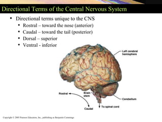
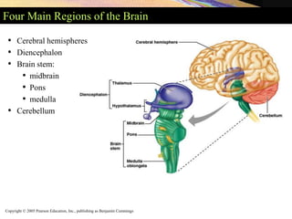
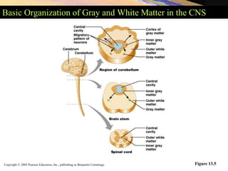
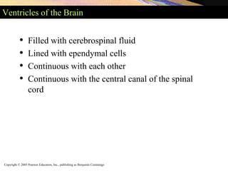
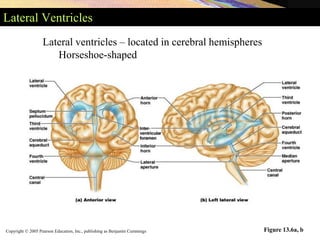
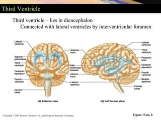
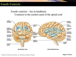
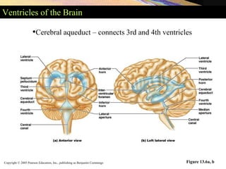
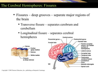
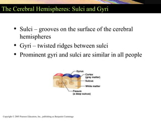
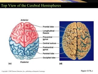
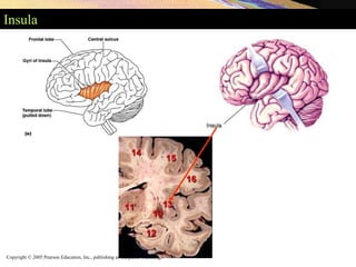
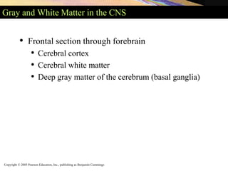
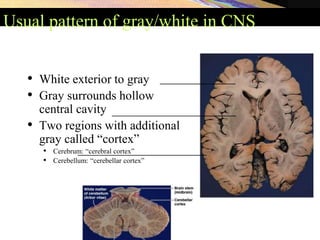
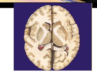
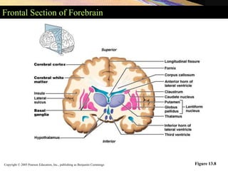
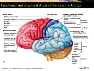
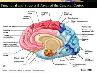
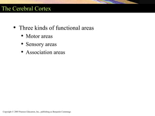
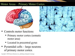
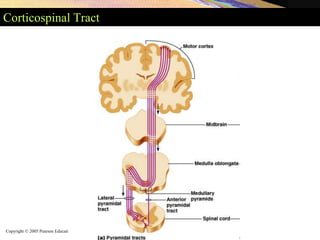
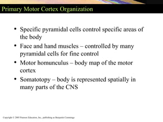
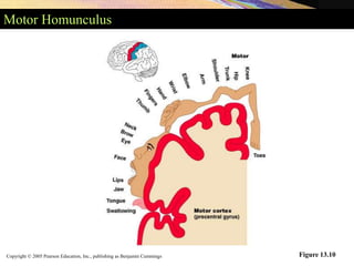
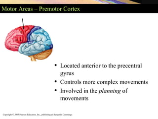
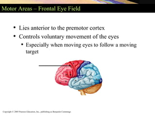
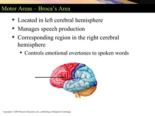
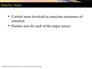
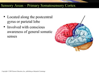
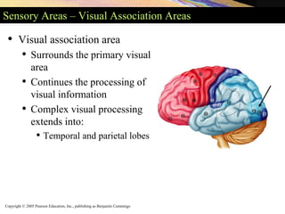
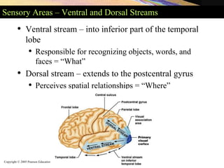
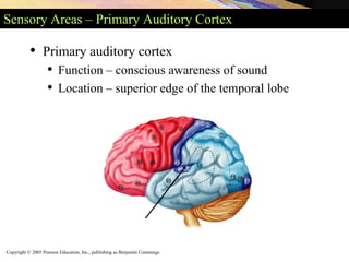
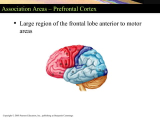
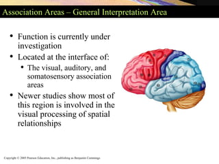
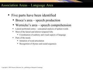
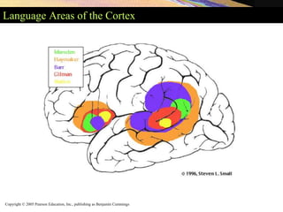
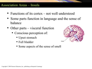
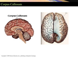
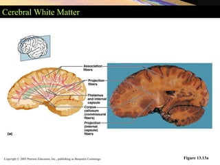
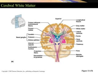
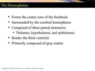
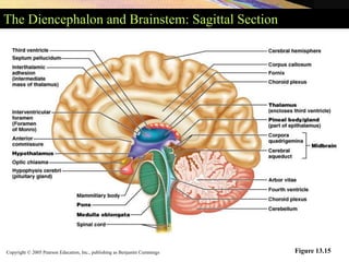
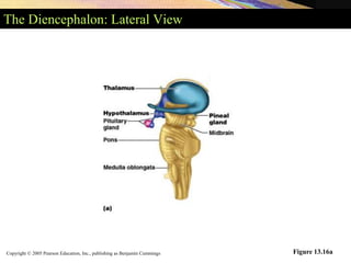
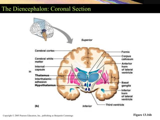
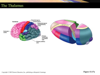
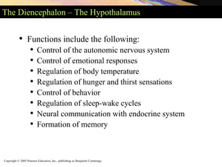
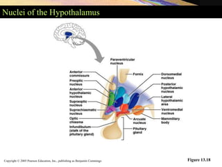
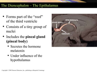
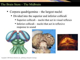
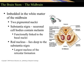
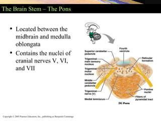
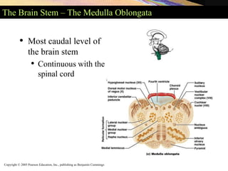
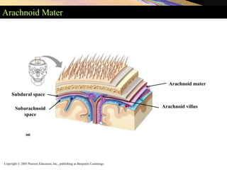
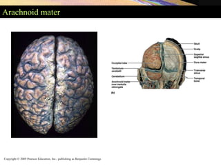
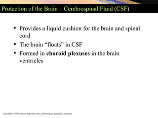
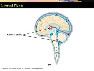
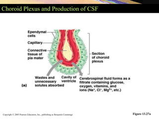
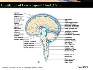
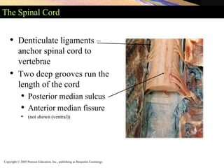
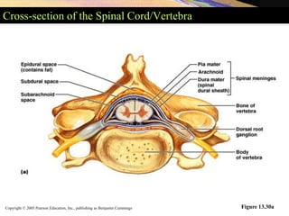
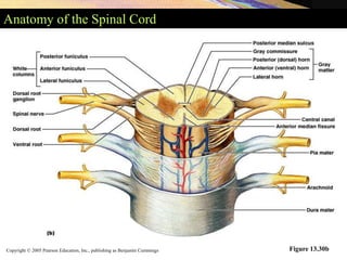
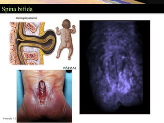
Copyright © 2005 Pearson Education, Inc., publishing as Benjamin Cummings
Central Nervous System
• Brain
• Spinal Cord
Copyright © 2005 Pearson Education, Inc., publishing as Benjamin Cummings
Directional Terms of the Central Nervous System
• Directional terms unique to the CNS
• Rostral – toward the nose (anterior)
• Caudal – toward the tail (posterior)
• Dorsal – superior
• Ventral - inferior
Copyright © 2005 Pearson Education, Inc., publishing as Benjamin Cummings
Four Main Regions of the Brain
• Cerebral hemispheres
• Diencephalon
• Brain stem:
• midbrain
• Pons
• medulla
• Cerebellum
Copyright © 2005 Pearson Education, Inc., publishing as Benjamin Cummings
Basic Organization of Gray and White Matter in the CNS
Figure 13.5
Copyright © 2005 Pearson Education, Inc., publishing as Benjamin Cummings
Ventricles of the Brain
• Filled with cerebrospinal fluid
• Lined with ependymal cells
• Continuous with each other
• Continuous with the central canal of the spinal
cord
Copyright © 2005 Pearson Education, Inc., publishing as Benjamin Cummings
Lateral Ventricles
Figure 13.6a, b
Lateral ventricles – located in cerebral hemispheres
Horseshoe-shaped
Copyright © 2005 Pearson Education, Inc., publishing as Benjamin Cummings
Third Ventricle
Figure 13.6a, b
Third ventricle – lies in diencephalon
Connected with lateral ventricles by interventricular foramen
Copyright © 2005 Pearson Education, Inc., publishing as Benjamin Cummings
Fourth Ventricle
Figure 13.6a, b
Fourth ventricle – lies in hindbrain
Connects to the central canal of the spinal cord
Copyright © 2005 Pearson Education, Inc., publishing as Benjamin Cummings
Ventricles of the Brain
Figure 13.6a, b
•Cerebral aqueduct – connects 3rd and 4th ventricles
Copyright © 2005 Pearson Education, Inc., publishing as Benjamin Cummings
The Cerebral Hemispheres: Fissures
• Fissures – deep grooves – separate major regions of
the brain
• Transverse fissure – separates cerebrum and
cerebellum
• Longitudinal fissure – separates cerebral
hemispheres
Copyright © 2005 Pearson Education, Inc., publishing as Benjamin Cummings
The Cerebral Hemispheres: Sulci and Gyri
• Sulci – grooves on the surface of the cerebral
hemispheres
• Gyri – twisted ridges between sulci
• Prominent gyri and sulci are similar in all people
Copyright © 2005 Pearson Education, Inc., publishing as Benjamin Cummings
The Cerebral Lobes
• Deeper sulci divide cerebrum into lobes
• Lobes are named for the skull bones overlying
them
Copyright © 2005 Pearson Education, Inc., publishing as Benjamin Cummings
Frontal Lobe
• Anterior/rostral to central sulcus
• Superior to transverse/lateral fissure
Copyright © 2005 Pearson Education, Inc., publishing as Benjamin Cummings
Parietal Lobe
• Posterior to central sulcus
• Superior to lateral fissure
• Anterior to parieto-occipital sulcus
Copyright © 2005 Pearson Education, Inc., publishing as Benjamin Cummings
Temporal Lobe
• Inferior to lateral fissure (sulcus)
• Anterior to occipital lobe
Copyright © 2005 Pearson Education, Inc., publishing as Benjamin Cummings
Occipital Lobe
• Posterior & inferior to parieto-occipital sulcus
• Posterior to temporal lobe
Copyright © 2005 Pearson Education, Inc., publishing as Benjamin Cummings
Top View of the Cerebral Hemispheres
Figure 13.7b, c
Copyright © 2005 Pearson Education, Inc., publishing as Benjamin Cummings
Insula
Copyright © 2005 Pearson Education, Inc., publishing as Benjamin Cummings
Gray and White Matter in the CNS
• Frontal section through forebrain
• Cerebral cortex
• Cerebral white matter
• Deep gray matter of the cerebrum (basal ganglia)
Usual pattern of gray/white in CNS
• White exterior to gray
• Gray surrounds hollow
central cavity
• Two regions with additional
gray called “cortex”
• Cerebrum: “cerebral cortex”
• Cerebellum: “cerebellar cortex”
_________________
____________________________
_____________________________
Copyright © 2005 Pearson Education, Inc., publishing as Benjamin Cummings
Frontal Section of Forebrain
Figure 13.8
Copyright © 2005 Pearson Education, Inc., publishing as Benjamin Cummings
The Cerebral Cortex
• Composed of gray matter 2 – 4 mm thick
• Neuronal cell bodies, dendrites, and short axons
• Folds in cortex – triples its size
• Approximately 40% of brain’s mass
• Brodmann areas – 52 structurally distinct areas
(we will not cover these by number, only name)
Copyright © 2005 Pearson Education, Inc., publishing as Benjamin Cummings
Functional and Structural Areas of the Cerebral Cortex
Figure 13.11a
Copyright © 2005 Pearson Education, Inc., publishing as Benjamin Cummings
Functional and Structural Areas of the Cerebral Cortex
Figure 13.11b
Copyright © 2005 Pearson Education, Inc., publishing as Benjamin Cummings
The Cerebral Cortex
• Three kinds of functional areas
• Motor areas
• Sensory areas
• Association areas
Copyright © 2005 Pearson Education, Inc., publishing as Benjamin Cummings
Motor Areas – Primary Motor Cortex
• Controls motor functions
• Primary motor cortex (somatic
motor area)
• Located in precentral gyrus
• Pyramidal cells – large neurons
of primary motor cortex
Copyright © 2005 Pearson Education, Inc., publishing as Benjamin Cummings
Motor Areas – Corticospinal Tract (Pyramidal Tract)
• Corticospinal tracts descend through brainstem
and spinal cord
• Axons signal motor neurons to control fine skilled
movements
• Contralateral – pyramidal axons cross over to
opposite side of the brain
Copyright © 2005 Pearson Education, Inc., publishing as Benjamin Cummings
Corticospinal Tract
Copyright © 2005 Pearson Education, Inc., publishing as Benjamin Cummings
Primary Motor Cortex Organization
• Specific pyramidal cells control specific areas of
the body
• Face and hand muscles – controlled by many
pyramidal cells for fine control
• Motor homunculus – body map of the motor
cortex
• Somatotopy – body is represented spatially in
many parts of the CNS
Copyright © 2005 Pearson Education, Inc., publishing as Benjamin Cummings
Motor Homunculus
Figure 13.10
Copyright © 2005 Pearson Education, Inc., publishing as Benjamin Cummings
Motor Areas – Premotor Cortex
• Located anterior to the precentral
gyrus
• Controls more complex movements
• Involved in the planning of
movements
Copyright © 2005 Pearson Education, Inc., publishing as Benjamin Cummings
Motor Areas – Frontal Eye Field
• Lies anterior to the premotor cortex
• Controls voluntary movement of the eyes
• Especially when moving eyes to follow a moving
target
Copyright © 2005 Pearson Education, Inc., publishing as Benjamin Cummings
Motor Areas – Broca’s Area
• Located in left cerebral hemisphere
• Manages speech production
• Corresponding region in the right cerebral
hemisphere
• Controls emotional overtones to spoken words
Copyright © 2005 Pearson Education, Inc., publishing as Benjamin Cummings
Sensory Areas
• Cortical areas involved in conscious awareness of
sensation
• Distinct area for each of the major senses
Copyright © 2005 Pearson Education, Inc., publishing as Benjamin Cummings
Sensory Areas – Primary Somatosensory Cortex
• Located along the postcentral
gyrus or parietal lobe
• Involved with conscious
awareness of general somatic
senses
Copyright © 2005 Pearson Education, Inc., publishing as Benjamin Cummings
Sensory Areas – Primary Somatosensory Cortex
• Projection is contralateral
• Cerebral hemispheres
• Receive sensory input from the opposite side of the
body
• Sensory homunculus – a body map of the sensory
cortex
Copyright © 2005 Pearson Education, Inc., publishing as Benjamin Cummings
Sensory Areas – Sensory Homunculus
Figure 13.10
Copyright © 2005 Pearson Education, Inc., publishing as Benjamin Cummings
Sensory Areas – Somatosensory Association Area
• Lies posterior to the primary
somatosensory cortex
• Integrates different sensory
inputs
• Touch, pressure, and others
• Draws upon stored memories
of past sensory experiences
Copyright © 2005 Pearson Education, Inc., publishing as Benjamin Cummings
Sensory Areas – Primary Visual Cortex
• Primary visual cortex
• Located deep within the
calcarine sulcus
• On the posterior and medial
part of the occipital lobe
• Receives visual information
that originates on the retina
• First of a series of areas that
interprets visual input
Copyright © 2005 Pearson Education, Inc., publishing as Benjamin Cummings
Sensory Areas – Visual Association Areas
• Visual association area
• Surrounds the primary visual
area
• Continues the processing of
visual information
• Complex visual processing
extends into:
• Temporal and parietal lobes
Copyright © 2005 Pearson Education, Inc., publishing as Benjamin Cummings
Visual Association Areas continued
• Approximately 30 cortical areas have been
identified
• Visual information proceeds in two streams
Copyright © 2005 Pearson Education, Inc., publishing as Benjamin Cummings
Sensory Areas – Ventral and Dorsal Streams
• Ventral stream – into inferior part of the temporal
lobe
• Responsible for recognizing objects, words, and
faces = “What”
• Dorsal stream – extends to the postcentral gyrus
• Perceives spatial relationships = “Where”
Copyright © 2005 Pearson Education, Inc., publishing as Benjamin Cummings
Sensory Areas – Primary Auditory Cortex
• Primary auditory cortex
• Function – conscious awareness of sound
• Location – superior edge of the temporal lobe
Copyright © 2005 Pearson Education, Inc., publishing as Benjamin Cummings
Sensory Areas – Auditory Association Areas
• Auditory association area
• Lies posterior to the
primary auditory cortex
• Permits evaluation of
different sounds
Copyright © 2005 Pearson Education, Inc., publishing as Benjamin Cummings
Sensory Areas – Gustatory Cortex
• Involved in the conscious
awareness of taste stimuli
• Located on the “roof” of the
lateral sulcus
Copyright © 2005 Pearson Education, Inc., publishing as Benjamin Cummings
Sensory Areas – Vestibular Cortex
• Located in the posterior
part of the insula
• Deep to the lateral
sulcus
Copyright © 2005 Pearson Education, Inc., publishing as Benjamin Cummings
Sensory Areas – Olfactory Cortex
• Lies on the medial aspect
of the cerebrum
• Located in a region called
the piriform lobe
• Olfactory nerves transmit
impulses to the olfactory
cortex
• Provides conscious
awareness of smells
Copyright © 2005 Pearson Education, Inc., publishing as Benjamin Cummings
Sensory Areas – Olfactory Cortex
• Connects the brain to the limbic system
• Explains why smells trigger emotions
• Orbitofrontal cortex
• Involved with consciously identifying and recalling
specific smells
Copyright © 2005 Pearson Education, Inc., publishing as Benjamin Cummings
Association Areas
• Make associations between different types of
sensory information
• Associate new sensory input with memories of
past experiences
• New name for association areas – higher order
processing areas
Copyright © 2005 Pearson Education, Inc., publishing as Benjamin Cummings
Association Areas – Prefrontal Cortex
• Large region of the frontal lobe anterior to motor
areas
Copyright © 2005 Pearson Education, Inc., publishing as Benjamin Cummings
Association Areas – Prefrontal Cortex
• Performs cognitive functions
• All aspects of thinking and perceiving
• Remembering and recalling information
• Problem solving
• Also related to mood
• Has close links to the limbic part of the forebrain
Copyright © 2005 Pearson Education, Inc., publishing as Benjamin Cummings
Association Areas – General Interpretation Area
• Function is currently under
investigation
• Located at the interface of:
• The visual, auditory, and
somatosensory association
areas
• Newer studies show most of
this region is involved in the
visual processing of spatial
relationships
Copyright © 2005 Pearson Education, Inc., publishing as Benjamin Cummings
Association Areas – Language Area
• Five parts have been identified
• Broca’s area – speech production
• Wernicke’s area – speech comprehension
• Lateral prefrontal cortex – conceptual analysis of spoken words
• Most of the lateral and inferior temporal lobe
• Coordination of auditory and visual aspects of language
• Parts of the insula
• Initiation of word articulation
• Recognition of rhymes and sound sequences
Copyright © 2005 Pearson Education, Inc., publishing as Benjamin Cummings
Language Areas of the Cortex
Copyright © 2005 Pearson Education, Inc., publishing as Benjamin Cummings
Association Areas – Insula
• Functions of its cortex – not well understood
• Some parts function in language and the sense of
balance
• Other parts – visceral function
• Conscious perception of:
• Upset stomach
• Full bladder
• Some aspects of the sense of smell
Copyright © 2005 Pearson Education, Inc., publishing as Benjamin Cummings
Lateralization of Cortical Functioning
• The two hemispheres control opposite sides of the
body
• Hemispheres are specialized for different
cognitive functions
Copyright © 2005 Pearson Education, Inc., publishing as Benjamin Cummings
Lateralization of Cortical Functioning
• Left cerebral hemisphere – more control over:
• Language abilities, math, and logic
• Right cerebral hemisphere – more involved with:
• Visual-spatial skills
• Reading facial expressions
• Intuition, emotion, artistic and musical skills
Copyright © 2005 Pearson Education, Inc., publishing as Benjamin Cummings
Cerebral White Matter
• Different areas of the cerebral cortex
communicate:
• With each other
• With the brainstem and spinal cord
• Fibers are usually myelinated and bundled into
tracts
Copyright © 2005 Pearson Education, Inc., publishing as Benjamin Cummings
Cerebral White Matter
• Types of tracts
• Commissures – composed of commissural fibers
• Allows communication between cerebral
hemispheres
• Corpus callosum – the largest commissure
• Association fibers
• Connect different parts of the same hemisphere
Copyright © 2005 Pearson Education, Inc., publishing as Benjamin Cummings
Corpus Callosum
Copyright © 2005 Pearson Education, Inc., publishing as Benjamin Cummings
Cerebral White Matter
Figure 13.13a
Copyright © 2005 Pearson Education, Inc., publishing as Benjamin Cummings
Cerebral White Matter
• Types of tracts (continued)
• Projection fibers – run vertically
• Descend from the cerebral cortex
• Ascend to the cortex from lower regions
Copyright © 2005 Pearson Education, Inc., publishing as Benjamin Cummings
Cerebral White Matter
Figure 13.13b
Copyright © 2005 Pearson Education, Inc., publishing as Benjamin Cummings
White Matter: MRI
Copyright © 2005 Pearson Education, Inc., publishing as Benjamin Cummings
White Matter: Horizontal Section
Copyright © 2005 Pearson Education, Inc., publishing as Benjamin Cummings
Deep Gray Matter of the Cerebrum
• Consists of:
• Basal forebrain nuclei – associated with memory
• Basal ganglia – invovled in motor control
• Claustrum – a nucleus of unknown function
• Amygdala – located in cerebrum but is considered
part of the of the limbic system
Copyright © 2005 Pearson Education, Inc., publishing as Benjamin Cummings
Basal Ganglia
• A group of nuclei deep within the cerebral white
matter
• Caudate nucleus – arches over the thalamus
• Lentiform nucleus – “lens shaped”
• Together, these nuclei are called the corpus
striatum
Copyright © 2005 Pearson Education, Inc., publishing as Benjamin Cummings
Basal Ganglia
• Lentiform nucleus
• Divided into two parts
• Globus pallidus
• Putamen
Copyright © 2005 Pearson Education, Inc., publishing as Benjamin Cummings
Basal Ganglia
Figure 13.13b
Copyright © 2005 Pearson Education, Inc., publishing as Benjamin Cummings Figure 13.14a
Basal Ganglia
• Corpus striatum – combination of the lentiform
and caudate nuclei
Copyright © 2005 Pearson Education, Inc., publishing as Benjamin Cummings
Basal Ganglia
• Cooperate with the cerebral cortex in controlling
movements
• Receive input from many cortical areas
• Evidence shows that they:
• Start, stop, and regulate intensity of voluntary
movements
• In some way estimate the passage of time
Copyright © 2005 Pearson Education, Inc., publishing as Benjamin Cummings
The Diencephalon
• Forms the center core of the forebrain
• Surrounded by the cerebral hemispheres
• Composed of three paired structures:
• Thalamus, hypothalamus, and epithalamus
• Border the third ventricle
• Primarily composed of gray matter
Copyright © 2005 Pearson Education, Inc., publishing as Benjamin Cummings
The Diencephalon and Brainstem: Sagittal Section
Figure 13.15
Copyright © 2005 Pearson Education, Inc., publishing as Benjamin Cummings
The Diencephalon: Lateral View
Figure 13.16a
Copyright © 2005 Pearson Education, Inc., publishing as Benjamin Cummings
The Diencephalon: Coronal Section
Figure 13.16b
Copyright © 2005 Pearson Education, Inc., publishing as Benjamin Cummings
The Diencephalon – The Thalamus
• Makes up 80% of the diencephalon
• Contains approximately a dozen major nuclei
• Send axons to regions of the cerebral cortex
• Nuclei act as relay stations for incoming sensory
messages
Copyright © 2005 Pearson Education, Inc., publishing as Benjamin Cummings
The Diencephalon – The Thalamus
• Afferent impulses converge on the thalamus
• Synapse in at least one of its nuclei
• Is the “gateway” to the cerebral cortex
• Nuclei organize and amplify or tone down signals
Copyright © 2005 Pearson Education, Inc., publishing as Benjamin Cummings
The Thalamus
Figure 13.17a
Copyright © 2005 Pearson Education, Inc., publishing as Benjamin Cummings
The Diencephalon – The Hypothalamus
• Lies between the optic chiasm and the mammillary
bodies
• Pituitary gland projects inferiorly
• Anterior pituitary
• Posterior pituitary
• Contains approximately a dozen nuclei
• Main visceral control center of the body
Copyright © 2005 Pearson Education, Inc., publishing as Benjamin Cummings
The Diencephalon – The Hypothalamus
• Functions include the following:
• Control of the autonomic nervous system
• Control of emotional responses
• Regulation of body temperature
• Regulation of hunger and thirst sensations
• Control of behavior
• Regulation of sleep-wake cycles
• Neural communication with endocrine system
• Formation of memory
Copyright © 2005 Pearson Education, Inc., publishing as Benjamin Cummings
Nuclei of the Hypothalamus
Figure 13.18
Copyright © 2005 Pearson Education, Inc., publishing as Benjamin Cummings
The Diencephalon – The Epithalamus
• Forms part of the “roof”
of the third ventricle
• Consists of a tiny group of
nuclei
• Includes the pineal gland
(pineal body)
• Secretes the hormone
melatonin
• Under influence of the
hypothalamus
Copyright © 2005 Pearson Education, Inc., publishing as Benjamin Cummings
The Brain Stem
• Includes the midbrain, pons, and medulla
oblongata
Copyright © 2005 Pearson Education, Inc., publishing as Benjamin Cummings
Ventral View of the Brain – Brain Stem and Diencephalon
Figure 13.19
Copyright © 2005 Pearson Education, Inc., publishing as Benjamin Cummings
The Brain Stem: General Functions
• Produces automatic behaviors necessary for
survival
• Passageway for all fiber tracts running between the
cerebrum and spinal cord
• Heavily involved with the innervation of the face
and head
• 10 of the 12 pairs of cranial nerves attach to it
Copyright © 2005 Pearson Education, Inc., publishing as Benjamin Cummings
The Brain Stem – The Midbrain
• Lies between the diencephalon
and the pons
• Central cavity – the cerebral
aqueduct
• Cerebral peduncles located on
the ventral surface of the brain
• Contain pyramidal
(corticospinal) tracts
• Superior cerebellar peduncles
• Connect midbrain to the
cerebellum
Copyright © 2005 Pearson Education, Inc., publishing as Benjamin Cummings Figure 13.21a
The Brain Stem – The Midbrain
• Periaqueductal gray
matter surrounds the
cerebral aqueduct
• Involved in two
related functions
• Fright-and-flight
reaction
• Mediates response
to visceral pain
Copyright © 2005 Pearson Education, Inc., publishing as Benjamin Cummings
The Brain Stem – The Midbrain
• Corpora quadrigemina – the largest nuclei
• Divided into the superior and inferior colliculi
• Superior colliculi – nuclei that act in visual reflexes
• Inferior colliculi – nuclei that act in reflexive
response to sound
Copyright © 2005 Pearson Education, Inc., publishing as Benjamin Cummings
The Brain Stem – The Midbrain
• Imbedded in the white matter
of the midbrain
• Two pigmented nuclei
• Substantia nigra – neuronal
cell bodies contain melanin
• Functionally linked to the
basal nuclei
• Red nucleus – lies deep to the
substantia nigra
• Largest nucleus of the
reticular formation
Copyright © 2005 Pearson Education, Inc., publishing as Benjamin Cummings
The Brain Stem – The Pons
• Located between the
midbrain and medulla
oblongata
• Contains the nuclei of
cranial nerves V, VI,
and VII
Copyright © 2005 Pearson Education, Inc., publishing as Benjamin Cummings
The Brain Stem – The Medulla Oblongata
• Most caudal level of
the brain stem
• Continuous with the
spinal cord
Copyright © 2005 Pearson Education, Inc., publishing as Benjamin Cummings
The Brain Stem – The Medulla Oblongata
• Choroid plexus lies in the
roof of the fourth ventricle
• Pyramids of the medulla –
lie on its ventral surface
• Decussation of the
pyramids – crossing over
of motor tracts
• Cranial nerves VIII–XII
attach to the medulla
Copyright © 2005 Pearson Education, Inc., publishing as Benjamin Cummings
The Brain Stem – The Medulla Oblongata
• The core of the medulla contains:
• Much of the reticular formation
• Nuclei influence autonomic functions
• Visceral centers of the reticular formation include:
• Cardiac center
• Vasomotor center
• The medullary respiratory center
• Centers for hiccupping, sneezing, swallowing, and
coughing
Copyright © 2005 Pearson Education, Inc., publishing as Benjamin Cummings
The Cerebellum
• Located dorsal to the pons and medulla
• Smoothes and coordinates body movements
• Helps maintain equilibrium
Copyright © 2005 Pearson Education, Inc., publishing as Benjamin Cummings
The Cerebellum
• Consists of two cerebellar hemispheres
• Surface folded into ridges called folia
• Separated by fissures
• Hemispheres each subdivided into:
• Anterior lobe
• Posterior lobe
• Flocculonodular lobe
Copyright © 2005 Pearson Education, Inc., publishing as Benjamin Cummings
Cerebellum
Copyright © 2005 Pearson Education, Inc., publishing as Benjamin Cummings
Cerebellum: Posterior View
Copyright © 2005 Pearson Education, Inc., publishing as Benjamin Cummings
The Cerebellum: Frontal Section
• Composed of three regions
• Cortex – gray matter
• Internal white matter
• Deep cerebellar nuclei – deeply situated gray
matter
Copyright © 2005 Pearson Education, Inc., publishing as Benjamin Cummings
Cerebellum: Sagittal Section
Copyright © 2005 Pearson Education, Inc., publishing as Benjamin Cummings
The Cerebellum
• Cerebellum must receive information
• On equilibrium
• On current movements of limbs, neck, and trunk
• From the cerebral cortex
Copyright © 2005 Pearson Education, Inc., publishing as Benjamin Cummings
The Cerebellum – Cerebellar Peduncles
• Thick tracts connecting the
cerebellum to the brain stem
• Superior cerebellar peduncles
• Middle cerebellar peduncles
• Inferior cerebellar peduncles
• Fibers to and from the
cerebellum are ipsilateral
• Run to and from the same
side of the body
Copyright © 2005 Pearson Education, Inc., publishing as Benjamin Cummings
Decussation of Superior Cerebellar Peduncle
1. Decussation of superior cerebellar peduncles 2. Superior cerebellar
peduncle 3. Superior medullary velum 4. Fastigial nucleus 5. Globose nuclei
6. Emboliform nucleus 7. White matter of vermis 8. Dentate nucleus
Copyright © 2005 Pearson Education, Inc., publishing as Benjamin Cummings
Functional Brain Systems
• Networks of neurons functioning together
• The limbic system – spread widely in the forebrain
• The reticular formation – spans the brain stem
Copyright © 2005 Pearson Education, Inc., publishing as Benjamin Cummings
Functional Brain Systems – The Limbic System
• Location
• Medial aspect of cerebral hemispheres
• Also within the diencephalon
• Composed of:
• Septal nuclei, cingulate gyrus, and hippocampal
formation
• Part of the amygdala
• The fornix and other tracts link the limbic system
together
Copyright © 2005 Pearson Education, Inc., publishing as Benjamin Cummings
Functional Brain Systems – The Limbic System
Figure 13.23
Copyright © 2005 Pearson Education, Inc., publishing as Benjamin Cummings
Functional Brain Systems – The Limbic System
• The “emotional brain”
• Cingulate gyrus
• Allows us to shift between thoughts
• Interprets pain as unpleasant
• Hippocampal formation
• Hippocampus and the parahippocampal gyrus
Copyright © 2005 Pearson Education, Inc., publishing as Benjamin Cummings
Functional Brain Systems – The Reticular Formation
• Runs through the central core of the medulla,
pons, and midbrain
• Forms three columns
• Midline raphe nuclei
• Medial nuclear group
• Lateral nuclear group
Copyright © 2005 Pearson Education, Inc., publishing as Benjamin Cummings
Functional Brain Systems – The Reticular Formation
Figure 13.21c
Copyright © 2005 Pearson Education, Inc., publishing as Benjamin Cummings
Functional Brain Systems – The Reticular Formation
• Widespread connections
• Ideal for arousal of the brain as a whole
• Reticular activating system (RAS)
• Maintains consciousness and alertness
• Functions in sleep and arousal from sleep
Copyright © 2005 Pearson Education, Inc., publishing as Benjamin Cummings
Functional Brain Systems – The Reticular Formation
Figure 13.24
Copyright © 2005 Pearson Education, Inc., publishing as Benjamin Cummings
Protection of the Brain
• The brain is protected from injury by
• The skull
• Meninges
• Cerebrospinal fluid
• Blood-brain barrier
Copyright © 2005 Pearson Education, Inc., publishing as Benjamin Cummings
Protection of the Brain – Meninges
• Three meninges
• Dura mater
• Arachnoid mater
• Pia mater
• Cover and protect the CNS
• Enclose and protect the vessels that supply the
CNS
• Contain the cerebrospinal fluid
Copyright © 2005 Pearson Education, Inc., publishing as Benjamin Cummings
The Dura Mater
• Strongest of the meninges
• Composed of two layers
• Periosteal layer
• Meningeal layer
• Two layers are fused except to enclose the dural
sinuses
Copyright © 2005 Pearson Education, Inc., publishing as Benjamin Cummings
The Dura Mater
Figure 13.25a
Periosteal dura
Meningeal dura
Superior sagittal
sinus
Copyright © 2005 Pearson Education, Inc., publishing as Benjamin Cummings
Dura mater
Copyright © 2005 Pearson Education, Inc., publishing as Benjamin Cummings
The Dura Mater
• Largest sinus – the
superior sagittal sinus
• Dura mater extends
inward to subdivide
the cranial cavity
Figure 13.26
Copyright © 2005 Pearson Education, Inc., publishing as Benjamin Cummings
Falx Cerebri
1. falx cerebri
2. location of inferior sagittal sinus
3. location of superior sagittal sinus
4. location of straight sinus
5. tentorium cerebelli
Copyright © 2005 Pearson Education, Inc., publishing as Benjamin Cummings
The Arachnoid Mater
• Located beneath the dura mater
• Subdural space
• Potential space between dura and arachnoid mater
• Subarachnoid space
• Filled with CSF
• Contains the blood vessels that supply the brain
• Arachnoid villi
• Allow CSF to pass into the dural blood sinuses
Copyright © 2005 Pearson Education, Inc., publishing as Benjamin Cummings
Arachnoid Mater
Arachnoid mater
Arachnoid villus
Subdural space
Subarachnoid
space
Copyright © 2005 Pearson Education, Inc., publishing as Benjamin Cummings
Arachnoid mater
Copyright © 2005 Pearson Education, Inc., publishing as Benjamin Cummings
The Pia Mater
• Delicate connective tissue
• Clings tightly to the surface of the brain
• Follows all convolutions of the cortex
Copyright © 2005 Pearson Education, Inc., publishing as Benjamin Cummings
The Pia Mater
Figure 13.25a
Copyright © 2005 Pearson Education, Inc., publishing as Benjamin Cummings
Protection of the Brain – Cerebrospinal Fluid (CSF)
• Provides a liquid cushion for the brain and spinal
cord
• The brain “floats” in CSF
• Formed in choroid plexuses in the brain
ventricles
Copyright © 2005 Pearson Education, Inc., publishing as Benjamin Cummings
Choroid Plexus
Choroid plexus
Copyright © 2005 Pearson Education, Inc., publishing as Benjamin Cummings
Choroid Plexus and Production of CSF
Figure 13.27a
Copyright © 2005 Pearson Education, Inc., publishing as Benjamin Cummings
Circulation of Cerebrospinal Fluid (CSF)
Figure 13.27b
Copyright © 2005 Pearson Education, Inc., publishing as Benjamin Cummings
Protection of the Brain – Blood-Brain Barrier
• Prevents most blood-borne toxins from entering
the brain
• Impermeable capillaries
• Not an absolute barrier
• Nutrients such as oxygen pass through
• Allows alcohol, nicotine, and anesthetics through
Copyright © 2005 Pearson Education, Inc., publishing as Benjamin Cummings
Blood-Brain Barrier
Copyright © 2005 Pearson Education, Inc., publishing as Benjamin Cummings
The Spinal Cord
• Runs through the
vertebral canal
• Extends from the
foramen magnum to the
level of the vertebra L1 or
L2
Copyright © 2005 Pearson Education, Inc., publishing as Benjamin Cummings
Spinal Cord: Cervical, Thoracic, Lumbar, and Sacral
Copyright © 2005 Pearson Education, Inc., publishing as Benjamin Cummings
The Spinal Cord Meninges
• Protected by bone,
meninges, and CSF
• Dura mater of the spinal
cord
• The spinal dural sheath –
only one layer
Copyright © 2005 Pearson Education, Inc., publishing as Benjamin Cummings
Spinal Cord Meninges
Dura mater
Arachnoid mater
Copyright © 2005 Pearson Education, Inc., publishing as Benjamin Cummings
Cervical and Lumbar Enlargements
Where nerves for upper and lower
limbs arise
Copyright © 2005 Pearson Education, Inc., publishing as Benjamin Cummings
The Spinal Cord
• Conus medullaris – the inferior end of the spinal
cord
• Filum terminale – long filament of connective
tissue
• Attaches to the coccyx inferiorly
• Cauda equina – collection of nerve roots
Copyright © 2005 Pearson Education, Inc., publishing as Benjamin Cummings
The Spinal Cord
Figure 13.29a
Copyright © 2005 Pearson Education, Inc., publishing as Benjamin Cummings
The Spinal Cord
• Denticulate ligaments –
anchor spinal cord to
vertebrae
• Two deep grooves run the
length of the cord
• Posterior median sulcus
• Anterior median fissure
• (not shown (ventral))
Copyright © 2005 Pearson Education, Inc., publishing as Benjamin Cummings
Cross-section of the Spinal Cord/Vertebra
Figure 13.30a
Copyright © 2005 Pearson Education, Inc., publishing as Benjamin Cummings
Anatomy of the Spinal Cord
Figure 13.30b
Copyright © 2005 Pearson Education, Inc., publishing as Benjamin Cummings
Gray Matter of the Spinal Cord and Spinal Roots
• Shaped like the letter H
• Gray commissure – contains the central canal
• Anterior horns – contain cell bodies of motor
neurons
• Posterior horns – consist of interneurons
• Gray matter – divided according to somatic and
visceral regions
Copyright © 2005 Pearson Education, Inc., publishing as Benjamin Cummings
Gray Matter of the Spinal Cord and Spinal Roots
Figure 13.32
Copyright © 2005 Pearson Education, Inc., publishing as Benjamin Cummings
White Matter of the Spinal Cord
• Composed of myelinated and unmyelinated axons
• Three types of fibers
• Ascending
• Descending
• Commissural
Copyright © 2005 Pearson Education, Inc., publishing as Benjamin Cummings
White Matter of the Spinal Cord
Figure 13.33
Copyright © 2005 Pearson Education, Inc., publishing as Benjamin Cummings
Sensory and Motor Pathways
• Most motor pathways:
• Decussate at some point along their course
• Consist of a chain of two or three neurons
• Exhibit somatotopy
• Tracts arranged according to the body region they
supply
• All pathways are paired
• One of each on each side of the body
Copyright © 2005 Pearson Education, Inc., publishing as Benjamin Cummings
Ascending (Sensory) Pathways
• Conduct general somatic sensory impulses
• Chains of neurons composed of:
• First-, second-, and third-order neurons
• Four main ascending pathways
• Dorsal column pathway
• Spinothalamic pathway
• Posterior spinocerebellar pathway
• Anterior spinocerebellar pathway
Copyright © 2005 Pearson Education, Inc., publishing as Benjamin Cummings
Ascending Pathways
Figure 13.34a
Copyright © 2005 Pearson Education, Inc., publishing as Benjamin Cummings
Ascending Pathways
Figure 13.34b
Copyright © 2005 Pearson Education, Inc., publishing as Benjamin Cummings
Descending (Motor) Pathways
• Deliver motor instructions from the brain to the
spinal cord
• Divided into two groups
• Pyramidal, or corticospinal, tracts
• Other motor pathways
• Tectospinal tracts
• Vestibulospinal tract
• Rubrospinal tract
• Reticulospinal tract
Copyright © 2005 Pearson Education, Inc., publishing as Benjamin Cummings
Descending Motor Pathways
Figure 13.35a
Copyright © 2005 Pearson Education, Inc., publishing as Benjamin Cummings
Descending Motor Pathways
Figure 13.35b
Copyright © 2005 Pearson Education, Inc., publishing as Benjamin Cummings
Disorders of the Central Nervous System: Trauma
• Traumatic brain injuries
• Concussion – brain injury is slight
• Contusion – marked destruction of brain tissue
Copyright © 2005 Pearson Education, Inc., publishing as Benjamin Cummings
Disorders of the Central Nervous System: Stroke
Cerebrovascular accident (stroke)
• Hemorrhagic
• Ischemic
• Blockage or interruption of blood flow to a brain region
Copyright © 2005 Pearson Education, Inc., publishing as Benjamin Cummings
Disorders of the Central Nervous System
• Spinal cord damage
• Paralysis – loss of motor function
• Parasthesia – loss of sensation
• Paraplegia – injury to the spinal cord is between T1
and L2
• Paralysis of the lower limbs
• Quadriplegia – injury to the spinal cord in the
cervical region
• Paralysis of all four limbs
Copyright © 2005 Pearson Education, Inc., publishing as Benjamin Cummings
The Central Nervous System Throughout Life
• Congenital malformations
• Hydrocephalus
• Neural tube defects
• Anencephaly – cerebrum and cerebellum are absent
• Spina bifida – absence of vertebral lamina
• Cerebral palsy – voluntary muscles are poorly
controlled
• Results from damage to the motor cortex
Copyright © 2005 Pearson Education, Inc., publishing as Benjamin Cummings
Hydrocephalus
Copyright © 2005 Pearson Education, Inc., publishing as Benjamin Cummings
Anencephaly
Copyright © 2005 Pearson Education, Inc., publishing as Benjamin Cummings
Spina bifida

1715606398115_central nervous system.ppt

  • 1.
    Copyright © 2005Pearson Education, Inc., publishing as Benjamin Cummings Central Nervous System • Brain • Spinal Cord
  • 2.
    Copyright © 2005Pearson Education, Inc., publishing as Benjamin Cummings Directional Terms of the Central Nervous System • Directional terms unique to the CNS • Rostral – toward the nose (anterior) • Caudal – toward the tail (posterior) • Dorsal – superior • Ventral - inferior
  • 3.
    Copyright © 2005Pearson Education, Inc., publishing as Benjamin Cummings Four Main Regions of the Brain • Cerebral hemispheres • Diencephalon • Brain stem: • midbrain • Pons • medulla • Cerebellum
  • 4.
    Copyright © 2005Pearson Education, Inc., publishing as Benjamin Cummings Basic Organization of Gray and White Matter in the CNS Figure 13.5
  • 5.
    Copyright © 2005Pearson Education, Inc., publishing as Benjamin Cummings Ventricles of the Brain • Filled with cerebrospinal fluid • Lined with ependymal cells • Continuous with each other • Continuous with the central canal of the spinal cord
  • 6.
    Copyright © 2005Pearson Education, Inc., publishing as Benjamin Cummings Lateral Ventricles Figure 13.6a, b Lateral ventricles – located in cerebral hemispheres Horseshoe-shaped
  • 7.
    Copyright © 2005Pearson Education, Inc., publishing as Benjamin Cummings Third Ventricle Figure 13.6a, b Third ventricle – lies in diencephalon Connected with lateral ventricles by interventricular foramen
  • 8.
    Copyright © 2005Pearson Education, Inc., publishing as Benjamin Cummings Fourth Ventricle Figure 13.6a, b Fourth ventricle – lies in hindbrain Connects to the central canal of the spinal cord
  • 9.
    Copyright © 2005Pearson Education, Inc., publishing as Benjamin Cummings Ventricles of the Brain Figure 13.6a, b •Cerebral aqueduct – connects 3rd and 4th ventricles
  • 10.
    Copyright © 2005Pearson Education, Inc., publishing as Benjamin Cummings The Cerebral Hemispheres: Fissures • Fissures – deep grooves – separate major regions of the brain • Transverse fissure – separates cerebrum and cerebellum • Longitudinal fissure – separates cerebral hemispheres
  • 11.
    Copyright © 2005Pearson Education, Inc., publishing as Benjamin Cummings The Cerebral Hemispheres: Sulci and Gyri • Sulci – grooves on the surface of the cerebral hemispheres • Gyri – twisted ridges between sulci • Prominent gyri and sulci are similar in all people
  • 12.
    Copyright © 2005Pearson Education, Inc., publishing as Benjamin Cummings The Cerebral Lobes • Deeper sulci divide cerebrum into lobes • Lobes are named for the skull bones overlying them
  • 13.
    Copyright © 2005Pearson Education, Inc., publishing as Benjamin Cummings Frontal Lobe • Anterior/rostral to central sulcus • Superior to transverse/lateral fissure
  • 14.
    Copyright © 2005Pearson Education, Inc., publishing as Benjamin Cummings Parietal Lobe • Posterior to central sulcus • Superior to lateral fissure • Anterior to parieto-occipital sulcus
  • 15.
    Copyright © 2005Pearson Education, Inc., publishing as Benjamin Cummings Temporal Lobe • Inferior to lateral fissure (sulcus) • Anterior to occipital lobe
  • 16.
    Copyright © 2005Pearson Education, Inc., publishing as Benjamin Cummings Occipital Lobe • Posterior & inferior to parieto-occipital sulcus • Posterior to temporal lobe
  • 17.
    Copyright © 2005Pearson Education, Inc., publishing as Benjamin Cummings Top View of the Cerebral Hemispheres Figure 13.7b, c
  • 18.
    Copyright © 2005Pearson Education, Inc., publishing as Benjamin Cummings Insula
  • 19.
    Copyright © 2005Pearson Education, Inc., publishing as Benjamin Cummings Gray and White Matter in the CNS • Frontal section through forebrain • Cerebral cortex • Cerebral white matter • Deep gray matter of the cerebrum (basal ganglia)
  • 20.
    Usual pattern ofgray/white in CNS • White exterior to gray • Gray surrounds hollow central cavity • Two regions with additional gray called “cortex” • Cerebrum: “cerebral cortex” • Cerebellum: “cerebellar cortex” _________________ ____________________________ _____________________________
  • 22.
    Copyright © 2005Pearson Education, Inc., publishing as Benjamin Cummings Frontal Section of Forebrain Figure 13.8
  • 23.
    Copyright © 2005Pearson Education, Inc., publishing as Benjamin Cummings The Cerebral Cortex • Composed of gray matter 2 – 4 mm thick • Neuronal cell bodies, dendrites, and short axons • Folds in cortex – triples its size • Approximately 40% of brain’s mass • Brodmann areas – 52 structurally distinct areas (we will not cover these by number, only name)
  • 24.
    Copyright © 2005Pearson Education, Inc., publishing as Benjamin Cummings Functional and Structural Areas of the Cerebral Cortex Figure 13.11a
  • 25.
    Copyright © 2005Pearson Education, Inc., publishing as Benjamin Cummings Functional and Structural Areas of the Cerebral Cortex Figure 13.11b
  • 26.
    Copyright © 2005Pearson Education, Inc., publishing as Benjamin Cummings The Cerebral Cortex • Three kinds of functional areas • Motor areas • Sensory areas • Association areas
  • 27.
    Copyright © 2005Pearson Education, Inc., publishing as Benjamin Cummings Motor Areas – Primary Motor Cortex • Controls motor functions • Primary motor cortex (somatic motor area) • Located in precentral gyrus • Pyramidal cells – large neurons of primary motor cortex
  • 28.
    Copyright © 2005Pearson Education, Inc., publishing as Benjamin Cummings Motor Areas – Corticospinal Tract (Pyramidal Tract) • Corticospinal tracts descend through brainstem and spinal cord • Axons signal motor neurons to control fine skilled movements • Contralateral – pyramidal axons cross over to opposite side of the brain
  • 29.
    Copyright © 2005Pearson Education, Inc., publishing as Benjamin Cummings Corticospinal Tract
  • 30.
    Copyright © 2005Pearson Education, Inc., publishing as Benjamin Cummings Primary Motor Cortex Organization • Specific pyramidal cells control specific areas of the body • Face and hand muscles – controlled by many pyramidal cells for fine control • Motor homunculus – body map of the motor cortex • Somatotopy – body is represented spatially in many parts of the CNS
  • 31.
    Copyright © 2005Pearson Education, Inc., publishing as Benjamin Cummings Motor Homunculus Figure 13.10
  • 32.
    Copyright © 2005Pearson Education, Inc., publishing as Benjamin Cummings Motor Areas – Premotor Cortex • Located anterior to the precentral gyrus • Controls more complex movements • Involved in the planning of movements
  • 33.
    Copyright © 2005Pearson Education, Inc., publishing as Benjamin Cummings Motor Areas – Frontal Eye Field • Lies anterior to the premotor cortex • Controls voluntary movement of the eyes • Especially when moving eyes to follow a moving target
  • 34.
    Copyright © 2005Pearson Education, Inc., publishing as Benjamin Cummings Motor Areas – Broca’s Area • Located in left cerebral hemisphere • Manages speech production • Corresponding region in the right cerebral hemisphere • Controls emotional overtones to spoken words
  • 35.
    Copyright © 2005Pearson Education, Inc., publishing as Benjamin Cummings Sensory Areas • Cortical areas involved in conscious awareness of sensation • Distinct area for each of the major senses
  • 36.
    Copyright © 2005Pearson Education, Inc., publishing as Benjamin Cummings Sensory Areas – Primary Somatosensory Cortex • Located along the postcentral gyrus or parietal lobe • Involved with conscious awareness of general somatic senses
  • 37.
    Copyright © 2005Pearson Education, Inc., publishing as Benjamin Cummings Sensory Areas – Primary Somatosensory Cortex • Projection is contralateral • Cerebral hemispheres • Receive sensory input from the opposite side of the body • Sensory homunculus – a body map of the sensory cortex
  • 38.
    Copyright © 2005Pearson Education, Inc., publishing as Benjamin Cummings Sensory Areas – Sensory Homunculus Figure 13.10
  • 39.
    Copyright © 2005Pearson Education, Inc., publishing as Benjamin Cummings Sensory Areas – Somatosensory Association Area • Lies posterior to the primary somatosensory cortex • Integrates different sensory inputs • Touch, pressure, and others • Draws upon stored memories of past sensory experiences
  • 40.
    Copyright © 2005Pearson Education, Inc., publishing as Benjamin Cummings Sensory Areas – Primary Visual Cortex • Primary visual cortex • Located deep within the calcarine sulcus • On the posterior and medial part of the occipital lobe • Receives visual information that originates on the retina • First of a series of areas that interprets visual input
  • 41.
    Copyright © 2005Pearson Education, Inc., publishing as Benjamin Cummings Sensory Areas – Visual Association Areas • Visual association area • Surrounds the primary visual area • Continues the processing of visual information • Complex visual processing extends into: • Temporal and parietal lobes
  • 42.
    Copyright © 2005Pearson Education, Inc., publishing as Benjamin Cummings Visual Association Areas continued • Approximately 30 cortical areas have been identified • Visual information proceeds in two streams
  • 43.
    Copyright © 2005Pearson Education, Inc., publishing as Benjamin Cummings Sensory Areas – Ventral and Dorsal Streams • Ventral stream – into inferior part of the temporal lobe • Responsible for recognizing objects, words, and faces = “What” • Dorsal stream – extends to the postcentral gyrus • Perceives spatial relationships = “Where”
  • 44.
    Copyright © 2005Pearson Education, Inc., publishing as Benjamin Cummings Sensory Areas – Primary Auditory Cortex • Primary auditory cortex • Function – conscious awareness of sound • Location – superior edge of the temporal lobe
  • 45.
    Copyright © 2005Pearson Education, Inc., publishing as Benjamin Cummings Sensory Areas – Auditory Association Areas • Auditory association area • Lies posterior to the primary auditory cortex • Permits evaluation of different sounds
  • 46.
    Copyright © 2005Pearson Education, Inc., publishing as Benjamin Cummings Sensory Areas – Gustatory Cortex • Involved in the conscious awareness of taste stimuli • Located on the “roof” of the lateral sulcus
  • 47.
    Copyright © 2005Pearson Education, Inc., publishing as Benjamin Cummings Sensory Areas – Vestibular Cortex • Located in the posterior part of the insula • Deep to the lateral sulcus
  • 48.
    Copyright © 2005Pearson Education, Inc., publishing as Benjamin Cummings Sensory Areas – Olfactory Cortex • Lies on the medial aspect of the cerebrum • Located in a region called the piriform lobe • Olfactory nerves transmit impulses to the olfactory cortex • Provides conscious awareness of smells
  • 49.
    Copyright © 2005Pearson Education, Inc., publishing as Benjamin Cummings Sensory Areas – Olfactory Cortex • Connects the brain to the limbic system • Explains why smells trigger emotions • Orbitofrontal cortex • Involved with consciously identifying and recalling specific smells
  • 50.
    Copyright © 2005Pearson Education, Inc., publishing as Benjamin Cummings Association Areas • Make associations between different types of sensory information • Associate new sensory input with memories of past experiences • New name for association areas – higher order processing areas
  • 51.
    Copyright © 2005Pearson Education, Inc., publishing as Benjamin Cummings Association Areas – Prefrontal Cortex • Large region of the frontal lobe anterior to motor areas
  • 52.
    Copyright © 2005Pearson Education, Inc., publishing as Benjamin Cummings Association Areas – Prefrontal Cortex • Performs cognitive functions • All aspects of thinking and perceiving • Remembering and recalling information • Problem solving • Also related to mood • Has close links to the limbic part of the forebrain
  • 53.
    Copyright © 2005Pearson Education, Inc., publishing as Benjamin Cummings Association Areas – General Interpretation Area • Function is currently under investigation • Located at the interface of: • The visual, auditory, and somatosensory association areas • Newer studies show most of this region is involved in the visual processing of spatial relationships
  • 54.
    Copyright © 2005Pearson Education, Inc., publishing as Benjamin Cummings Association Areas – Language Area • Five parts have been identified • Broca’s area – speech production • Wernicke’s area – speech comprehension • Lateral prefrontal cortex – conceptual analysis of spoken words • Most of the lateral and inferior temporal lobe • Coordination of auditory and visual aspects of language • Parts of the insula • Initiation of word articulation • Recognition of rhymes and sound sequences
  • 55.
    Copyright © 2005Pearson Education, Inc., publishing as Benjamin Cummings Language Areas of the Cortex
  • 56.
    Copyright © 2005Pearson Education, Inc., publishing as Benjamin Cummings Association Areas – Insula • Functions of its cortex – not well understood • Some parts function in language and the sense of balance • Other parts – visceral function • Conscious perception of: • Upset stomach • Full bladder • Some aspects of the sense of smell
  • 57.
    Copyright © 2005Pearson Education, Inc., publishing as Benjamin Cummings Lateralization of Cortical Functioning • The two hemispheres control opposite sides of the body • Hemispheres are specialized for different cognitive functions
  • 58.
    Copyright © 2005Pearson Education, Inc., publishing as Benjamin Cummings Lateralization of Cortical Functioning • Left cerebral hemisphere – more control over: • Language abilities, math, and logic • Right cerebral hemisphere – more involved with: • Visual-spatial skills • Reading facial expressions • Intuition, emotion, artistic and musical skills
  • 59.
    Copyright © 2005Pearson Education, Inc., publishing as Benjamin Cummings Cerebral White Matter • Different areas of the cerebral cortex communicate: • With each other • With the brainstem and spinal cord • Fibers are usually myelinated and bundled into tracts
  • 60.
    Copyright © 2005Pearson Education, Inc., publishing as Benjamin Cummings Cerebral White Matter • Types of tracts • Commissures – composed of commissural fibers • Allows communication between cerebral hemispheres • Corpus callosum – the largest commissure • Association fibers • Connect different parts of the same hemisphere
  • 61.
    Copyright © 2005Pearson Education, Inc., publishing as Benjamin Cummings Corpus Callosum
  • 62.
    Copyright © 2005Pearson Education, Inc., publishing as Benjamin Cummings Cerebral White Matter Figure 13.13a
  • 63.
    Copyright © 2005Pearson Education, Inc., publishing as Benjamin Cummings Cerebral White Matter • Types of tracts (continued) • Projection fibers – run vertically • Descend from the cerebral cortex • Ascend to the cortex from lower regions
  • 64.
    Copyright © 2005Pearson Education, Inc., publishing as Benjamin Cummings Cerebral White Matter Figure 13.13b
  • 65.
    Copyright © 2005Pearson Education, Inc., publishing as Benjamin Cummings White Matter: MRI
  • 66.
    Copyright © 2005Pearson Education, Inc., publishing as Benjamin Cummings White Matter: Horizontal Section
  • 67.
    Copyright © 2005Pearson Education, Inc., publishing as Benjamin Cummings Deep Gray Matter of the Cerebrum • Consists of: • Basal forebrain nuclei – associated with memory • Basal ganglia – invovled in motor control • Claustrum – a nucleus of unknown function • Amygdala – located in cerebrum but is considered part of the of the limbic system
  • 68.
    Copyright © 2005Pearson Education, Inc., publishing as Benjamin Cummings Basal Ganglia • A group of nuclei deep within the cerebral white matter • Caudate nucleus – arches over the thalamus • Lentiform nucleus – “lens shaped” • Together, these nuclei are called the corpus striatum
  • 69.
    Copyright © 2005Pearson Education, Inc., publishing as Benjamin Cummings Basal Ganglia • Lentiform nucleus • Divided into two parts • Globus pallidus • Putamen
  • 70.
    Copyright © 2005Pearson Education, Inc., publishing as Benjamin Cummings Basal Ganglia Figure 13.13b
  • 71.
    Copyright © 2005Pearson Education, Inc., publishing as Benjamin Cummings Figure 13.14a Basal Ganglia • Corpus striatum – combination of the lentiform and caudate nuclei
  • 72.
    Copyright © 2005Pearson Education, Inc., publishing as Benjamin Cummings Basal Ganglia • Cooperate with the cerebral cortex in controlling movements • Receive input from many cortical areas • Evidence shows that they: • Start, stop, and regulate intensity of voluntary movements • In some way estimate the passage of time
  • 73.
    Copyright © 2005Pearson Education, Inc., publishing as Benjamin Cummings The Diencephalon • Forms the center core of the forebrain • Surrounded by the cerebral hemispheres • Composed of three paired structures: • Thalamus, hypothalamus, and epithalamus • Border the third ventricle • Primarily composed of gray matter
  • 74.
    Copyright © 2005Pearson Education, Inc., publishing as Benjamin Cummings The Diencephalon and Brainstem: Sagittal Section Figure 13.15
  • 75.
    Copyright © 2005Pearson Education, Inc., publishing as Benjamin Cummings The Diencephalon: Lateral View Figure 13.16a
  • 76.
    Copyright © 2005Pearson Education, Inc., publishing as Benjamin Cummings The Diencephalon: Coronal Section Figure 13.16b
  • 77.
    Copyright © 2005Pearson Education, Inc., publishing as Benjamin Cummings The Diencephalon – The Thalamus • Makes up 80% of the diencephalon • Contains approximately a dozen major nuclei • Send axons to regions of the cerebral cortex • Nuclei act as relay stations for incoming sensory messages
  • 78.
    Copyright © 2005Pearson Education, Inc., publishing as Benjamin Cummings The Diencephalon – The Thalamus • Afferent impulses converge on the thalamus • Synapse in at least one of its nuclei • Is the “gateway” to the cerebral cortex • Nuclei organize and amplify or tone down signals
  • 79.
    Copyright © 2005Pearson Education, Inc., publishing as Benjamin Cummings The Thalamus Figure 13.17a
  • 80.
    Copyright © 2005Pearson Education, Inc., publishing as Benjamin Cummings The Diencephalon – The Hypothalamus • Lies between the optic chiasm and the mammillary bodies • Pituitary gland projects inferiorly • Anterior pituitary • Posterior pituitary • Contains approximately a dozen nuclei • Main visceral control center of the body
  • 81.
    Copyright © 2005Pearson Education, Inc., publishing as Benjamin Cummings The Diencephalon – The Hypothalamus • Functions include the following: • Control of the autonomic nervous system • Control of emotional responses • Regulation of body temperature • Regulation of hunger and thirst sensations • Control of behavior • Regulation of sleep-wake cycles • Neural communication with endocrine system • Formation of memory
  • 82.
    Copyright © 2005Pearson Education, Inc., publishing as Benjamin Cummings Nuclei of the Hypothalamus Figure 13.18
  • 83.
    Copyright © 2005Pearson Education, Inc., publishing as Benjamin Cummings The Diencephalon – The Epithalamus • Forms part of the “roof” of the third ventricle • Consists of a tiny group of nuclei • Includes the pineal gland (pineal body) • Secretes the hormone melatonin • Under influence of the hypothalamus
  • 84.
    Copyright © 2005Pearson Education, Inc., publishing as Benjamin Cummings The Brain Stem • Includes the midbrain, pons, and medulla oblongata
  • 85.
    Copyright © 2005Pearson Education, Inc., publishing as Benjamin Cummings Ventral View of the Brain – Brain Stem and Diencephalon Figure 13.19
  • 86.
    Copyright © 2005Pearson Education, Inc., publishing as Benjamin Cummings The Brain Stem: General Functions • Produces automatic behaviors necessary for survival • Passageway for all fiber tracts running between the cerebrum and spinal cord • Heavily involved with the innervation of the face and head • 10 of the 12 pairs of cranial nerves attach to it
  • 87.
    Copyright © 2005Pearson Education, Inc., publishing as Benjamin Cummings The Brain Stem – The Midbrain • Lies between the diencephalon and the pons • Central cavity – the cerebral aqueduct • Cerebral peduncles located on the ventral surface of the brain • Contain pyramidal (corticospinal) tracts • Superior cerebellar peduncles • Connect midbrain to the cerebellum
  • 88.
    Copyright © 2005Pearson Education, Inc., publishing as Benjamin Cummings Figure 13.21a The Brain Stem – The Midbrain • Periaqueductal gray matter surrounds the cerebral aqueduct • Involved in two related functions • Fright-and-flight reaction • Mediates response to visceral pain
  • 89.
    Copyright © 2005Pearson Education, Inc., publishing as Benjamin Cummings The Brain Stem – The Midbrain • Corpora quadrigemina – the largest nuclei • Divided into the superior and inferior colliculi • Superior colliculi – nuclei that act in visual reflexes • Inferior colliculi – nuclei that act in reflexive response to sound
  • 90.
    Copyright © 2005Pearson Education, Inc., publishing as Benjamin Cummings The Brain Stem – The Midbrain • Imbedded in the white matter of the midbrain • Two pigmented nuclei • Substantia nigra – neuronal cell bodies contain melanin • Functionally linked to the basal nuclei • Red nucleus – lies deep to the substantia nigra • Largest nucleus of the reticular formation
  • 91.
    Copyright © 2005Pearson Education, Inc., publishing as Benjamin Cummings The Brain Stem – The Pons • Located between the midbrain and medulla oblongata • Contains the nuclei of cranial nerves V, VI, and VII
  • 92.
    Copyright © 2005Pearson Education, Inc., publishing as Benjamin Cummings The Brain Stem – The Medulla Oblongata • Most caudal level of the brain stem • Continuous with the spinal cord
  • 93.
    Copyright © 2005Pearson Education, Inc., publishing as Benjamin Cummings The Brain Stem – The Medulla Oblongata • Choroid plexus lies in the roof of the fourth ventricle • Pyramids of the medulla – lie on its ventral surface • Decussation of the pyramids – crossing over of motor tracts • Cranial nerves VIII–XII attach to the medulla
  • 94.
    Copyright © 2005Pearson Education, Inc., publishing as Benjamin Cummings The Brain Stem – The Medulla Oblongata • The core of the medulla contains: • Much of the reticular formation • Nuclei influence autonomic functions • Visceral centers of the reticular formation include: • Cardiac center • Vasomotor center • The medullary respiratory center • Centers for hiccupping, sneezing, swallowing, and coughing
  • 95.
    Copyright © 2005Pearson Education, Inc., publishing as Benjamin Cummings The Cerebellum • Located dorsal to the pons and medulla • Smoothes and coordinates body movements • Helps maintain equilibrium
  • 96.
    Copyright © 2005Pearson Education, Inc., publishing as Benjamin Cummings The Cerebellum • Consists of two cerebellar hemispheres • Surface folded into ridges called folia • Separated by fissures • Hemispheres each subdivided into: • Anterior lobe • Posterior lobe • Flocculonodular lobe
  • 97.
    Copyright © 2005Pearson Education, Inc., publishing as Benjamin Cummings Cerebellum
  • 98.
    Copyright © 2005Pearson Education, Inc., publishing as Benjamin Cummings
  • 99.
    Copyright © 2005Pearson Education, Inc., publishing as Benjamin Cummings Cerebellum: Posterior View
  • 100.
    Copyright © 2005Pearson Education, Inc., publishing as Benjamin Cummings The Cerebellum: Frontal Section • Composed of three regions • Cortex – gray matter • Internal white matter • Deep cerebellar nuclei – deeply situated gray matter
  • 101.
    Copyright © 2005Pearson Education, Inc., publishing as Benjamin Cummings Cerebellum: Sagittal Section
  • 102.
    Copyright © 2005Pearson Education, Inc., publishing as Benjamin Cummings The Cerebellum • Cerebellum must receive information • On equilibrium • On current movements of limbs, neck, and trunk • From the cerebral cortex
  • 103.
    Copyright © 2005Pearson Education, Inc., publishing as Benjamin Cummings The Cerebellum – Cerebellar Peduncles • Thick tracts connecting the cerebellum to the brain stem • Superior cerebellar peduncles • Middle cerebellar peduncles • Inferior cerebellar peduncles • Fibers to and from the cerebellum are ipsilateral • Run to and from the same side of the body
  • 104.
    Copyright © 2005Pearson Education, Inc., publishing as Benjamin Cummings Decussation of Superior Cerebellar Peduncle 1. Decussation of superior cerebellar peduncles 2. Superior cerebellar peduncle 3. Superior medullary velum 4. Fastigial nucleus 5. Globose nuclei 6. Emboliform nucleus 7. White matter of vermis 8. Dentate nucleus
  • 105.
    Copyright © 2005Pearson Education, Inc., publishing as Benjamin Cummings Functional Brain Systems • Networks of neurons functioning together • The limbic system – spread widely in the forebrain • The reticular formation – spans the brain stem
  • 106.
    Copyright © 2005Pearson Education, Inc., publishing as Benjamin Cummings Functional Brain Systems – The Limbic System • Location • Medial aspect of cerebral hemispheres • Also within the diencephalon • Composed of: • Septal nuclei, cingulate gyrus, and hippocampal formation • Part of the amygdala • The fornix and other tracts link the limbic system together
  • 107.
    Copyright © 2005Pearson Education, Inc., publishing as Benjamin Cummings Functional Brain Systems – The Limbic System Figure 13.23
  • 108.
    Copyright © 2005Pearson Education, Inc., publishing as Benjamin Cummings Functional Brain Systems – The Limbic System • The “emotional brain” • Cingulate gyrus • Allows us to shift between thoughts • Interprets pain as unpleasant • Hippocampal formation • Hippocampus and the parahippocampal gyrus
  • 109.
    Copyright © 2005Pearson Education, Inc., publishing as Benjamin Cummings Functional Brain Systems – The Reticular Formation • Runs through the central core of the medulla, pons, and midbrain • Forms three columns • Midline raphe nuclei • Medial nuclear group • Lateral nuclear group
  • 110.
    Copyright © 2005Pearson Education, Inc., publishing as Benjamin Cummings Functional Brain Systems – The Reticular Formation Figure 13.21c
  • 111.
    Copyright © 2005Pearson Education, Inc., publishing as Benjamin Cummings Functional Brain Systems – The Reticular Formation • Widespread connections • Ideal for arousal of the brain as a whole • Reticular activating system (RAS) • Maintains consciousness and alertness • Functions in sleep and arousal from sleep
  • 112.
    Copyright © 2005Pearson Education, Inc., publishing as Benjamin Cummings Functional Brain Systems – The Reticular Formation Figure 13.24
  • 113.
    Copyright © 2005Pearson Education, Inc., publishing as Benjamin Cummings Protection of the Brain • The brain is protected from injury by • The skull • Meninges • Cerebrospinal fluid • Blood-brain barrier
  • 114.
    Copyright © 2005Pearson Education, Inc., publishing as Benjamin Cummings Protection of the Brain – Meninges • Three meninges • Dura mater • Arachnoid mater • Pia mater • Cover and protect the CNS • Enclose and protect the vessels that supply the CNS • Contain the cerebrospinal fluid
  • 115.
    Copyright © 2005Pearson Education, Inc., publishing as Benjamin Cummings The Dura Mater • Strongest of the meninges • Composed of two layers • Periosteal layer • Meningeal layer • Two layers are fused except to enclose the dural sinuses
  • 116.
    Copyright © 2005Pearson Education, Inc., publishing as Benjamin Cummings The Dura Mater Figure 13.25a Periosteal dura Meningeal dura Superior sagittal sinus
  • 117.
    Copyright © 2005Pearson Education, Inc., publishing as Benjamin Cummings Dura mater
  • 118.
    Copyright © 2005Pearson Education, Inc., publishing as Benjamin Cummings The Dura Mater • Largest sinus – the superior sagittal sinus • Dura mater extends inward to subdivide the cranial cavity Figure 13.26
  • 119.
    Copyright © 2005Pearson Education, Inc., publishing as Benjamin Cummings Falx Cerebri 1. falx cerebri 2. location of inferior sagittal sinus 3. location of superior sagittal sinus 4. location of straight sinus 5. tentorium cerebelli
  • 120.
    Copyright © 2005Pearson Education, Inc., publishing as Benjamin Cummings The Arachnoid Mater • Located beneath the dura mater • Subdural space • Potential space between dura and arachnoid mater • Subarachnoid space • Filled with CSF • Contains the blood vessels that supply the brain • Arachnoid villi • Allow CSF to pass into the dural blood sinuses
  • 121.
    Copyright © 2005Pearson Education, Inc., publishing as Benjamin Cummings Arachnoid Mater Arachnoid mater Arachnoid villus Subdural space Subarachnoid space
  • 122.
    Copyright © 2005Pearson Education, Inc., publishing as Benjamin Cummings Arachnoid mater
  • 123.
    Copyright © 2005Pearson Education, Inc., publishing as Benjamin Cummings The Pia Mater • Delicate connective tissue • Clings tightly to the surface of the brain • Follows all convolutions of the cortex
  • 124.
    Copyright © 2005Pearson Education, Inc., publishing as Benjamin Cummings The Pia Mater Figure 13.25a
  • 125.
    Copyright © 2005Pearson Education, Inc., publishing as Benjamin Cummings Protection of the Brain – Cerebrospinal Fluid (CSF) • Provides a liquid cushion for the brain and spinal cord • The brain “floats” in CSF • Formed in choroid plexuses in the brain ventricles
  • 126.
    Copyright © 2005Pearson Education, Inc., publishing as Benjamin Cummings Choroid Plexus Choroid plexus
  • 127.
    Copyright © 2005Pearson Education, Inc., publishing as Benjamin Cummings Choroid Plexus and Production of CSF Figure 13.27a
  • 128.
    Copyright © 2005Pearson Education, Inc., publishing as Benjamin Cummings Circulation of Cerebrospinal Fluid (CSF) Figure 13.27b
  • 129.
    Copyright © 2005Pearson Education, Inc., publishing as Benjamin Cummings Protection of the Brain – Blood-Brain Barrier • Prevents most blood-borne toxins from entering the brain • Impermeable capillaries • Not an absolute barrier • Nutrients such as oxygen pass through • Allows alcohol, nicotine, and anesthetics through
  • 130.
    Copyright © 2005Pearson Education, Inc., publishing as Benjamin Cummings Blood-Brain Barrier
  • 131.
    Copyright © 2005Pearson Education, Inc., publishing as Benjamin Cummings The Spinal Cord • Runs through the vertebral canal • Extends from the foramen magnum to the level of the vertebra L1 or L2
  • 132.
    Copyright © 2005Pearson Education, Inc., publishing as Benjamin Cummings Spinal Cord: Cervical, Thoracic, Lumbar, and Sacral
  • 133.
    Copyright © 2005Pearson Education, Inc., publishing as Benjamin Cummings The Spinal Cord Meninges • Protected by bone, meninges, and CSF • Dura mater of the spinal cord • The spinal dural sheath – only one layer
  • 134.
    Copyright © 2005Pearson Education, Inc., publishing as Benjamin Cummings Spinal Cord Meninges Dura mater Arachnoid mater
  • 135.
    Copyright © 2005Pearson Education, Inc., publishing as Benjamin Cummings Cervical and Lumbar Enlargements Where nerves for upper and lower limbs arise
  • 136.
    Copyright © 2005Pearson Education, Inc., publishing as Benjamin Cummings The Spinal Cord • Conus medullaris – the inferior end of the spinal cord • Filum terminale – long filament of connective tissue • Attaches to the coccyx inferiorly • Cauda equina – collection of nerve roots
  • 137.
    Copyright © 2005Pearson Education, Inc., publishing as Benjamin Cummings The Spinal Cord Figure 13.29a
  • 138.
    Copyright © 2005Pearson Education, Inc., publishing as Benjamin Cummings The Spinal Cord • Denticulate ligaments – anchor spinal cord to vertebrae • Two deep grooves run the length of the cord • Posterior median sulcus • Anterior median fissure • (not shown (ventral))
  • 139.
    Copyright © 2005Pearson Education, Inc., publishing as Benjamin Cummings Cross-section of the Spinal Cord/Vertebra Figure 13.30a
  • 140.
    Copyright © 2005Pearson Education, Inc., publishing as Benjamin Cummings Anatomy of the Spinal Cord Figure 13.30b
  • 141.
    Copyright © 2005Pearson Education, Inc., publishing as Benjamin Cummings Gray Matter of the Spinal Cord and Spinal Roots • Shaped like the letter H • Gray commissure – contains the central canal • Anterior horns – contain cell bodies of motor neurons • Posterior horns – consist of interneurons • Gray matter – divided according to somatic and visceral regions
  • 142.
    Copyright © 2005Pearson Education, Inc., publishing as Benjamin Cummings Gray Matter of the Spinal Cord and Spinal Roots Figure 13.32
  • 143.
    Copyright © 2005Pearson Education, Inc., publishing as Benjamin Cummings White Matter of the Spinal Cord • Composed of myelinated and unmyelinated axons • Three types of fibers • Ascending • Descending • Commissural
  • 144.
    Copyright © 2005Pearson Education, Inc., publishing as Benjamin Cummings White Matter of the Spinal Cord Figure 13.33
  • 145.
    Copyright © 2005Pearson Education, Inc., publishing as Benjamin Cummings Sensory and Motor Pathways • Most motor pathways: • Decussate at some point along their course • Consist of a chain of two or three neurons • Exhibit somatotopy • Tracts arranged according to the body region they supply • All pathways are paired • One of each on each side of the body
  • 146.
    Copyright © 2005Pearson Education, Inc., publishing as Benjamin Cummings Ascending (Sensory) Pathways • Conduct general somatic sensory impulses • Chains of neurons composed of: • First-, second-, and third-order neurons • Four main ascending pathways • Dorsal column pathway • Spinothalamic pathway • Posterior spinocerebellar pathway • Anterior spinocerebellar pathway
  • 147.
    Copyright © 2005Pearson Education, Inc., publishing as Benjamin Cummings Ascending Pathways Figure 13.34a
  • 148.
    Copyright © 2005Pearson Education, Inc., publishing as Benjamin Cummings Ascending Pathways Figure 13.34b
  • 149.
    Copyright © 2005Pearson Education, Inc., publishing as Benjamin Cummings Descending (Motor) Pathways • Deliver motor instructions from the brain to the spinal cord • Divided into two groups • Pyramidal, or corticospinal, tracts • Other motor pathways • Tectospinal tracts • Vestibulospinal tract • Rubrospinal tract • Reticulospinal tract
  • 150.
    Copyright © 2005Pearson Education, Inc., publishing as Benjamin Cummings Descending Motor Pathways Figure 13.35a
  • 151.
    Copyright © 2005Pearson Education, Inc., publishing as Benjamin Cummings Descending Motor Pathways Figure 13.35b
  • 152.
    Copyright © 2005Pearson Education, Inc., publishing as Benjamin Cummings Disorders of the Central Nervous System: Trauma • Traumatic brain injuries • Concussion – brain injury is slight • Contusion – marked destruction of brain tissue
  • 153.
    Copyright © 2005Pearson Education, Inc., publishing as Benjamin Cummings Disorders of the Central Nervous System: Stroke Cerebrovascular accident (stroke) • Hemorrhagic • Ischemic • Blockage or interruption of blood flow to a brain region
  • 154.
    Copyright © 2005Pearson Education, Inc., publishing as Benjamin Cummings Disorders of the Central Nervous System • Spinal cord damage • Paralysis – loss of motor function • Parasthesia – loss of sensation • Paraplegia – injury to the spinal cord is between T1 and L2 • Paralysis of the lower limbs • Quadriplegia – injury to the spinal cord in the cervical region • Paralysis of all four limbs
  • 155.
    Copyright © 2005Pearson Education, Inc., publishing as Benjamin Cummings The Central Nervous System Throughout Life • Congenital malformations • Hydrocephalus • Neural tube defects • Anencephaly – cerebrum and cerebellum are absent • Spina bifida – absence of vertebral lamina • Cerebral palsy – voluntary muscles are poorly controlled • Results from damage to the motor cortex
  • 156.
    Copyright © 2005Pearson Education, Inc., publishing as Benjamin Cummings Hydrocephalus
  • 157.
    Copyright © 2005Pearson Education, Inc., publishing as Benjamin Cummings Anencephaly
  • 158.
    Copyright © 2005Pearson Education, Inc., publishing as Benjamin Cummings Spina bifida