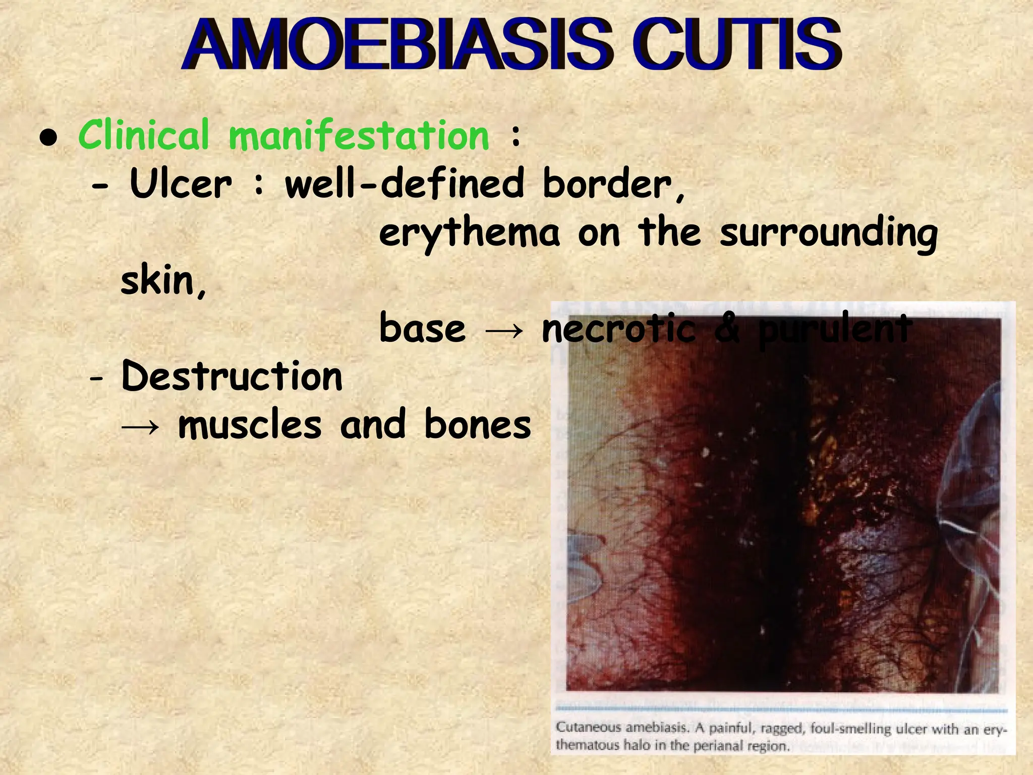
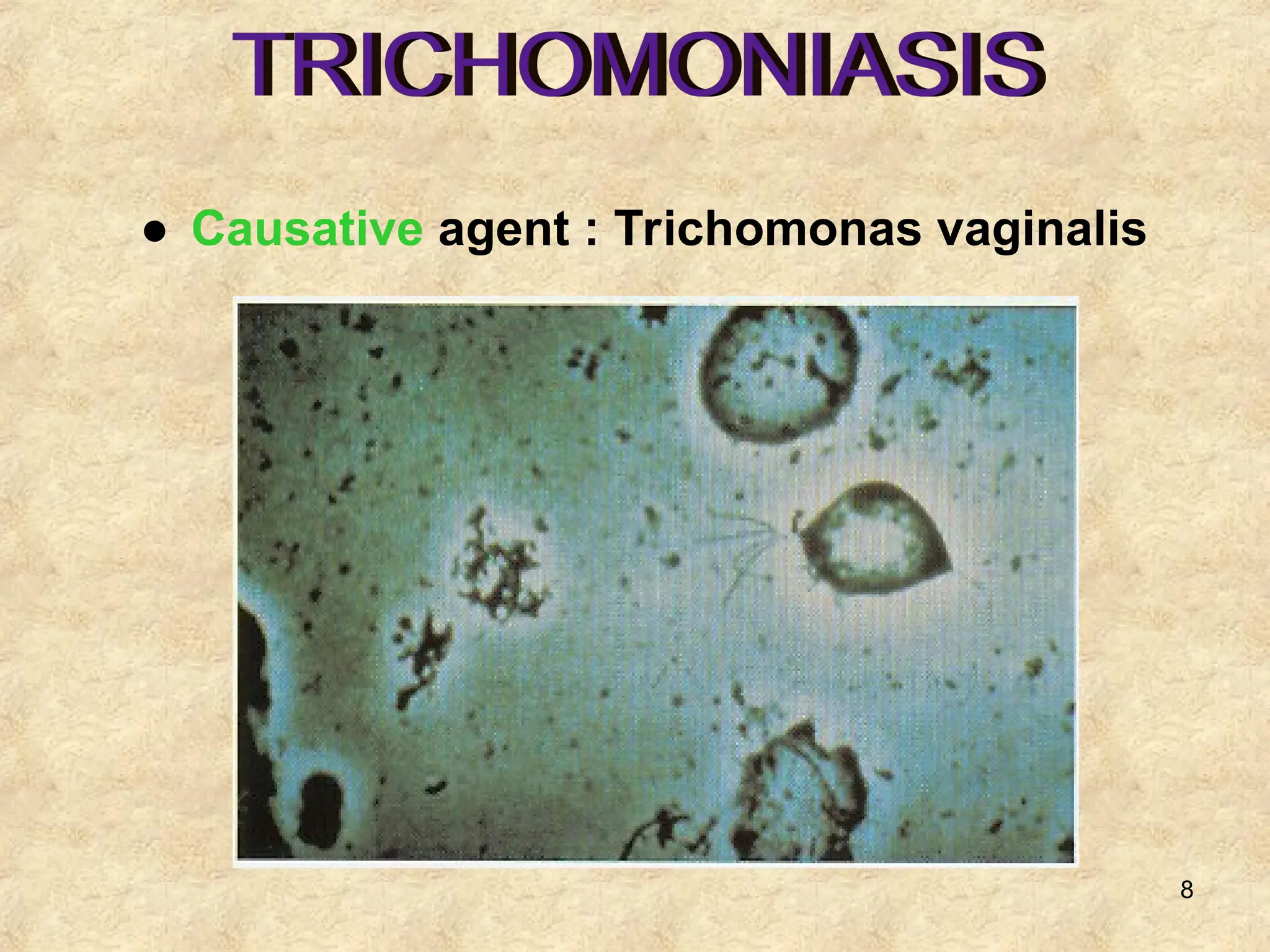
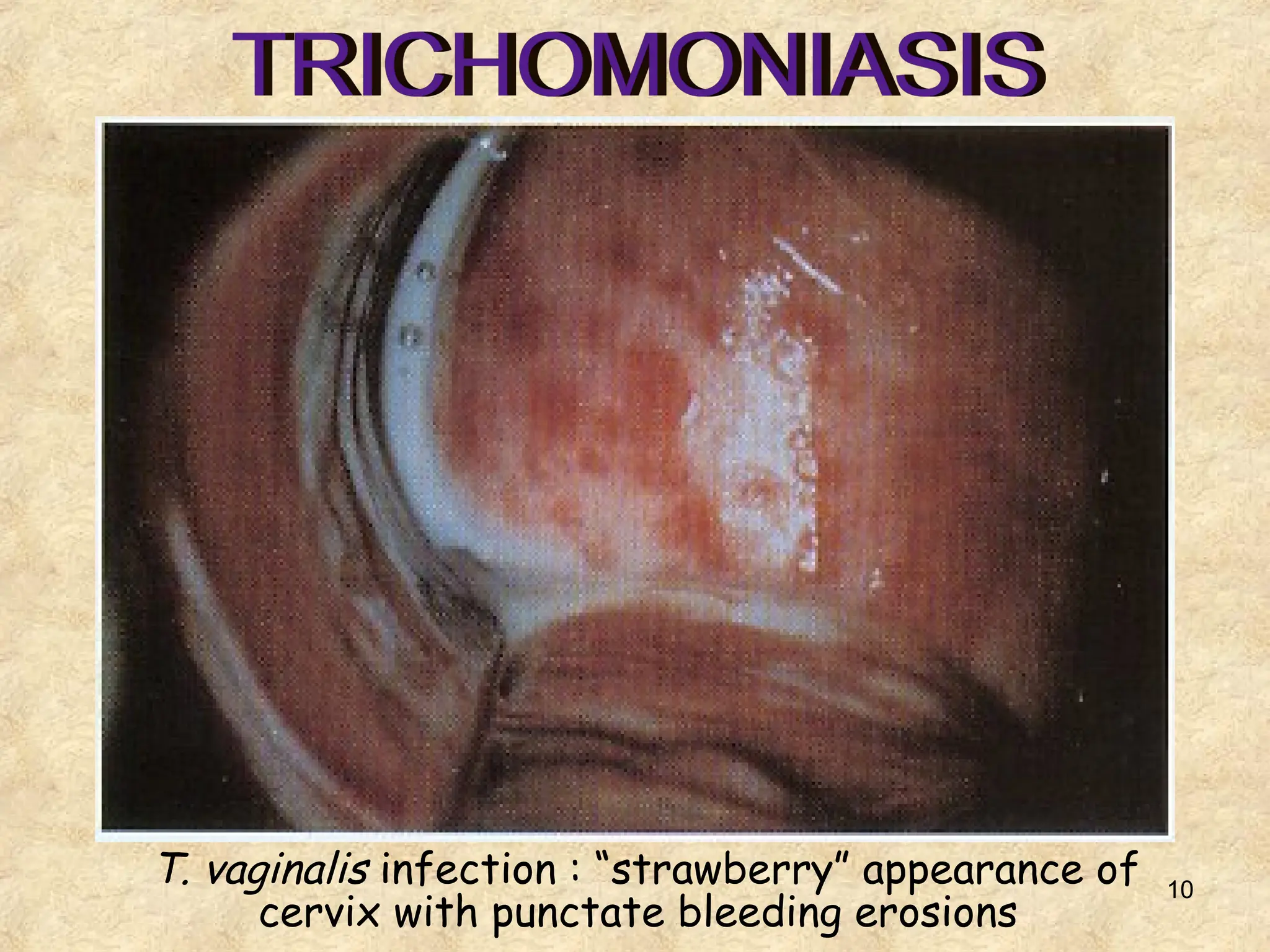
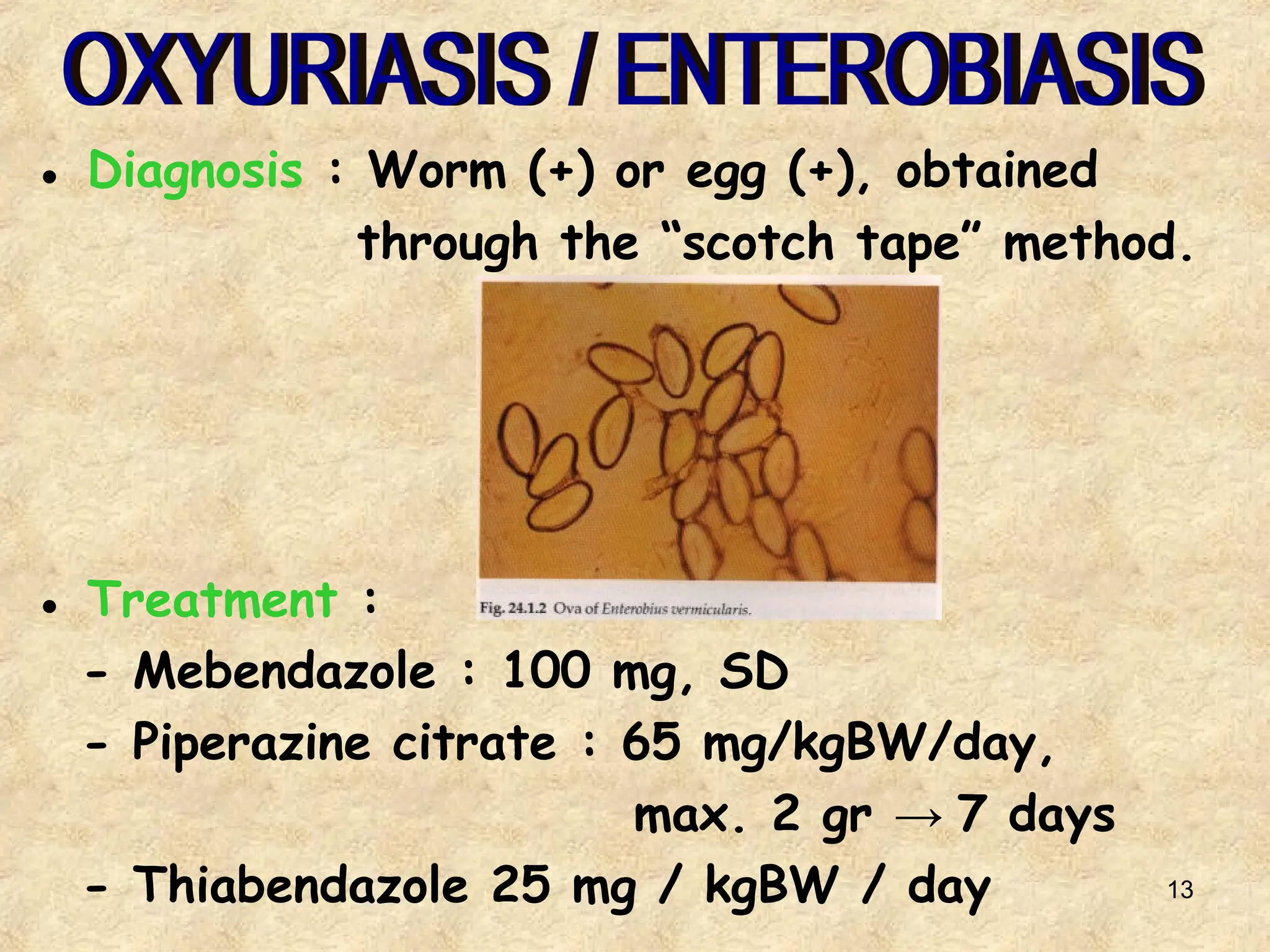
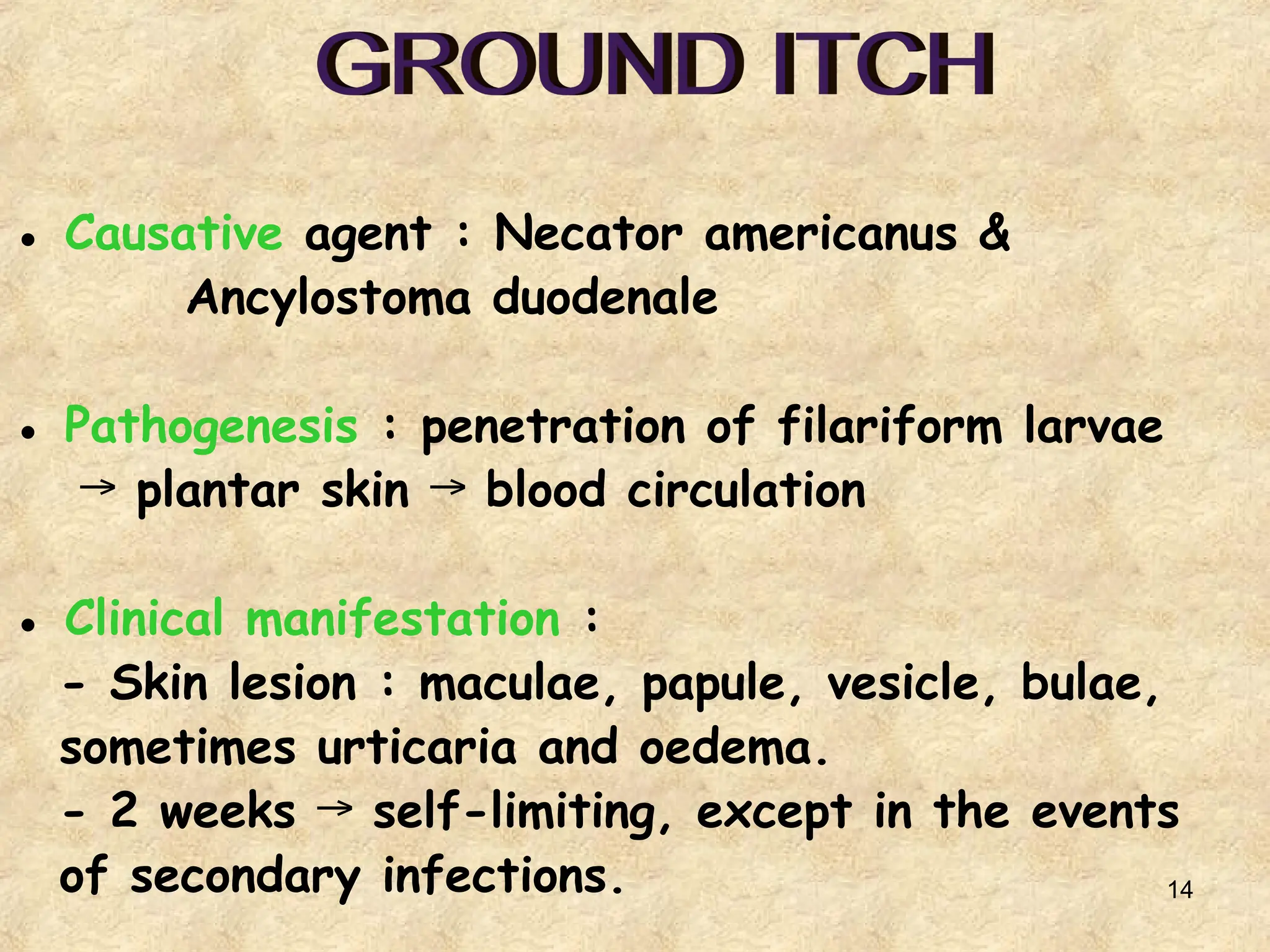
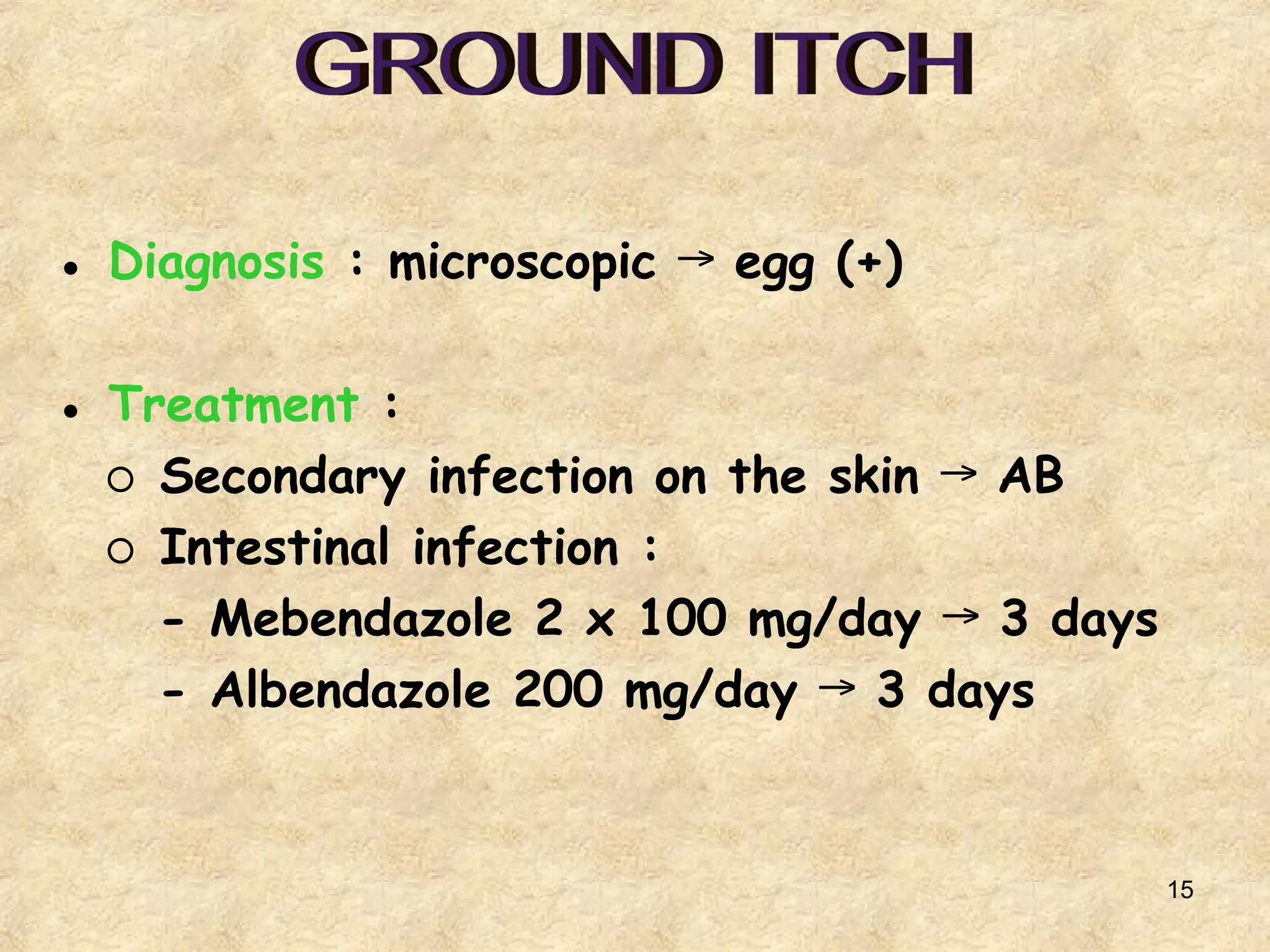
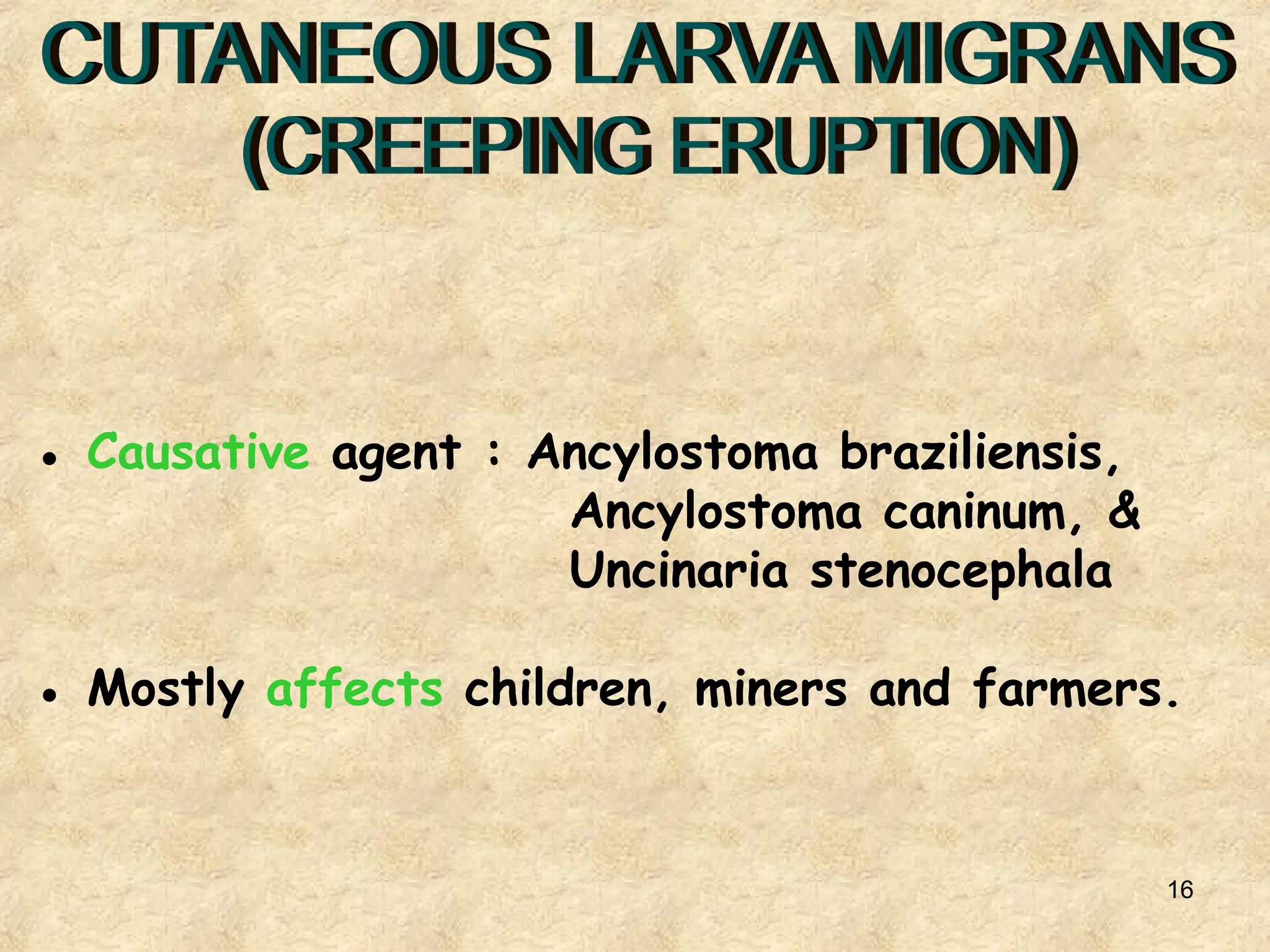
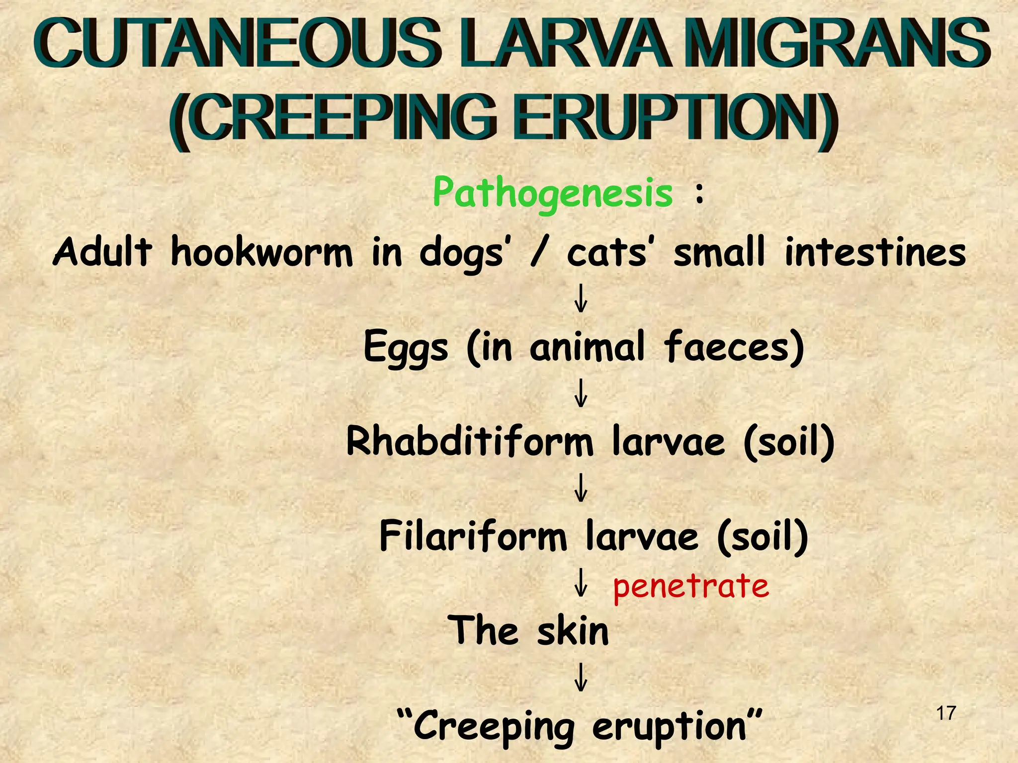
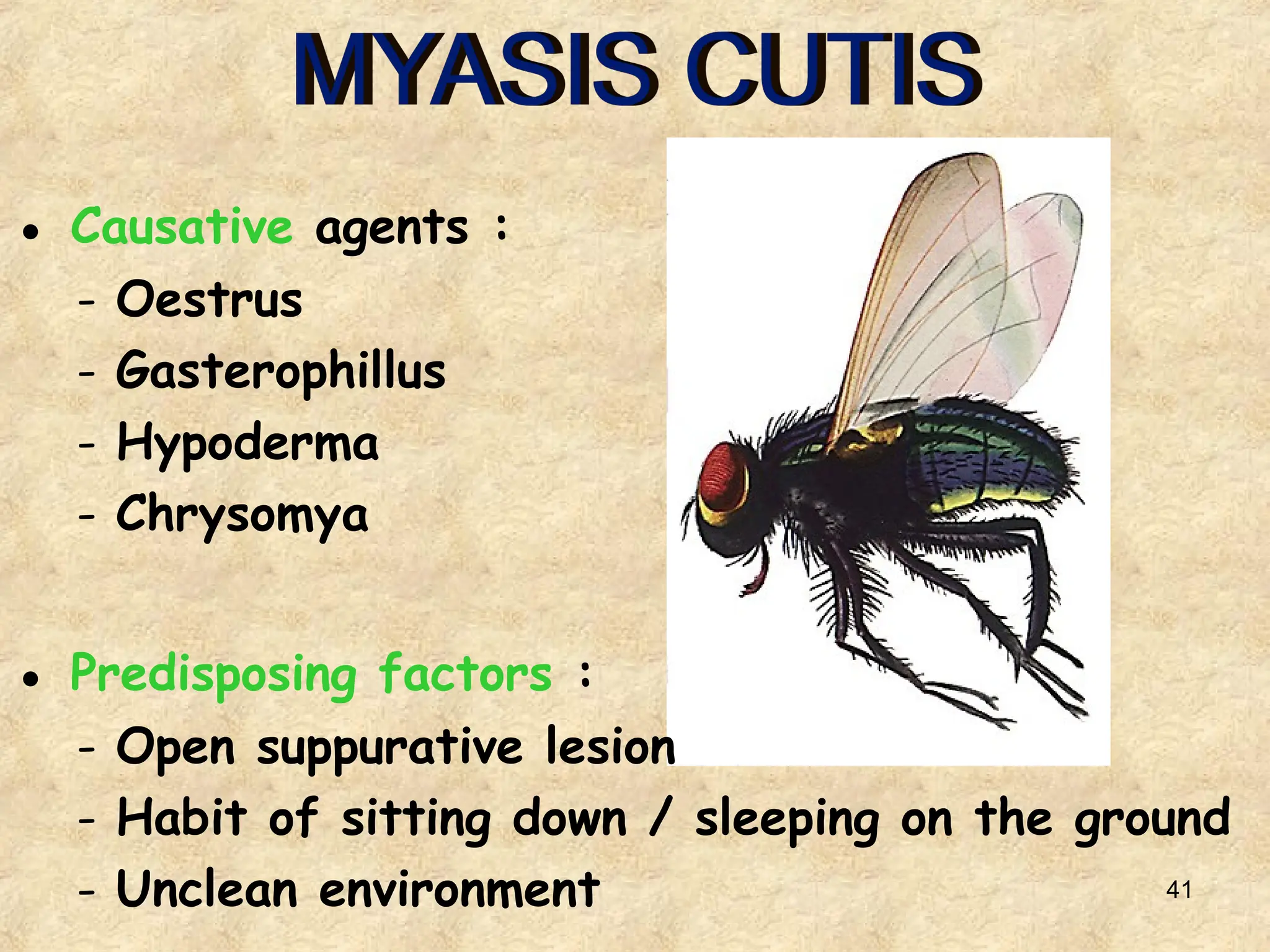
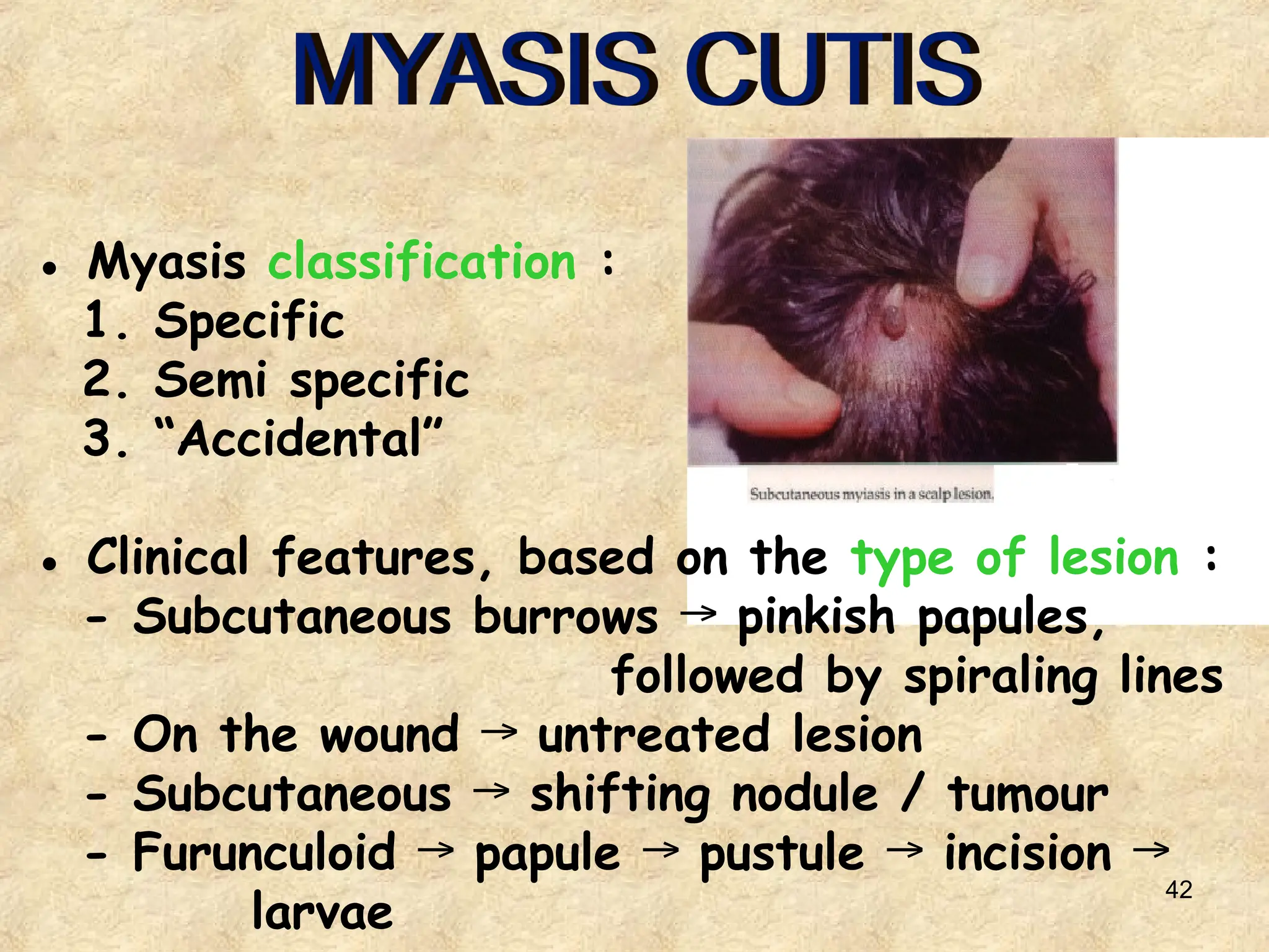
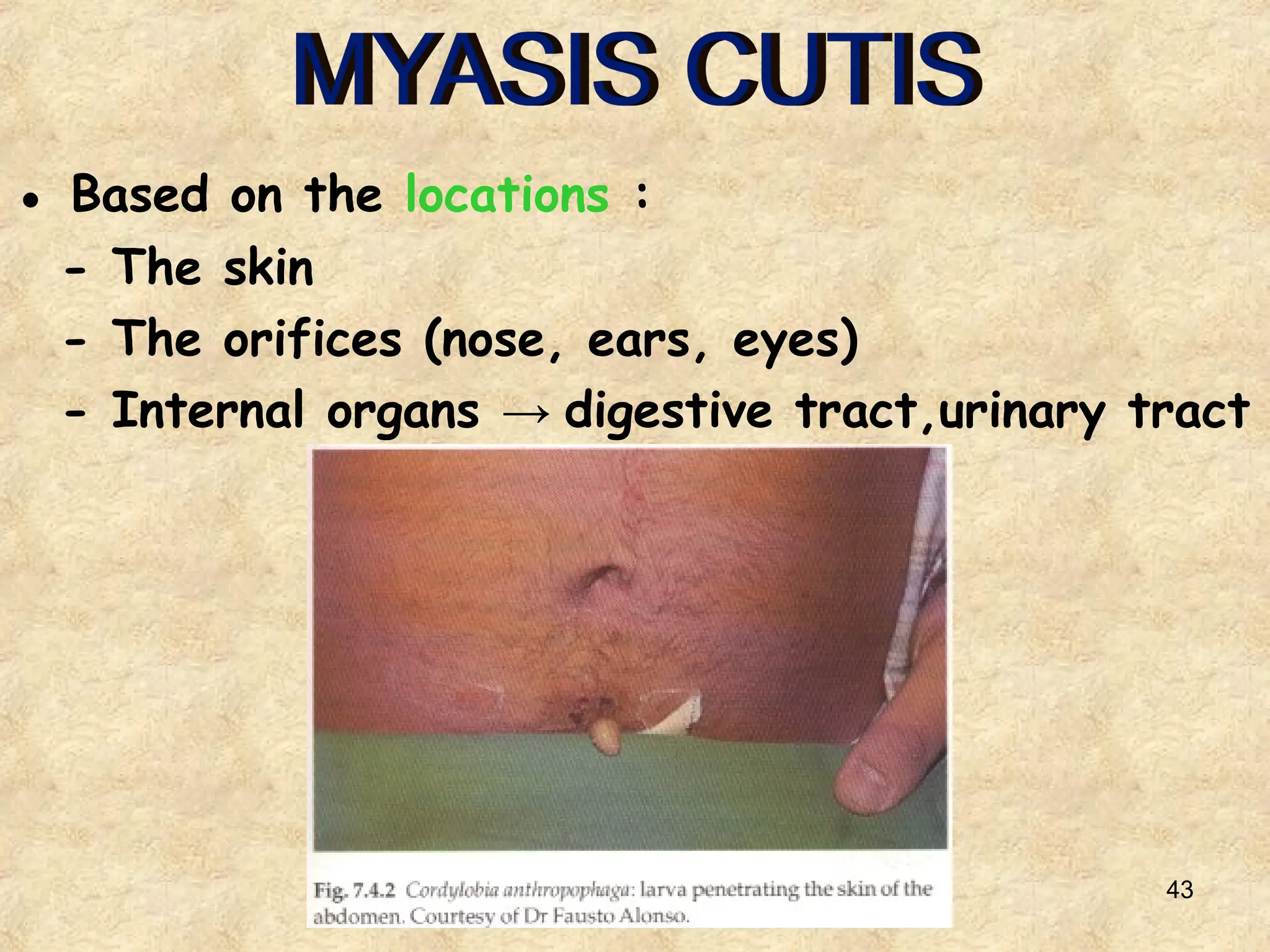
1
KULIAH
REGULER 2016
2
ZOONOSIS
Dr. LIZA AFRILIANA, SpKK (K)
Department of dermatovenereology
Faculty of medicine, Diponegoro University
3
● Zoonosis : skin disease caused by
various parasites
● This disease is often found in :
o Crowded areas
o Low socio-economic condition
o Poor sanitation and hygiene
● In the eradication effort, these attempts
are needed:
◦ Early diagnosis and prompt treatment
◦ Public health education about :
- Prevention
- Proper hygiene
- Reservoir-host-vector control
4
The causes of this disease are divided into
3 major groups :
1. PROTOZOA : - Amoebiasis
- Trichomoniasis
2. NEMATODA : - Oxyuriasis / enterobiasis
- “Ground itch”
- Cutaneous larva migrans
- Current larva
- Filariasis
- Dracunculiasis
3. ARTROPODA : - Scabies
- Pediculosis
5
● Causative agent : Entamoeba histolytica
● The amoeba may affect the skin through
these mechanisms :
1. Direct invasion of intestinal amoeba on the
surrounding skin
2. directly from hepatic abscess
3. Direct implantation of trophozoit on skin
with / without lesion
6
● Clinical manifestation :
- Ulcer : well-defined border,
erythema on the surrounding
skin,
base → necrotic & purulent
- Destruction
→ muscles and bones
7
● Diagnosis :
– Material scrapes and biopsy taken from
→
the edge of the ulcer, including
the base of ulcer, necrotic border,
& some surrounding skin
– Microscopic motile trophozoit
→
● Treatment :
– Metronidazole :
3 x 750 mg / day 10 days
→
– Dihydroemetin :
1,5 mg/kgBW/day IM 10 days
→
8
● Causative agent : Trichomonas vaginalis
9
● Transmission : sexual contact.
● Clinical manifestation :
- Vaginal discharge seropurulent,
→
yellow / greenish yellow,
foul-smelling & fizzy.
- Vulva pruritus, irritant dermatitis.
→
- Strawberry appearance the vaginal wall is
→
red and swollen.
- Infected men : are asymptomatic.
10
T. vaginalis infection : “strawberry” appearance of
cervix with punctate bleeding erosions
11
● Diagnosis : vaginal discharge / urine + NaCl
→ microscopic
→ movement of T.vaginalis
● Treatment :
- Metronidazole 3 x 250 mg 7 – 10 days
→
- Metronidazole single dose 2 gr
12
● Causative agent : Enterobius vermicularis
● Often affects children aged 5 – 14 years old.
● The transmission is through :
- food / beverage
- egg-infested hands
● Clinical manifestation :
- Perianal/perineal pruritus, especially at night
- Perineal intertrigo → excoriation &
superinfection
13
● Diagnosis : Worm (+) or egg (+), obtained
through the “scotch tape” method.
● Treatment :
- Mebendazole : 100 mg, SD
- Piperazine citrate : 65 mg/kgBW/day,
max. 2 gr → 7 days
- Thiabendazole 25 mg / kgBW / day
14
● Causative agent : Necator americanus &
Ancylostoma duodenale
● Pathogenesis : penetration of filariform larvae
→ plantar skin blood circulation
→
● Clinical manifestation :
- Skin lesion : maculae, papule, vesicle, bulae,
sometimes urticaria and oedema.
- 2 weeks self-limiting, except in the events
→
of secondary infections.
15
● Diagnosis : microscopic egg (+)
→
● Treatment :
○ Secondary infection on the skin AB
→
○ Intestinal infection :
- Mebendazole 2 x 100 mg/day 3 days
→
- Albendazole 200 mg/day 3 days
→
16
● Causative agent : Ancylostoma braziliensis,
Ancylostoma caninum, &
Uncinaria stenocephala
● Mostly affects children, miners and farmers.
17
Pathogenesis :
Adult hookworm in dogs’ / cats’ small intestines
↓
Eggs (in animal faeces)
↓
Rhabditiform larvae (soil)
↓
Filariform larvae (soil)
↓ penetrate
The skin
↓
“Creeping eruption”
18
Clinical features :
- Common locations are on the buttocks,
feet, & hands.
- The diameter of the lesion is 1 – 4 mm,
red in color, a bit raised,
like a coiled thread.
19
Typical track of CLM located on plantar aspect of foot.
20
Vesiculobullous lesion of CLM.
21
● Diagnosis : typical lesion manifestation
● Treatment :
○ Topical :
- Classic : Chlorethyl spray, CO2, liquid N2
- Thiabendazole 10%
- Albendazole 2%
○ Systemic :
- Thiabendazole 25 mg/kgBW/day 2 days
→
- Albendazole 50 mg/kgBW/once a week
22
● Causative agent :
Strongyloides
stercoralis
● Method of
transmission :
autoinfection
23
● Clinical features :
- Lesion urtica ribbon, quickly lengthening,
→
10 cm / hour
● Location : anus, spreading to the buttocks,
abdomen, or thighs.
● Diagnosis :
- Typical lesion
- Larva in the feces (microscopic)
● Treatment :
- Thiabendazole 25 mg/kgBW/day 5 days
→
- Albendazole / Mebendazole
24
● Causative agent : Wuchereria bancrofti
Brugia malayi
● Vector : anopheles, culex, aedes, & mansonia
mosquitos.
● Method of transmission :
Wuchereria bancrofti (adult form)
↓ lymphatic glands
Microfilaria (peripheral lymphatic glands /
blood vessels)
↓
Mosquito (infective)
↓
Human
25
● Clinical features :
1. Asymptomatic stage :
- Incubation period 8 – 12 months
- Lab : microfilaria in the blood,
eosinophilia
2. Acute inflammation stage :
- Lymphangitis on the extremities/scrotum,
pain, rigid, glossy skin, heat.
- Lymphadenitis
- Orchitis, funiculitis, epididimitis
26
3. Chronic obstructive stage :
- Repetitive inflammations & fibrosis of the
lymphatic glands obstruction
→
- Obstruction disturbance of the
→
lymphatic drainage liquid accumulation
→
in the tissue and lymphatic glands →
progressive in nature due to repetitive
inflammations.
– Manifestations : varicose lymph &
lymphedema
– Chronic edema connective tissues
→ →
deformity & elephantiasis
● Clinical features :
27
● Diagnosis :
- Examination of the
peripheral blood vessels
at night-time →
microfilaria
- Examination of the
hydrocele’s liquid or
urine
● Treatment :
Diethylcarbamazine
2 mg/kgBW/day 3 weeks
→
Lymphatic elephantiasis secondary
to Wuchereria bancrofti
28
● Causative agent : Dracunculus medinensis
● Intermediate host :
Crustacea species from the Cyclops genus
Consumption of Cyclops-infested
water / beverage
↓
Intestinal walls & retroperitoneal tissues
(adult worms)
↓
Sub-cutaneous tissues (body part that has
contact with the water / feet)
↓
Larvae
29
Clinical features :
Erythema, urticaria, itch, several hours →
papule vesicle bullae
→ → →
erosion / ulceration
30
● Diagnosis :
Radiology examination →
calcification of the dead worms
● Treatment :
- Thiabendazole 50–100 mg/kgBW 2 days
→
- Metronidazole 30–40 mg/kgBW/day 3 days
→
31
● Method of transmission :
- Direct handshake, sexual contact
→
- Indirect through objects
→
● Etiology : Sarcoptes scabiei var hominis
Life cycle : Female mites in stratum corneum
↓
Eggs
↓ 3–4 days
Larvae 10–14
↓ days
Mites
32
Clinical features :
- Predilection : in-between fingers,
flexor of the wrists,
genitalia, axillae folds,
lower abdomen, buttocks.
- Lesion papule, vesicle,
→
excoriation/secondary infection,
sometimes forming burrows.
33
Clinical variations :
 “Incognito” scabies
 Scabies in infants &
children
 Noduler scabies
 Scabies transmitted
by animals
 Scabies “in a clean”
 Norwegian scabies
34
35
● Additional examinations :
- Microscopic mites, eggs, faeces
→
- Burrows tetracycline
→
- Skin biopsy
● Diagnosis :
- Itch, especially at night-time
(nocturnal itching)
- History of infection on members of the
family / people living under the same roof
- Characteristic distribution of lesion
- Characteristic lesion burrows
→
- Definite diagnosis mites, eggs, faeces
→
- Tx antiscabies improvement
→
36
● Treatment :
– Gamexan 1%
– Crotamiton 10%
– Sulfur 5 – 10%
– Benzoil benzoate 20 – 35%
– Permethrin 5%
● To achieve treatment success :
- Treat every contact person
- Correct drug administration
- Washing clothes and towels with hot water,
air mattress under the sun
- Avoid excess treatment
37
• P. capitis → P. humanus var capitis
• P. corporis → P. humanus var corporis
• P. pubis → Phtirus pubis
Pathogenesis :
- Direct contact
- Indirect contact
38
P. CAPITIS
● Clinical manifestation : often affects children
● Symptoms :
- itch, especially on the occipital &
temporal parts excoriation, erosion /
→
secondary infection
- swelling of the lymph glands
P. CORPORIS
● Clinical features :
- hemorrhagic macules / papules with
punctum in the middle
- urtica
39
P. PUBIS
● Mites pubic hair, eyebrows, eyelashes,
→
axillae region, sometimes body hair
● Clinical features :
- itch excoriation / secondary infection
→
- Characteristic “maculae cerulae”
→
40
Diagnosis :
P. capitis : shiny eggs on hair, mites
P. corporis : eggs / mites on clothes’ folds
P. pubis : eggs / mites on pubic hair,
eyelashes, body hair
Treatment :
P. capitis : - gamexan 1% shampoo
- permethrin 1% cream
- crotamiton 10% cream / lotion
P. corporis : - gamexan 1%
- Washing clothes / bed sheets
using hot water / ironing them
P. pubis : - petrolatum
- physostigmine 0,025% eye oint
41
● Causative agents :
- Oestrus
- Gasterophillus
- Hypoderma
- Chrysomya
● Predisposing factors :
- Open suppurative lesion
- Habit of sitting down / sleeping on the ground
- Unclean environment
42
● Myasis classification :
1. Specific
2. Semi specific
3. “Accidental”
● Clinical features, based on the type of lesion :
- Subcutaneous burrows pinkish papules,
→
followed by spiraling lines
- On the wound untreated lesion
→
- Subcutaneous shifting nodule / tumour
→
- Furunculoid papule pustule incision
→ → → →
larvae
43
● Based on the locations :
- The skin
- The orifices (nose, ears, eyes)
- Internal organs → digestive tract,urinary tract
44
● Diagnosis : Larva on the lesion (+)
● Treatment :
- Furunculoid :
○ Seal with petrolatum / paraffin,
then press larvae will be squeezed out
→
- On the wound : irrigation with chloroform /
ether
- Classic : fish the larvae out with
clover liquid
45
● Causative agents : Cimex lectularis
Cimex hemipterus
● Clinical features :
- Mite bite papule with punctum
→
- Hemorrhagic bulla
→
46
● Treatment :
- Eradication of mites with malathion / DDT
- Lesion with antipruritic lotion
47
TERIMA
KASIH

13. Zoonosis management and therapy algorithm

  • 1.
  • 2.
    2 ZOONOSIS Dr. LIZA AFRILIANA,SpKK (K) Department of dermatovenereology Faculty of medicine, Diponegoro University
  • 3.
    3 ● Zoonosis :skin disease caused by various parasites ● This disease is often found in : o Crowded areas o Low socio-economic condition o Poor sanitation and hygiene ● In the eradication effort, these attempts are needed: ◦ Early diagnosis and prompt treatment ◦ Public health education about : - Prevention - Proper hygiene - Reservoir-host-vector control
  • 4.
    4 The causes ofthis disease are divided into 3 major groups : 1. PROTOZOA : - Amoebiasis - Trichomoniasis 2. NEMATODA : - Oxyuriasis / enterobiasis - “Ground itch” - Cutaneous larva migrans - Current larva - Filariasis - Dracunculiasis 3. ARTROPODA : - Scabies - Pediculosis
  • 5.
    5 ● Causative agent: Entamoeba histolytica ● The amoeba may affect the skin through these mechanisms : 1. Direct invasion of intestinal amoeba on the surrounding skin 2. directly from hepatic abscess 3. Direct implantation of trophozoit on skin with / without lesion
  • 6.
    6 ● Clinical manifestation: - Ulcer : well-defined border, erythema on the surrounding skin, base → necrotic & purulent - Destruction → muscles and bones
  • 7.
    7 ● Diagnosis : –Material scrapes and biopsy taken from → the edge of the ulcer, including the base of ulcer, necrotic border, & some surrounding skin – Microscopic motile trophozoit → ● Treatment : – Metronidazole : 3 x 750 mg / day 10 days → – Dihydroemetin : 1,5 mg/kgBW/day IM 10 days →
  • 8.
    8 ● Causative agent: Trichomonas vaginalis
  • 9.
    9 ● Transmission :sexual contact. ● Clinical manifestation : - Vaginal discharge seropurulent, → yellow / greenish yellow, foul-smelling & fizzy. - Vulva pruritus, irritant dermatitis. → - Strawberry appearance the vaginal wall is → red and swollen. - Infected men : are asymptomatic.
  • 10.
    10 T. vaginalis infection: “strawberry” appearance of cervix with punctate bleeding erosions
  • 11.
    11 ● Diagnosis :vaginal discharge / urine + NaCl → microscopic → movement of T.vaginalis ● Treatment : - Metronidazole 3 x 250 mg 7 – 10 days → - Metronidazole single dose 2 gr
  • 12.
    12 ● Causative agent: Enterobius vermicularis ● Often affects children aged 5 – 14 years old. ● The transmission is through : - food / beverage - egg-infested hands ● Clinical manifestation : - Perianal/perineal pruritus, especially at night - Perineal intertrigo → excoriation & superinfection
  • 13.
    13 ● Diagnosis :Worm (+) or egg (+), obtained through the “scotch tape” method. ● Treatment : - Mebendazole : 100 mg, SD - Piperazine citrate : 65 mg/kgBW/day, max. 2 gr → 7 days - Thiabendazole 25 mg / kgBW / day
  • 14.
    14 ● Causative agent: Necator americanus & Ancylostoma duodenale ● Pathogenesis : penetration of filariform larvae → plantar skin blood circulation → ● Clinical manifestation : - Skin lesion : maculae, papule, vesicle, bulae, sometimes urticaria and oedema. - 2 weeks self-limiting, except in the events → of secondary infections.
  • 15.
    15 ● Diagnosis :microscopic egg (+) → ● Treatment : ○ Secondary infection on the skin AB → ○ Intestinal infection : - Mebendazole 2 x 100 mg/day 3 days → - Albendazole 200 mg/day 3 days →
  • 16.
    16 ● Causative agent: Ancylostoma braziliensis, Ancylostoma caninum, & Uncinaria stenocephala ● Mostly affects children, miners and farmers.
  • 17.
    17 Pathogenesis : Adult hookwormin dogs’ / cats’ small intestines ↓ Eggs (in animal faeces) ↓ Rhabditiform larvae (soil) ↓ Filariform larvae (soil) ↓ penetrate The skin ↓ “Creeping eruption”
  • 18.
    18 Clinical features : -Common locations are on the buttocks, feet, & hands. - The diameter of the lesion is 1 – 4 mm, red in color, a bit raised, like a coiled thread.
  • 19.
    19 Typical track ofCLM located on plantar aspect of foot.
  • 20.
  • 21.
    21 ● Diagnosis :typical lesion manifestation ● Treatment : ○ Topical : - Classic : Chlorethyl spray, CO2, liquid N2 - Thiabendazole 10% - Albendazole 2% ○ Systemic : - Thiabendazole 25 mg/kgBW/day 2 days → - Albendazole 50 mg/kgBW/once a week
  • 22.
    22 ● Causative agent: Strongyloides stercoralis ● Method of transmission : autoinfection
  • 23.
    23 ● Clinical features: - Lesion urtica ribbon, quickly lengthening, → 10 cm / hour ● Location : anus, spreading to the buttocks, abdomen, or thighs. ● Diagnosis : - Typical lesion - Larva in the feces (microscopic) ● Treatment : - Thiabendazole 25 mg/kgBW/day 5 days → - Albendazole / Mebendazole
  • 24.
    24 ● Causative agent: Wuchereria bancrofti Brugia malayi ● Vector : anopheles, culex, aedes, & mansonia mosquitos. ● Method of transmission : Wuchereria bancrofti (adult form) ↓ lymphatic glands Microfilaria (peripheral lymphatic glands / blood vessels) ↓ Mosquito (infective) ↓ Human
  • 25.
    25 ● Clinical features: 1. Asymptomatic stage : - Incubation period 8 – 12 months - Lab : microfilaria in the blood, eosinophilia 2. Acute inflammation stage : - Lymphangitis on the extremities/scrotum, pain, rigid, glossy skin, heat. - Lymphadenitis - Orchitis, funiculitis, epididimitis
  • 26.
    26 3. Chronic obstructivestage : - Repetitive inflammations & fibrosis of the lymphatic glands obstruction → - Obstruction disturbance of the → lymphatic drainage liquid accumulation → in the tissue and lymphatic glands → progressive in nature due to repetitive inflammations. – Manifestations : varicose lymph & lymphedema – Chronic edema connective tissues → → deformity & elephantiasis ● Clinical features :
  • 27.
    27 ● Diagnosis : -Examination of the peripheral blood vessels at night-time → microfilaria - Examination of the hydrocele’s liquid or urine ● Treatment : Diethylcarbamazine 2 mg/kgBW/day 3 weeks → Lymphatic elephantiasis secondary to Wuchereria bancrofti
  • 28.
    28 ● Causative agent: Dracunculus medinensis ● Intermediate host : Crustacea species from the Cyclops genus Consumption of Cyclops-infested water / beverage ↓ Intestinal walls & retroperitoneal tissues (adult worms) ↓ Sub-cutaneous tissues (body part that has contact with the water / feet) ↓ Larvae
  • 29.
    29 Clinical features : Erythema,urticaria, itch, several hours → papule vesicle bullae → → → erosion / ulceration
  • 30.
    30 ● Diagnosis : Radiologyexamination → calcification of the dead worms ● Treatment : - Thiabendazole 50–100 mg/kgBW 2 days → - Metronidazole 30–40 mg/kgBW/day 3 days →
  • 31.
    31 ● Method oftransmission : - Direct handshake, sexual contact → - Indirect through objects → ● Etiology : Sarcoptes scabiei var hominis Life cycle : Female mites in stratum corneum ↓ Eggs ↓ 3–4 days Larvae 10–14 ↓ days Mites
  • 32.
    32 Clinical features : -Predilection : in-between fingers, flexor of the wrists, genitalia, axillae folds, lower abdomen, buttocks. - Lesion papule, vesicle, → excoriation/secondary infection, sometimes forming burrows.
  • 33.
    33 Clinical variations : “Incognito” scabies  Scabies in infants & children  Noduler scabies  Scabies transmitted by animals  Scabies “in a clean”  Norwegian scabies
  • 34.
  • 35.
    35 ● Additional examinations: - Microscopic mites, eggs, faeces → - Burrows tetracycline → - Skin biopsy ● Diagnosis : - Itch, especially at night-time (nocturnal itching) - History of infection on members of the family / people living under the same roof - Characteristic distribution of lesion - Characteristic lesion burrows → - Definite diagnosis mites, eggs, faeces → - Tx antiscabies improvement →
  • 36.
    36 ● Treatment : –Gamexan 1% – Crotamiton 10% – Sulfur 5 – 10% – Benzoil benzoate 20 – 35% – Permethrin 5% ● To achieve treatment success : - Treat every contact person - Correct drug administration - Washing clothes and towels with hot water, air mattress under the sun - Avoid excess treatment
  • 37.
    37 • P. capitis→ P. humanus var capitis • P. corporis → P. humanus var corporis • P. pubis → Phtirus pubis Pathogenesis : - Direct contact - Indirect contact
  • 38.
    38 P. CAPITIS ● Clinicalmanifestation : often affects children ● Symptoms : - itch, especially on the occipital & temporal parts excoriation, erosion / → secondary infection - swelling of the lymph glands P. CORPORIS ● Clinical features : - hemorrhagic macules / papules with punctum in the middle - urtica
  • 39.
    39 P. PUBIS ● Mitespubic hair, eyebrows, eyelashes, → axillae region, sometimes body hair ● Clinical features : - itch excoriation / secondary infection → - Characteristic “maculae cerulae” →
  • 40.
    40 Diagnosis : P. capitis: shiny eggs on hair, mites P. corporis : eggs / mites on clothes’ folds P. pubis : eggs / mites on pubic hair, eyelashes, body hair Treatment : P. capitis : - gamexan 1% shampoo - permethrin 1% cream - crotamiton 10% cream / lotion P. corporis : - gamexan 1% - Washing clothes / bed sheets using hot water / ironing them P. pubis : - petrolatum - physostigmine 0,025% eye oint
  • 41.
    41 ● Causative agents: - Oestrus - Gasterophillus - Hypoderma - Chrysomya ● Predisposing factors : - Open suppurative lesion - Habit of sitting down / sleeping on the ground - Unclean environment
  • 42.
    42 ● Myasis classification: 1. Specific 2. Semi specific 3. “Accidental” ● Clinical features, based on the type of lesion : - Subcutaneous burrows pinkish papules, → followed by spiraling lines - On the wound untreated lesion → - Subcutaneous shifting nodule / tumour → - Furunculoid papule pustule incision → → → → larvae
  • 43.
    43 ● Based onthe locations : - The skin - The orifices (nose, ears, eyes) - Internal organs → digestive tract,urinary tract
  • 44.
    44 ● Diagnosis :Larva on the lesion (+) ● Treatment : - Furunculoid : ○ Seal with petrolatum / paraffin, then press larvae will be squeezed out → - On the wound : irrigation with chloroform / ether - Classic : fish the larvae out with clover liquid
  • 45.
    45 ● Causative agents: Cimex lectularis Cimex hemipterus ● Clinical features : - Mite bite papule with punctum → - Hemorrhagic bulla →
  • 46.
    46 ● Treatment : -Eradication of mites with malathion / DDT - Lesion with antipruritic lotion
  • 47.