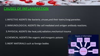
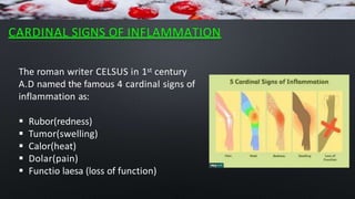
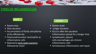
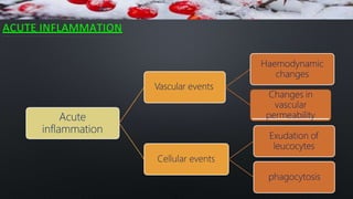
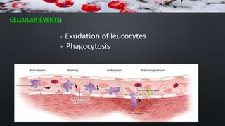
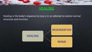
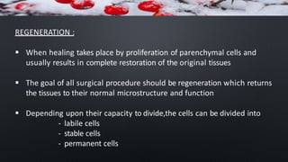
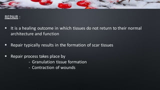
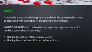
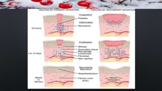
INFLAMMATION
AND WOUND HEALING
CONTENTS
Basic mechanism involved in the process of inflammation and repair:
Introduction
Clinical signs of inflammation
Different types of Inflammation
Mechanism of Inflammation – Alteration in vascular permeability and blood
flow, migration of WBC’s
Mediators of inflammation
Basic principles of wound healing in the skin
INTRODUCTION
INFLAMMATION (derived from LATIN word inflammare, (To set on
fire)is a localized protective response elicited by injury or destruction
of tissues which serves to destroy ,dilute the wall of both the injurious
agent and injured tissues
DEFINITION
 Inflammation is defined as the local response of living mammalian
tissues to injury due to any agent .It is a body defence reaction in order
to eliminate or limit the spread of injurious agent as well as to remove
the consequent necrosed cells and tissues
John hunter (1973) stated “inflammation is not a disease
but a specific response that has a salutary effect on its host“
CAUSES OF INFLAMMATION
1.INFECTIVE AGENTS like bacteria ,viruses,and their toxins,fungi,parasites.
2.IMMUNOLOGICAL AGENTS like cell mediated and antigen antibody reactions.
3.PHYSICAL AGENTS like heat,cold,radiation,mechanical trauma
4.CHEMICAL AGENTS like organic and inorganic poisons
5.INERT MATERIALS such as foreign bodies
CARDINAL SIGNS OF INFLAMMATION
The roman writer CELSUS in 1st century
A.D named the famous 4 cardinal signs of
inflammation as:
 Rubor(redness)
 Tumor(swelling)
 Calor(heat)
 Dolar(pain)
 Functio laesa (loss of function)
TYPES OF INFLAMMATION
 Slower onset
 Longer duration
 Occurs after the causative
inflammation persist for a longer time
 Lymphocytes ,plasma
cells,macrophages are chronic
inflammatory cells
 Simultaneous inflammation and repair
ACUTE
 Rapid onset
 Slow duration
 Accumulation of fluids and plasma
at the affected site
 Polymorphonuclear neutrophils as
inflammatory cells
 Represents early body reactions
followed by repair
CHRONIC
ACUTE INFLAMMATION
Acute
inflammation
Haemodynamic
changes
Vascular events
Changes in
vascular
permeability
Exudation of
leucocytes
Cellular events
phagocytosis
HAEMODYNAMIC CHANGES
Transient
vasoconstriction
Persistant progressive
vasodilatation
Local hydrostatic
pressure
Slowing and stasis
Leucocyte migration
LEWIS EXPERIMENT
 Red line (vasodilation of micro
vasculature)
 Flare (bright reddish appearance
surrounding the red line due to
vasodilation of adjacent
arterioles)
 wheal(swelling or oedema due
to transudation of fluid)
CHANGES IN VASCULAR PERMEABILITY
The appearance of Inflammatory oedema due to increased
vascular permeability of microvasculature bed is explained on
the basis of “STARLINGS HYPOTHESIS”
In normal circumstances ,the fluid balance is maintained by two
sets of forces
1)Forces that causes outward movement of fluid from
microcirculation are INTRAVASCULAR HYDROSTATIC PRESSURE and
OSMOTIC PRESSURE OF INTERSTITIAL FLUID
2)Forces that causes inward movement of fluid into circulation
are INTRAVASCULAR OSMOTIC PRESSURE and HYDROSTATIC PRESSURE
OF INTERSTITIAL FLUID
 Left over fluid in the interstitial compartment is drained away by
lymphatics and thus no oedema results normally in inflamed tissue
 Endothelial lining of microvasculature becomes more leaky
 Consequently ,intravascular osmotic pressure decreases and
osmotic pressure of interstitial fluid increases resulting in
excessive out flow of fluid into the interstitial compartment which
is exudative inflammatory oedema
MECHANISM OF INCREASED VASCULAR PERMEABILITY
1) ENDOTHELIAL CELL CONTRACTION
• Microvasculature;
• Venules response type; immediate (15 - 30min)
pathogenesis;
•By mediators like histamins,bradikinins,others
example; mild thermal injury
ENDOTHELIAL CELL RETRACTION
• Microvasculature;
• Venules response type ;delayed prolonged path;
Cytokines - IL-1.TNF ;
• 4-6 hours
DIRECT ENDOTHELIAL CELL INJURY
• Cell necrosis and detachment;
• All microvasculature;
• Increase in permeability might be immediately after injury and last for severa
hours or days.
example; moderate to severe burns,bacterial infection,etc
LEUCOCYTE MEDIATED ENDOTHELIAL INJURY
Microvasculature ;venules response type ;
activated leucocytes(proteolytic enzymes and toxic 02 species)
delayed prolonged pathogenesis;
Example; pulmanary venules and capilaries
LEAKINESS IN NEOVASCULARISATION
Newly formed capillaries
Vascular endothelial growth factor
During repair and in tumuors
CELLULAR EVENTS:
- Exudation of leucocytes
- Phagocytosis
EXUDATION OF LEUCOCYTES
1) MARGINATION AND PAVEMENTING
MARGINATION : Cells line up against the endothelium.
PAVEMENTING : Sticking of white blood cells to the
linings of the finest blood vessels (capillaries) when
inflammation occurs. WBC’s gather along the
endothelium, like bricks paving a road). From here the
leukocytes crawl between the endothelial cells and
enter the inflamed connective tissue.
2)ADHESION OR ROLLING :
Adhesion – connecting to the endothelial wall.
ROLLING: neutrophils roll over endothelial cells
- Selectins
- Integrins
- Immunoglobulin gene super family adhesion
molecules
3)EMIGRATION – Diapedesis
Neutrophils undergo lateral migration or
crawling on endothelial cells to find a permissive
site for transmigration. Movement of leukocytes
out of the circulatory system and towards the
site of tissue damage or infection.
 CHEMOTAXIS –
Chemotaxis is a fundamental
biological process in which a
cell migrates following the
direction of a spatial cue. This
spatial cue is provided in a
form of a gradient of
chemoattractants.
Chemotaxis of
leukocytes, a requisite process
for neutrophil extravasation from
the blood into tissues, is a critical
step for initiating and maintaining
inflammation in both acute and
chronic inflammation.
PHAGOCYTOSIS
Phagocytosis is defined as the process of engulfment of solid particulate
material by the cells(cell eating) The cells performing this function are called
phagocytes.
There are two types of phagocytic cells
- PMNs
- Macrophages
Neutrophils and macrophages in the tissue space produce several proteolytic
enzymes such as lysosymes, proteases, collagenase, elastase, lipase,
proteinase,gelatinase and acid hydrolases.
These enzymes degrade collagen and extra cellular matrix.
STAGES IN PHAGOCYTOSIS
1) ATTACHMENT STAGE
•Opsonised microbes attaches to phagocytic surface receptors
•Opsonin are naturally occurring factors in the serum
•Two main opsonins present in the serum are IgG opsonin and
C3b opsonin and their corresponding receptors on the surface of
phagocytes
2)ENGULFMENT STAGE
Cytoplasmic extensions surround and engulf the bound opsonised
microbe by formation of pseudopods around the particle ,enveloping it
in a phagocytic vacuole or phagosome.
•This phagosome fuses with the lysosome
of cell to form phagolysosome
3)KILLING (OR) DEGRADATION STAGE
• In this stage the microbes embedded in phagosome is killed by
antimicrobial or bactericidal substances present in granules of
phagocytes and degraded by hydrolytic enzymes synthesised by
phagocytes.
CHEMICAL MEDIATORS OF INFLAMMATION
VASOACTIVE AMINES
A)HISTAMINE:
Histamine is stored in the granules of mast cells,basophils,and platelets
Histamine is released from these cells by various agents like:
a)Stimuli or substances inducing acute inflammation eg; heat ,cold,trauma
irritant chemicals etc
b) Histamine releasing factors from neutrophils,monocytes,and platelets,
c) Interleukins
The main action of histamine are vasodilation,increase vascular
permeability,itching and pain
B) 5-HYDROXYTRYPTAMINE(5-HT OR SERATONIN )
It is present in tissues like chromaffin cells of GIT,spleen,nervous
tissues,mast cells,and platelets
The action of Serotonin is similar to histamine but it is a less potent
mediator of increased permeability and vasodilation than histamine
C) NEUROPEPTIDES:These are the small peptides produced in CNS and PNS
(substance p, neurokinin etc)
Actions:- 1) Increase vascular permeability
2) pain
3) mast cell degranulation
ARACHIDONIC ACID METABOLITES
Arachidonic acid is a fatty acids and is a constituent of phospholipid cell
membrane besides obtaining through diet.(essential fatty acids – linoleic
acid – arachidonic acid)
 Arachidonic acid metabolites are most potent inflammatory mediators
than oxygen free radicals
LYSOSOMAL COMPONENTS
The inflammatory cells contains lysosomal granules which on release
elaborate a variety of mediators of Inflammation,They are as follows
a)Granules of neutrophils
b)Granules of monocytes and tissue macrophages
a)GRANULES OF NEUTROPHILS
 primary granules (myeloperoxidase)
 secondary granules
(lactoferrin,lysozyme, alkaline
phosphatase,collagenase )
b)GRANULES OF MONOCYTES AND TISSUE
MACROPHAGES
 Releases mediators like acid
proteases,collagenase,elastase, and
plasminogen
PLATELET ACTIVATING FACTOR(PAF)
It is released from IgE sensitised basophils,or mast cells .Actions of
PAF as mediators of inflammation are
a)increased vascular permeability
b)vasodilation in low concentration and vasoconstriction in higher
concentration
c)Bronchoconstriction
d)adhesion of leukocytes toendothelium
e)chemotaxis
CYTOKINES
 Cytokines are lower molecular weight regulatory proteins or
glycoproteins(polypeptide substances) secreted by activated lymphocytes
(lymphokines) and Monocytes(monokines)
 Main cytokines acting as a mediators of inflammation are as follows
Interleukin-1(IL-1); Tumour necrosis factor( alpha.beta);
Interferon(gama) and chemokines(IL-8.PF-4)
OXYGEN METABOLITES
1) They are released from activated neutrophils and macrophages
2)Includes superoxide,H2O2,toxic NO products are few oxygen
metabolites
3) Role of oxygen metabolites as inflammatory mediators includes
- vascular permeability by endothelial cell damage,
- activation of protease,
- damage of other cells,
NITRIC OXIDE METABOLITES
 Nitric oxide was originally described as vascular relaxation factor
,it has recently been included as a mediators of inflammation
 Nitric oxide has been shown to have fungicidal and anti parasitic
action
 Role of Nitric oxide in the inflammation are vasodilation,
antiplatelet activating agent, microbicidal action, etc
PLASMA DERIVED MEDIATORS (plasma proteases)
These include various products derived from activation and interaction
of 4 inter linked systems as follows:
a)The Kinin system
b) The Clotting system
c)The fibrinolytic system
d)The complement system
Effects of bradykinin are:
- Smooth muscle Contraction,
- vasodilation(hypotension),
-Increases vascular Permeability(edema),
&
- Involved in mechanism of pain.
Action of fibrinopeptides in
inflammation are:
a)Increased vascular
permeability b)chemotaxis for
leucocytes
Healing is the body’s response to injury in an attempt to restore normal
structure and function
HEALING
REGENERATION
HEALING
REPAIR
REGENERATION :
 When healing takes place by proliferation of parenchymal cells and
usually results in complete restoration of the original tissues
 The goal of all surgical procedure should be regeneration which returns
the tissues to their normal microstructure and function
 Depending upon their capacity to divide,the cells can be divided into
- labile cells
- stable cells
- permanent cells
REPAIR :
 It is a healing outcome in which tissues do not return to their normal
architecture and function
 Repair typically results in the formation of scar tissues
 Repair process takes place by
- Granulation tissue formation
- Contraction of wounds
WOUND HEALING
WOUND:
A wound is a break in the integrity of the skin or tissue often which may
be associated with disruption of the structure and function
WOUND HEALING is a combination of repair and regeneration which
can be accomplished in two ways:
 Healing by first intention(primary union)
 Healing by second intention(secondary union)
 Wound is clean and uninfected
 Without much loss of cells and
tissues
 Edges of wound are
approximated by surgical
sutures
 Wound is open with large
tissue defect,at times infected
 Having extensive loss of
tissues
 The wound is not
approximated by surgical
sutures but is left open
PRIMARY UNION SECONDARY UNION
HEALING BY FIRST INTENSION (PRIMARY UNION)
1) INITIAL HAEMORRHAGE
 Immediately after injury,the space between the approximated
surfaces of incised wound is filled with blood
 The filled blood clots seals the wound against infection
2) ACUTE INFLAMMATORY RESPONSE
 This occurs within 24 hrs with appearance of PMN from the
margins of incised wound
 By 3rd day ,polymorphs are replaced by macrophages
3)EPITHELIAL CHANGES
 The basal cells of epidermis from both the cut margins start
proliferating and migrating towards incisional space in the form of
epithelial spurs
 These migrated epithelial cells separate the under lying viable dermis
from the overlying necrotic material and clot forming a SCAB
 By 5th day ,a multilayered new epidermis is formed which is
differentiated into superficial and deep layers
 A well approximated wound is covered by a layer of epithelium in
48hours
ORGANISATION:
 By 3rd day ,fibroblast also invade the wound area.
 By 5th day ,new collagen fibrils start forming which dominate till healing
is completed
 By 4th week,the scar tissue with scanty cellular , vascular elements with
few inflammatory cells and epithelialised surface is formed
HEALING BY SECONDARY INTENTION(SECONDARY UNION)
1)INITIAL HAEMORRHAGE
As a result of injury ,the wound space is filled with blood and
fibrin clot which dries
2)INFLAMMATORY PHASE
There is an initial acute inflammatory response followed by
appearence of macrophages which clear of the debris as in primary
union
EPITHELIAL CHANGES
 As in the primary healing, the epidermal cells from both the margins
of the wound proliferate and migrate into the form of epithelial spurs
till they meet in the middle and reepithelialise the gap completely
 These proliferated epithelial cells do not cover the surface fully until
granulation tissue from the base has started filling the wound space
 After these epithelial cells meet in the middle they separate the
underlying viable tissues from necrotic tissue at the surface forming
scab
GRANULATION TISSUE FORMATION
 The main bulk of secondary healing is by granulation
 Granulation tissue is formed by proliferation of fibroblasts and
neovascularisation from the adjoining viable elements
 The newly formed granulation tissue is deep red,granular and very
fragile
 The specialised structure of skin like hair follicle and sweat glands are
not replaced unless their viable residues remain which may regenerate
WOUND CONTRACTION
 Contraction of wound is an important feature of secondary healing
,and not seen in primary healing
 Wound contraction occurs at a time when active granulation tissues
is being formed(approximately 4 to 5 days after wounding )
 Due to the action of myofibroblasts present in granulation tissue,the
wound contracts to one third of its original size
 Maximal contraction occurs for 12 to15 days although it will
continue if wound remain opens
COMPLICATION OF WOUND HEALING
1)Infection
2)Pigmentation
3)Deficient scar formation
4)Incisional hernia
5) Hypertrophied scar and keloid formation
6)Excessive contraction of wound
7)Epidermal cyst formation
REFERENCE : TEXT BOOK OF PATHOPHYSIOLOGY BY DR.
MADAN KAUSHIK; Page no:42-76
NO SELF STUDY TOPIC FROM
THIS CHAPTER

1.2 Inflammation & wound healing.pptx

  • 1.
  • 2.
    CONTENTS Basic mechanism involvedin the process of inflammation and repair: Introduction Clinical signs of inflammation Different types of Inflammation Mechanism of Inflammation – Alteration in vascular permeability and blood flow, migration of WBC’s Mediators of inflammation Basic principles of wound healing in the skin
  • 3.
    INTRODUCTION INFLAMMATION (derived fromLATIN word inflammare, (To set on fire)is a localized protective response elicited by injury or destruction of tissues which serves to destroy ,dilute the wall of both the injurious agent and injured tissues
  • 4.
    DEFINITION  Inflammation isdefined as the local response of living mammalian tissues to injury due to any agent .It is a body defence reaction in order to eliminate or limit the spread of injurious agent as well as to remove the consequent necrosed cells and tissues John hunter (1973) stated “inflammation is not a disease but a specific response that has a salutary effect on its host“
  • 5.
    CAUSES OF INFLAMMATION 1.INFECTIVEAGENTS like bacteria ,viruses,and their toxins,fungi,parasites. 2.IMMUNOLOGICAL AGENTS like cell mediated and antigen antibody reactions. 3.PHYSICAL AGENTS like heat,cold,radiation,mechanical trauma 4.CHEMICAL AGENTS like organic and inorganic poisons 5.INERT MATERIALS such as foreign bodies
  • 6.
    CARDINAL SIGNS OFINFLAMMATION The roman writer CELSUS in 1st century A.D named the famous 4 cardinal signs of inflammation as:  Rubor(redness)  Tumor(swelling)  Calor(heat)  Dolar(pain)  Functio laesa (loss of function)
  • 7.
    TYPES OF INFLAMMATION Slower onset  Longer duration  Occurs after the causative inflammation persist for a longer time  Lymphocytes ,plasma cells,macrophages are chronic inflammatory cells  Simultaneous inflammation and repair ACUTE  Rapid onset  Slow duration  Accumulation of fluids and plasma at the affected site  Polymorphonuclear neutrophils as inflammatory cells  Represents early body reactions followed by repair CHRONIC
  • 8.
    ACUTE INFLAMMATION Acute inflammation Haemodynamic changes Vascular events Changesin vascular permeability Exudation of leucocytes Cellular events phagocytosis
  • 9.
  • 10.
    LEWIS EXPERIMENT  Redline (vasodilation of micro vasculature)  Flare (bright reddish appearance surrounding the red line due to vasodilation of adjacent arterioles)  wheal(swelling or oedema due to transudation of fluid)
  • 11.
    CHANGES IN VASCULARPERMEABILITY The appearance of Inflammatory oedema due to increased vascular permeability of microvasculature bed is explained on the basis of “STARLINGS HYPOTHESIS” In normal circumstances ,the fluid balance is maintained by two sets of forces 1)Forces that causes outward movement of fluid from microcirculation are INTRAVASCULAR HYDROSTATIC PRESSURE and OSMOTIC PRESSURE OF INTERSTITIAL FLUID 2)Forces that causes inward movement of fluid into circulation are INTRAVASCULAR OSMOTIC PRESSURE and HYDROSTATIC PRESSURE OF INTERSTITIAL FLUID
  • 12.
     Left overfluid in the interstitial compartment is drained away by lymphatics and thus no oedema results normally in inflamed tissue  Endothelial lining of microvasculature becomes more leaky  Consequently ,intravascular osmotic pressure decreases and osmotic pressure of interstitial fluid increases resulting in excessive out flow of fluid into the interstitial compartment which is exudative inflammatory oedema
  • 13.
    MECHANISM OF INCREASEDVASCULAR PERMEABILITY 1) ENDOTHELIAL CELL CONTRACTION • Microvasculature; • Venules response type; immediate (15 - 30min) pathogenesis; •By mediators like histamins,bradikinins,others example; mild thermal injury
  • 14.
    ENDOTHELIAL CELL RETRACTION •Microvasculature; • Venules response type ;delayed prolonged path; Cytokines - IL-1.TNF ; • 4-6 hours DIRECT ENDOTHELIAL CELL INJURY • Cell necrosis and detachment; • All microvasculature; • Increase in permeability might be immediately after injury and last for severa hours or days. example; moderate to severe burns,bacterial infection,etc
  • 15.
    LEUCOCYTE MEDIATED ENDOTHELIALINJURY Microvasculature ;venules response type ; activated leucocytes(proteolytic enzymes and toxic 02 species) delayed prolonged pathogenesis; Example; pulmanary venules and capilaries LEAKINESS IN NEOVASCULARISATION Newly formed capillaries Vascular endothelial growth factor During repair and in tumuors
  • 16.
    CELLULAR EVENTS: - Exudationof leucocytes - Phagocytosis
  • 17.
    EXUDATION OF LEUCOCYTES 1)MARGINATION AND PAVEMENTING MARGINATION : Cells line up against the endothelium. PAVEMENTING : Sticking of white blood cells to the linings of the finest blood vessels (capillaries) when inflammation occurs. WBC’s gather along the endothelium, like bricks paving a road). From here the leukocytes crawl between the endothelial cells and enter the inflamed connective tissue. 2)ADHESION OR ROLLING : Adhesion – connecting to the endothelial wall. ROLLING: neutrophils roll over endothelial cells - Selectins - Integrins - Immunoglobulin gene super family adhesion molecules
  • 18.
    3)EMIGRATION – Diapedesis Neutrophilsundergo lateral migration or crawling on endothelial cells to find a permissive site for transmigration. Movement of leukocytes out of the circulatory system and towards the site of tissue damage or infection.  CHEMOTAXIS – Chemotaxis is a fundamental biological process in which a cell migrates following the direction of a spatial cue. This spatial cue is provided in a form of a gradient of chemoattractants. Chemotaxis of leukocytes, a requisite process for neutrophil extravasation from the blood into tissues, is a critical step for initiating and maintaining inflammation in both acute and chronic inflammation.
  • 19.
    PHAGOCYTOSIS Phagocytosis is definedas the process of engulfment of solid particulate material by the cells(cell eating) The cells performing this function are called phagocytes. There are two types of phagocytic cells - PMNs - Macrophages Neutrophils and macrophages in the tissue space produce several proteolytic enzymes such as lysosymes, proteases, collagenase, elastase, lipase, proteinase,gelatinase and acid hydrolases. These enzymes degrade collagen and extra cellular matrix.
  • 20.
    STAGES IN PHAGOCYTOSIS 1)ATTACHMENT STAGE •Opsonised microbes attaches to phagocytic surface receptors •Opsonin are naturally occurring factors in the serum •Two main opsonins present in the serum are IgG opsonin and C3b opsonin and their corresponding receptors on the surface of phagocytes
  • 21.
    2)ENGULFMENT STAGE Cytoplasmic extensionssurround and engulf the bound opsonised microbe by formation of pseudopods around the particle ,enveloping it in a phagocytic vacuole or phagosome. •This phagosome fuses with the lysosome of cell to form phagolysosome
  • 22.
    3)KILLING (OR) DEGRADATIONSTAGE • In this stage the microbes embedded in phagosome is killed by antimicrobial or bactericidal substances present in granules of phagocytes and degraded by hydrolytic enzymes synthesised by phagocytes.
  • 23.
  • 24.
    VASOACTIVE AMINES A)HISTAMINE: Histamine isstored in the granules of mast cells,basophils,and platelets Histamine is released from these cells by various agents like: a)Stimuli or substances inducing acute inflammation eg; heat ,cold,trauma irritant chemicals etc b) Histamine releasing factors from neutrophils,monocytes,and platelets, c) Interleukins The main action of histamine are vasodilation,increase vascular permeability,itching and pain
  • 25.
    B) 5-HYDROXYTRYPTAMINE(5-HT ORSERATONIN ) It is present in tissues like chromaffin cells of GIT,spleen,nervous tissues,mast cells,and platelets The action of Serotonin is similar to histamine but it is a less potent mediator of increased permeability and vasodilation than histamine C) NEUROPEPTIDES:These are the small peptides produced in CNS and PNS (substance p, neurokinin etc) Actions:- 1) Increase vascular permeability 2) pain 3) mast cell degranulation
  • 26.
    ARACHIDONIC ACID METABOLITES Arachidonicacid is a fatty acids and is a constituent of phospholipid cell membrane besides obtaining through diet.(essential fatty acids – linoleic acid – arachidonic acid)  Arachidonic acid metabolites are most potent inflammatory mediators than oxygen free radicals
  • 27.
    LYSOSOMAL COMPONENTS The inflammatorycells contains lysosomal granules which on release elaborate a variety of mediators of Inflammation,They are as follows a)Granules of neutrophils b)Granules of monocytes and tissue macrophages a)GRANULES OF NEUTROPHILS  primary granules (myeloperoxidase)  secondary granules (lactoferrin,lysozyme, alkaline phosphatase,collagenase ) b)GRANULES OF MONOCYTES AND TISSUE MACROPHAGES  Releases mediators like acid proteases,collagenase,elastase, and plasminogen
  • 28.
    PLATELET ACTIVATING FACTOR(PAF) Itis released from IgE sensitised basophils,or mast cells .Actions of PAF as mediators of inflammation are a)increased vascular permeability b)vasodilation in low concentration and vasoconstriction in higher concentration c)Bronchoconstriction d)adhesion of leukocytes toendothelium e)chemotaxis
  • 29.
    CYTOKINES  Cytokines arelower molecular weight regulatory proteins or glycoproteins(polypeptide substances) secreted by activated lymphocytes (lymphokines) and Monocytes(monokines)  Main cytokines acting as a mediators of inflammation are as follows Interleukin-1(IL-1); Tumour necrosis factor( alpha.beta); Interferon(gama) and chemokines(IL-8.PF-4)
  • 30.
    OXYGEN METABOLITES 1) Theyare released from activated neutrophils and macrophages 2)Includes superoxide,H2O2,toxic NO products are few oxygen metabolites 3) Role of oxygen metabolites as inflammatory mediators includes - vascular permeability by endothelial cell damage, - activation of protease, - damage of other cells,
  • 31.
    NITRIC OXIDE METABOLITES Nitric oxide was originally described as vascular relaxation factor ,it has recently been included as a mediators of inflammation  Nitric oxide has been shown to have fungicidal and anti parasitic action  Role of Nitric oxide in the inflammation are vasodilation, antiplatelet activating agent, microbicidal action, etc
  • 32.
    PLASMA DERIVED MEDIATORS(plasma proteases) These include various products derived from activation and interaction of 4 inter linked systems as follows: a)The Kinin system b) The Clotting system c)The fibrinolytic system d)The complement system
  • 33.
    Effects of bradykininare: - Smooth muscle Contraction, - vasodilation(hypotension), -Increases vascular Permeability(edema), & - Involved in mechanism of pain.
  • 34.
    Action of fibrinopeptidesin inflammation are: a)Increased vascular permeability b)chemotaxis for leucocytes
  • 36.
    Healing is thebody’s response to injury in an attempt to restore normal structure and function HEALING REGENERATION HEALING REPAIR
  • 37.
    REGENERATION :  Whenhealing takes place by proliferation of parenchymal cells and usually results in complete restoration of the original tissues  The goal of all surgical procedure should be regeneration which returns the tissues to their normal microstructure and function  Depending upon their capacity to divide,the cells can be divided into - labile cells - stable cells - permanent cells
  • 38.
    REPAIR :  Itis a healing outcome in which tissues do not return to their normal architecture and function  Repair typically results in the formation of scar tissues  Repair process takes place by - Granulation tissue formation - Contraction of wounds
  • 39.
  • 40.
    WOUND: A wound isa break in the integrity of the skin or tissue often which may be associated with disruption of the structure and function WOUND HEALING is a combination of repair and regeneration which can be accomplished in two ways:  Healing by first intention(primary union)  Healing by second intention(secondary union)
  • 41.
     Wound isclean and uninfected  Without much loss of cells and tissues  Edges of wound are approximated by surgical sutures  Wound is open with large tissue defect,at times infected  Having extensive loss of tissues  The wound is not approximated by surgical sutures but is left open PRIMARY UNION SECONDARY UNION
  • 42.
    HEALING BY FIRSTINTENSION (PRIMARY UNION) 1) INITIAL HAEMORRHAGE  Immediately after injury,the space between the approximated surfaces of incised wound is filled with blood  The filled blood clots seals the wound against infection 2) ACUTE INFLAMMATORY RESPONSE  This occurs within 24 hrs with appearance of PMN from the margins of incised wound  By 3rd day ,polymorphs are replaced by macrophages
  • 43.
    3)EPITHELIAL CHANGES  Thebasal cells of epidermis from both the cut margins start proliferating and migrating towards incisional space in the form of epithelial spurs  These migrated epithelial cells separate the under lying viable dermis from the overlying necrotic material and clot forming a SCAB  By 5th day ,a multilayered new epidermis is formed which is differentiated into superficial and deep layers  A well approximated wound is covered by a layer of epithelium in 48hours
  • 44.
    ORGANISATION:  By 3rdday ,fibroblast also invade the wound area.  By 5th day ,new collagen fibrils start forming which dominate till healing is completed  By 4th week,the scar tissue with scanty cellular , vascular elements with few inflammatory cells and epithelialised surface is formed
  • 45.
    HEALING BY SECONDARYINTENTION(SECONDARY UNION) 1)INITIAL HAEMORRHAGE As a result of injury ,the wound space is filled with blood and fibrin clot which dries 2)INFLAMMATORY PHASE There is an initial acute inflammatory response followed by appearence of macrophages which clear of the debris as in primary union
  • 46.
    EPITHELIAL CHANGES  Asin the primary healing, the epidermal cells from both the margins of the wound proliferate and migrate into the form of epithelial spurs till they meet in the middle and reepithelialise the gap completely  These proliferated epithelial cells do not cover the surface fully until granulation tissue from the base has started filling the wound space  After these epithelial cells meet in the middle they separate the underlying viable tissues from necrotic tissue at the surface forming scab
  • 47.
    GRANULATION TISSUE FORMATION The main bulk of secondary healing is by granulation  Granulation tissue is formed by proliferation of fibroblasts and neovascularisation from the adjoining viable elements  The newly formed granulation tissue is deep red,granular and very fragile  The specialised structure of skin like hair follicle and sweat glands are not replaced unless their viable residues remain which may regenerate
  • 48.
    WOUND CONTRACTION  Contractionof wound is an important feature of secondary healing ,and not seen in primary healing  Wound contraction occurs at a time when active granulation tissues is being formed(approximately 4 to 5 days after wounding )  Due to the action of myofibroblasts present in granulation tissue,the wound contracts to one third of its original size  Maximal contraction occurs for 12 to15 days although it will continue if wound remain opens
  • 50.
    COMPLICATION OF WOUNDHEALING 1)Infection 2)Pigmentation 3)Deficient scar formation 4)Incisional hernia 5) Hypertrophied scar and keloid formation 6)Excessive contraction of wound 7)Epidermal cyst formation
  • 51.
    REFERENCE : TEXTBOOK OF PATHOPHYSIOLOGY BY DR. MADAN KAUSHIK; Page no:42-76 NO SELF STUDY TOPIC FROM THIS CHAPTER