
Recommended
More Related Content
Recently uploaded
Recently uploaded (20)
Featured
Featured (20)
10.1148-rg.2021200106Figure2.pptx.ppt
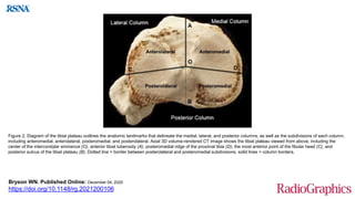
- 1. Figure 2. Diagram of the tibial plateau outlines the anatomic landmarks that delineate the medial, lateral, and posterior columns, as well as the subdivisions of each column, including anteromedial, anterolateral, posteromedial, and posterolateral. Axial 3D volume-rendered CT image shows the tibial plateau viewed from above, including the center of the intercondylar eminence (O), anterior tibial tuberosity (A), posteromedial ridge of the proximal tibia (D), the most anterior point of the fibular head (C), and posterior sulcus of the tibial plateau (B). Dotted line = border between posterolateral and posteromedial subdivisions, solid lines = column borders. Bryson WN. Published Online: December 04, 2020 https://doi.org/10.1148/rg.2021200106