Downloaded 46 times
![INTRODUCTION
1
INTRODUCTION
Hypospadias repair is one of the commonest operations done in
pediatric surgery centers, with an incidence of 1 in 200 to 1 in 300[1]
. The
term hypospadias stems from two Greek words: hypo, which means
‘‘below’’ and spadon, which means ‘‘hole’’. The anomaly is characterized
by a urethral meatus ectopically located proximal to the normal place on
the ventral side of the penis. Different anatomic presentations can be
observed. The position of the urethral meatus can be classified as anterior
or distal (glandular, coronal, or subcoronal; 60–65% of cases), middle
(midpenile; 20–30% of cases), or posterior or proximal (posterior penile,
penoscrotal, scrotal, or perineal; 10–15% of cases) [2]
.
Surgical techniques for hypospadias have evolved over the years
but the principles of any surgery remain the same; namely, to give the
child a straight penis with a terminal or near terminal meatus which is
cosmetically and functionally acceptable with minimal morbidity [3]
.
Hypospadias repair has a long and flat learning curve and requires
patience, experience, and great enthusiasm to achieve acceptable results.
The results published on the various operative techniques need to be
repeated and validated by other surgeons, and long-term results (up to
adulthood) are essential to justify operative methods and identify late
complications [4]
.
Proximal hypospadias defects represent the most challenging and
complex manifestations of this entity and might be successfully treated
with one of several one- or two-stage repairs[5]
. Each technique, whether
two stage or one-stage, has its advantages and drawbacks and not one
technique has gained widespread popularity. Heralding one technique as
the gold standard for posterior hypospadias is probably unrealistic and](https://image.slidesharecdn.com/0d76b122-16b8-4db3-a4a2-c2c7dd2f68b6-150812214802-lva1-app6892/85/02-All-Thesis-Dr-Aboubakr-Omar-FINAL1-1-320.jpg)
![INTRODUCTION
1
INTRODUCTION
Hypospadias repair is one of the commonest operations done in
pediatric surgery centers, with an incidence of 1 in 200 to 1 in 300[1]
. The
term hypospadias stems from two Greek words: hypo, which means
‘‘below’’ and spadon, which means ‘‘hole’’. The anomaly is characterized
by a urethral meatus ectopically located proximal to the normal place on
the ventral side of the penis. Different anatomic presentations can be
observed. The position of the urethral meatus can be classified as anterior
or distal (glandular, coronal, or subcoronal; 60–65% of cases), middle
(midpenile; 20–30% of cases), or posterior or proximal (posterior penile,
penoscrotal, scrotal, or perineal; 10–15% of cases) [2]
.
Surgical techniques for hypospadias have evolved over the years
but the principles of any surgery remain the same; namely, to give the
child a straight penis with a terminal or near terminal meatus which is
cosmetically and functionally acceptable with minimal morbidity [3]
.
Hypospadias repair has a long and flat learning curve and requires
patience, experience, and great enthusiasm to achieve acceptable results.
The results published on the various operative techniques need to be
repeated and validated by other surgeons, and long-term results (up to
adulthood) are essential to justify operative methods and identify late
complications [4]
.
Proximal hypospadias defects represent the most challenging and
complex manifestations of this entity and might be successfully treated
with one of several one- or two-stage repairs[5]
. Each technique, whether
two stage or one-stage, has its advantages and drawbacks and not one
technique has gained widespread popularity. Heralding one technique as
the gold standard for posterior hypospadias is probably unrealistic and](https://image.slidesharecdn.com/0d76b122-16b8-4db3-a4a2-c2c7dd2f68b6-150812214802-lva1-app6892/75/02-All-Thesis-Dr-Aboubakr-Omar-FINAL1-1-2048.jpg)
![INTRODUCTION
2
pediatric urologists have to master a variety of techniques because
various patient-related specifics can favor one or another technique [6]
.
The Koyanagi Nanamura procedure combines a meatal-based flap
and a pedicle island flap into single procedure. It allows for excision of
ventral midline chordee without jeopardizing the flap[7]
. The technique
was modified to improve the blood supply to the neourethral flaps. The
modified technique was found to achieve nearly normal phallic cosmetic
appearance with a low complication rate [8]
.](https://image.slidesharecdn.com/0d76b122-16b8-4db3-a4a2-c2c7dd2f68b6-150812214802-lva1-app6892/85/02-All-Thesis-Dr-Aboubakr-Omar-FINAL1-2-320.jpg)
![REVIEW OF LITERATURE
Embryology
4
EMBRYOLOGY
DEVELOPMENT OF THE GENITAL SYSTEM:
Development of the genital system is one phase in the overall
sexual differentiation of an individual. Sexual determination begins at
fertilization, when a Y chromosome or an additional X chromosome is
joined to the X chromosome already in the egg. This phase represents the
genetic determination of gender. Although the genetic gender of the
embryo is fixed at fertilization, the gross phenotypic gender of the
embryo is not manifested until the seventh week of development [9]
.
The phenotypic differentiation of gender is traditionally considered
to begin with the gonads and progresses with gonadal influences on the
sexual duct systems. Similar influences on the differentiation of the
external genitalia and finally on the development of the secondary sexual
characteristics (e.g., body configuration, breasts, hair patterns) complete
the events that constitute the overall process of sexual differentiation.
Sexual differentiation of the brain, which has an influence on behavior,
also occurs [9]
.
Under certain circumstances, an individual's genetic gender can be
overridden by environmental factors so that the genotypic sex and the
phenotypic sex do not correspond. An important general principle is that
the development of phenotypic maleness requires the action of substances
produced by the testis. In the absence of specific testicular influences or
the ability to respond to them, a female phenotype results. Based on
present information, the female phenotype is considered to be a baseline,
or default, condition, which must be acted on by male influences to
produce a male phenotype [9]
.](https://image.slidesharecdn.com/0d76b122-16b8-4db3-a4a2-c2c7dd2f68b6-150812214802-lva1-app6892/85/02-All-Thesis-Dr-Aboubakr-Omar-FINAL1-4-320.jpg)
![REVIEW OF LITERATURE
Embryology
5
Fig. [1] Male development [9]
.
GONADAL DIFFERENTIATION:
In human foetuses during the ambisexual stage, primordial germ
cells migrate beginning in the fifth week from their site of origin in the
hindgut/allantois into the genital ridges. Sex cords are formed within the
gonads in the sixth embryonic week, and the urogenital folds and
labioscrotal folds begin to form in the perineum. The Mullerian and
Wolffian ducts form around the seventh embryonic week [10]
.
Gonadal differentiation occurs from the 10th
through 12th
embryonic weeks. The Sex-determining Region of the Y chromosome
(SRY) gene is required to initiate signaling for male gonadal
differentiation. SRY gene expression results in differentiation of Sertoli
cells, which then produce Mullerian inhibiting substance (MIS)[10]
.](https://image.slidesharecdn.com/0d76b122-16b8-4db3-a4a2-c2c7dd2f68b6-150812214802-lva1-app6892/85/02-All-Thesis-Dr-Aboubakr-Omar-FINAL1-5-320.jpg)
![REVIEW OF LITERATURE
Embryology
6
Fig. [2] Gonadal differentiation [9]
.
DEVELOPMENT OF MALE DUCT SYSTEM:
During embryonic weeks 9and 10 SRY causes differentiation of
Leydig cells, which produce testosterone. In the presence of fetal
testicular androgens the Wolffian ducts persist and develop into the
epididymis, vas deferens, and seminal vesicles .Testosterone is also a
substrate for the enzyme, 5a-reductase, which converts testosterone to
dihydrotestosterone .This even more potent androgen drives growth of the
external genitalia and prostate [10]
.](https://image.slidesharecdn.com/0d76b122-16b8-4db3-a4a2-c2c7dd2f68b6-150812214802-lva1-app6892/85/02-All-Thesis-Dr-Aboubakr-Omar-FINAL1-6-320.jpg)
![REVIEW OF LITERATURE
Embryology
7
Fig. [3] Development of male duct system [9]
.
THE CLOACA:
The cloaca is an endoderm-lined cavity covered at its ventral
boundary by surface ectoderm. This boundary between the endoderm and
the ectoderm forms the cloacal membrane. During the fourth to the
seventh weeks of development, the cloaca divides into the urogenital
sinus anteriorly and the anal canal posteriorly[11]
.
A layer of mesoderm, the urorectal septum, separates the region
between the allantois and hindgut. This septum is derived from the
merging of mesoderm covering the yolk sac and surrounding the allantois.
As the embryo grows and caudal folding continues, the tip of the
urorectal septum comes to lie close to the cloacal membrane. At the end
of the seventh week, the cloacal membrane ruptures, creating the anal](https://image.slidesharecdn.com/0d76b122-16b8-4db3-a4a2-c2c7dd2f68b6-150812214802-lva1-app6892/85/02-All-Thesis-Dr-Aboubakr-Omar-FINAL1-7-320.jpg)
![REVIEW OF LITERATURE
Embryology
8
opening for the hindgut and a ventral opening for the urogenital sinus.
Between the two, the tip of the urorectal septum forms the perineal
body[11]
.
Three portions of the urogenital sinus can be distinguished. The
upper and the largest part is the urinary bladder .initially the bladder is
continuous with the allantois, but when the lumen of the allantois is
obliterated, a thick fibrous cord, the urachus, remains and connects the
apex of the bladder with the umbilicus. In the adult, it forms the median
umbilical ligament. The next part is a rather narrow canal, the pelvic part
of the urogenital sinus, which in the male give rise to the prostatic and the
membranous part of the urethra. The last part is the phallic part of the
urogenital sinusit is flattened from side to side, and as the genital tubercle
grows, this art of the sinus will be pulled ventrally. Development of the
phallic part of the urogenital sinus differs greatly between the two
sexes[11]
.
Fig [4] Division of the cloaca [11]
.
During differentiation of the cloaca, the caudal portions of the
mesonephric ducts are absorbed into the wall of the urinary bladder.
Consequently the ureters, initially outgrowths from the mesonephric
ducts, enter the bladder separately .As a result of ascent of the kidneys the
orifices of the ureters move farther cranially; those of the mesonephric
ducts move close together to enter the prostatic urethra and in the male
become the ejaculatory ducts[11]
.](https://image.slidesharecdn.com/0d76b122-16b8-4db3-a4a2-c2c7dd2f68b6-150812214802-lva1-app6892/85/02-All-Thesis-Dr-Aboubakr-Omar-FINAL1-8-320.jpg)
![REVIEW OF LITERATURE
Embryology
9
Since both the mesonephric ducts and ureters originate in the
mesoderm, the mucosa of the bladder formed by incorporation of the
ducts (the trigone of the bladder) is also mesodermal. With time, the
mesodermal lining of the trigone is replaced by endodermal epithelium,
so that finally, the inside of the bladder is completely lined with
endodermal epithelium[11]
.
Fig. [5] Relation of the ureter and the mesonephric duct during development[11]
.
The epithelium of the urethra in both sexes originates in the
endoderm; the surrounding connective and smooth muscle tissue is
derived from visceral mesoderm. At the end of the third month,
epithelium of the prostatic urethra begins to proliferate and forms a
number of outgrowths that penetrate the surrounding mesenchyme. In the
male, these buds form the prostate gland. In the female, the cranial part of
the urethra gives rise to the urethral and paraurethral glands[11]
.
SEX DIFFERENTIATION OF THE EXTERNAL GENITALIA:
Sex differentiation of the external genitalia occurs between the 7th
and 17th
weeks of gestation[12]
.
Development of male and female external genitalia begins with the
formation of structures constituting the ambisexual stage that in turn](https://image.slidesharecdn.com/0d76b122-16b8-4db3-a4a2-c2c7dd2f68b6-150812214802-lva1-app6892/85/02-All-Thesis-Dr-Aboubakr-Omar-FINAL1-9-320.jpg)
![REVIEW OF LITERATURE
Embryology
10
undergo sex differentiation to generate the male and female forms of
external genitalia. In the fifth embryonic week, the cloacal folds form
from mesenchymal cells migrating into the perineum. These
mesenchymal cells pile up in the midline and form an elevation in the
perineum called the genital tubercle[10]
.
The genital tubercle is located just cranial to the midline opening of
the endodermal urogenital sinus called the urogenital ostium. The
urogenital ostium is flanked laterally by the urogenital folds and the
genital swellings/labioscrotal folds. Endodermal epithelial cells from the
urogenital sinus are thought to invade into the genital tubercle to form the
solid midline epithelial urethral plate[10]
.
Formation of these structures occurs identically in male and female
fetuses in the ambisexual stage of external genitalia development through
a hormone-independent process. In humans this ambisexual stage of
development occurs between gestational weeks 8 and 12[10]
.
The genital tubercle elongates to become the penis in males under
the influence of fetal testicular androgens. In the absence of androgens in
females the genital tubercle exhibits minimal growth in size and becomes
the clitoris[10]
.
A portion of the cloacal folds becomes the urogenital folds, which
laterally bound the urogenital ostium with the labioscrotal folds
developing laterally. The labioscrotal folds fuse in the midline to form the
scrotum in males, but remain separate forming the labia majora in
females[10]
.
As the solid epithelial urethral plate elongates towards the tip of the
genital tubercle, it canalizes to form a groove on the ventral surface of the
genital tubercle bounded by urethral folds in males. These urethral folds
fuse in the midline converting the urethral groove into the penile urethra.](https://image.slidesharecdn.com/0d76b122-16b8-4db3-a4a2-c2c7dd2f68b6-150812214802-lva1-app6892/85/02-All-Thesis-Dr-Aboubakr-Omar-FINAL1-10-320.jpg)
![REVIEW OF LITERATURE
Embryology
11
Failure of fusion of the penile urethral folds from embryonic weeks
eleven to sixteen results in hypospadias, an abnormal opening of the
urethra proximal to its normal location at the tip of the penis[10]
.
Fig. [6] Sex differentiation of the external genitalia [12]
.
Fig. [7] Photograph of genitalia of a male at 12 weeks and female fetus at 11
weeks[11]
.](https://image.slidesharecdn.com/0d76b122-16b8-4db3-a4a2-c2c7dd2f68b6-150812214802-lva1-app6892/85/02-All-Thesis-Dr-Aboubakr-Omar-FINAL1-11-320.jpg)
![REVIEW OF LITERATURE
Anatomy
12
ANATOMY
THE HUMAN PENIS:
The human penis is made up of three columns of tissue. Two
corpora cavernosa are located next to each other on the dorsal side and
one corpus spongiosum lies between them on the ventral side[13]
.
The end of the corpus spongiosum is enlarged and forms the glans,
which supports the foreskin or prepuce, a loose fold of skin that in adults
can retract to expose the glans. The area on the underside of the penis,
where the foreskin is attached, is called the frenulum[13]
.
The urethra traverses the corpus spongiosum and its opening lies
on the tip of the glans. It is a passage for both urine and semen[13]
.
The raphe is the visible ridge between the lateral halves of the
penis, found underside of the penis running from the meatus (opening of
the urethra) across the scrotum to the perineum (area between scrotum
and anus)[13]
.
The glans penis is homologous to the clitoral glans, the corpora
cavernosa are homologous to the body of the clitoris and the corpus
spongiosum is homologous to the vestibular bulbs beneath the labia
minora. The raphe does not exist in females because there the two halves
are not connected[13]
.
THE MALE URETHRA:
The male urethra is divided into six parts: bladder neck, prostatic
urethra, membranous urethra surrounded by external sphincter, bulbous
urethra proximal to the ischiocavernosus muscle, penile/pendulous
urethra distal to the ischiocavernosus muscle, and the fossa navicularis](https://image.slidesharecdn.com/0d76b122-16b8-4db3-a4a2-c2c7dd2f68b6-150812214802-lva1-app6892/85/02-All-Thesis-Dr-Aboubakr-Omar-FINAL1-12-320.jpg)
![REVIEW OF LITERATURE
Anatomy
13
within the distal glans. The corpus spongiosum is erectile tissue akin to
corpora cavernosa, but with a thinner tunica albuginea. The penile and
bulbar urethra lie within the spongiosum. The penile urethra lies in a
central location within the spongiosum, whereas the bulbar urethra lies
eccentrically closer to the dorsal spongiosum prior to exiting dorsally to
become the membranous urethra to join the prostate. Whereas the
condition known as “chordee” or penile curvature was once believed to
result from fibrous bands near the urethra, no such fibrous tissue has been
found in the penile urethra, even in severe cases of hypospadias. Because
of anastomotic communications between the dorsal arteries and the
bulbourethral arteries, the urethra receives arterial supply from both distal
and proximal directions. This enables complete transaction of the urethra
without necrosis of the distal segment[12]
.
THE CORPUS SPONGIOSUM:
The corpus spongiosum is a midline structure nestled on the ventral
surface of the paired erectile bodies that are known as the corpora
cavernosa. Distally, the spongiosum expands to form the glans penis,
which serves to cap the cavernosa in a smooth and rounded shape that
should be maintained for ideal vaginal penetration [14]
.
Within the glans, the distal urethra is lined with stratified squamous
epithelium and because it widens here to form a small, boat-shaped fossa,
it is termed the fossa navicularis. More proximally, and throughout the
length of the penile shaft, the relatively narrow pendulous urethra is lined
with simple squamous cell epithelium [14]
.
The larger base of the spongiosum, or the bulb, contains the aptly
named bulbous urethra, which takes on a dorsal orientation. The distal
most point of the ischiocavernosus muscles marks the transition from
pendulous to bulbous urethra. These two muscles sweep anteromedially
from their lateral roots on the ischial rami. They fuse in the midline at a](https://image.slidesharecdn.com/0d76b122-16b8-4db3-a4a2-c2c7dd2f68b6-150812214802-lva1-app6892/85/02-All-Thesis-Dr-Aboubakr-Omar-FINAL1-13-320.jpg)
![REVIEW OF LITERATURE
Anatomy
14
point that is ventral to the bulbous urethra, facilitating its ability to empty.
The base of the spongiosum is more directly covered by the solitary
bulbospongiosus muscle, which is attached at its posterior aspect to the
perineal body, the central meeting point of eight muscles of the
perineum[14]
.
THE CORPORA CAVERNOSA:
The corpora cavernosa is in the pendulous portion of the penis, it is
endothelial-lined lacunar spaces, which are supported by a trabecular
architecture of smooth muscle and fibroelastic tissue, they are two in
communication. Proximally, however, the corpora cavernosa diverge into
two crura that are adherent to the ventral aspects of the inferior pubic
rami[14]
.
Fig. [8] Cross section in the mid shaft of the penis [15]
.
COVERINGS OF THE PENIS:
The tunica albuginea, a sheath of dense, fibroelastic tissue,
surrounds each of the corpora cavernosa and the corpus spongiosum. The
tunica is comprised of an outer longitudinal layer, which inserts
proximally on the inferior pubic rami, and an inner circular layer. Fibers
extend from the circular layer to form the vertical septum that separates](https://image.slidesharecdn.com/0d76b122-16b8-4db3-a4a2-c2c7dd2f68b6-150812214802-lva1-app6892/85/02-All-Thesis-Dr-Aboubakr-Omar-FINAL1-14-320.jpg)
![REVIEW OF LITERATURE
Anatomy
15
the two cavernosa. This septum and the tunica albuginea together
facilitate penile rigidity when erect[14]
.
Buck fascia surrounds both cavernosal bodies dorsally and splits to
surround the spongiosum ventrally. Elastic and collagenous fibers from
the rectus sheath blend with and surround Buck fascia as the fundiform
ligament of the penis. Deeper fibers from the pubis form the suspensory
ligament of the penis. In the perineum, Buck fascia fuses with the tunica
albuginea deep to the muscles of the erectile bodies . Distally, it fuses
with the base of the glans at the corona[15]
.
Buck’s fascia is immediately superficial to the deep dorsal vein of
the penis, the paired dorsal arteries of the penis, and branches of the
dorsal nerves of the penis, all of which directly overlie the tunica. The
bulbospongiosus and ischiocavernosus musculature are superficial to
Buck’s fascia. More superficial still is the areolar dartos fascia, or Colles’
fascia, which additionally invests the perineum and the scrotal contents,
extending to form the scrotal septum and the median raphe of the ventral
penis, the scrotum, and the perineum. A deeper layer of this fascia serves
to separate the scrotal contents from the superficial perineal pouch.
Colles’ fascia contains within it the superficial nerves and vasculature of
the penis as well as the muscle fibers responsible for the scrotal rugae[14]
.
The skin of the penile shaft is highly elastic and without
appendages (hair or glandular elements), except for the smegma-
producing glands at the base of the corona. It is devoid of fat and quite
mobile because of the loose attachment of its dartos backing to Buck
fascia. Distally, it folds over the glans as the foreskin and attaches firmly
below the corona. Its blood supply is independent of the erectile bodies
and is derived from the external pudendal branches of the femoral
vessels . These vessels enter the base of the penis to run longitudinally in
the dartos fascia as a richly anastomotic network. Thus penile skin may
be mobilized on a vascular pedicle as the ideal tissue for urethral](https://image.slidesharecdn.com/0d76b122-16b8-4db3-a4a2-c2c7dd2f68b6-150812214802-lva1-app6892/85/02-All-Thesis-Dr-Aboubakr-Omar-FINAL1-15-320.jpg)
![REVIEW OF LITERATURE
Anatomy
16
reconstruction. The skin of the glans is immobile as a result of its direct
attachment to the underlying, thin tunica albuginea[15]
.
PENILE NERVE SUPPLY:
Penile innervation consists of the dorsal, cavernosal, and perineal
nerves. Dorsal nerves arising from the pudendal nerves travel within
Buck’s fascia, together with the dorsal arteries and veins, to supply
sensation to penile skin[8]
. Despite its nomenclature, it is important to note
that the nerves do not lie directly in the dorsal midline, but rather extend
from the 11 and 1 o’clock positions laterally to the junction of the
cavernosa and spongiosum[9,10]
. These nerves do not send perforators
deep through the tunica albuginea to the corpora cavernosa[9]
. There is a
paucity of nerves at the 12 o’clock shaft position. Therefore, in correction
of penile curvature, plication at the 12 o’clock position is the area least
likely to result in nerve damage[12]
.
Like the dorsal nerves, the perineal nerves also arise from the
pudendal nerve to supply the ventral shaft skin, the frenulum, and the
bulbospongiosus muscle. The cavernosal nerves arise from the autonomic
pelvic plexus and travel along the periprostatic neurovascular bundle,
well known to urologists performing radical retropubic prostatectomies.
Underneath the pubic arch, the cavernosal nerves pierce through the
corpora cavernosa[12]
. Proximal to this point, the cavernosal and dorsal
nerves lie within close proximity at the penile hilum and are thought to
exchange signal communication, which may have implications on erectile
function. As well, there are interactions between perineal and dorsal
nerves laterally at the junction of the cavernosa and spongiosum along the
penis, which may also have implications on erection and ejaculation[12]
.
PENILE ARTERIAL SUPPLY:
There are three paired main arteries in the penis: cavernosal, dorsal,
and bulbourethral. All three arise from a shared branch of the internal
pudendal artery, which itself arises from the internal iliac artery. On each](https://image.slidesharecdn.com/0d76b122-16b8-4db3-a4a2-c2c7dd2f68b6-150812214802-lva1-app6892/85/02-All-Thesis-Dr-Aboubakr-Omar-FINAL1-16-320.jpg)
![REVIEW OF LITERATURE
Anatomy
17
side, the first branching occurs at the bulb of the spongiosum external to
the urogenital diaphragm forming the bulbourethral artery, which then
lies at the 9 and 3 o’clock positions of the corpus spongiosum. Then the
cavernous artery branches to penetrate the corpora cavernosa and the
remainder of the artery continues as the deep dorsal artery. The deep
dorsal artery causes glans enlargement during erection, whereas the
cavernosal arteries cause corporal enlargement. All three arteries
communicate distally near the glans to provide an extensive anastomotic
network. Penile skin derives its supply from a separate origin. Branches
of the external pudendal artery supply the dorsal and lateral aspects of the
penis, and branches of the internal pudendal artery supply the ventral
penis and scrotum via the posterior scrotal artery. These branches course
in the Dartos fascia and enable pedicled skin flaps to be used in urethral
reconstruction[12]
.
PENILE VENOUS DRAINAGE:
Venous drainage is not analogous to arterial supply, unlike many
other body systems. In contrast to the paired dorsal arterial system, there
exists only one deep dorsal vein that runs alongside the dorsal arteries and
nerves in Buck’s fascia above the tunica albuginea. The deep dorsal vein
receives drainage from the distal two-thirds of the corpora cavernosa via
emissary veins and the corpus spongiosum via circumflex veins.
Emissary veins are the veins that traverse obliquely through the tunica
albuginea, allowing them to be compressed during erections for penile
tumescence. The deep dorsal vein then drains to the periprostatic plexus.
Recently, a small pair of dorsal veins have been found that lie just deep to
the deep dorsal vein, but above the tunica albuginea, which independently
receive emissary vein drainage. These veins have been termed cavernosal
veins, but do not lie within the corpora cavernosa. Older literature refers
to the cavernosal veins as short veins located in the triangle between the
proximal crus that drain the proximal one-third of the corpora cavernosa.
These veins join with the bulbourethral veins (which drain the proximal](https://image.slidesharecdn.com/0d76b122-16b8-4db3-a4a2-c2c7dd2f68b6-150812214802-lva1-app6892/85/02-All-Thesis-Dr-Aboubakr-Omar-FINAL1-17-320.jpg)
![REVIEW OF LITERATURE
Anatomy
18
spongiosum) to lead into the internal pudendal vein. The penile skin
drains via the superficial dorsal vein, which drains into the saphenous
vein[12]
.](https://image.slidesharecdn.com/0d76b122-16b8-4db3-a4a2-c2c7dd2f68b6-150812214802-lva1-app6892/85/02-All-Thesis-Dr-Aboubakr-Omar-FINAL1-18-320.jpg)
![REVIEW OF LITERATURE
Hypospadias
19
HYPOSPADIAS
DEFINITION:
Hypospadias, a term derived from the Greek terms hypo (under)
and spadon (rent, fissure) In most cases, hypospadias in the male is
associated with three anomalies of the penis: (1) a ventral meatus that
may be located anywhere between the glans and the perineum, (2) ventral
deviation of the penis (chordee), and (3) the dorsal prepuce hood in
association with a ventral deficit of the prepuce. The second and third
abnormalities are not necessary for the diagnosis of hypospadias[4]
.
One particular form is hypospadias sine hypospadias, in which the
meatus is found in the glans, with a severe deviation of the penile shaft. A
further distinctive form is megalomeatus, in which the meatus is found in
a coronal position, the fossa navicularis is open, and the prepuce is
intact[4]
.
Hypospadiology is a term coined by John W. Duckett, Jr., the
former chief of the Division of Urology at the Children’s Hospital of
Philadelphia (CHOP) and a pioneer in hypospadias repairs.
Hypospadiology encompasses a continuously evolving and expanding
discipline. While modern experiments have only recently begun to yield a
deeper understanding of the genetic, hormonal, and environmental basis
of hypospadias, the quest for a surgical procedure that consistently results
in a straight penis with a normally placed glanular meatus has occupied
surgeons for over 2 centuries[16]
.](https://image.slidesharecdn.com/0d76b122-16b8-4db3-a4a2-c2c7dd2f68b6-150812214802-lva1-app6892/85/02-All-Thesis-Dr-Aboubakr-Omar-FINAL1-19-320.jpg)
![REVIEW OF LITERATURE
Hypospadias
20
EPIDEMIOLOGY:
Hypospadias is the most common congenital anomaly of the penis,
affecting 0.4–8.2 of 1000 live male babies[17]
.
ETIOLOGY:
It is considered to be a mild form of the 46, XY disorders of sex
development (DSD), In most cases, the degree of hypospadias is
relatively mild and a specific endocrine cause is not sought or is not
found. However, four main elements are involved in male genital
construction and may contribute to this malformation:
(1) The genetic and endocrine background of the child, principally the
genes of phallic development, gonadal steroid synthesis (mainly
testosterone and its 5α reduced form, dihydrotestosterone, DHT),
and the responsiveness to these hormones. The genital tubercle
thus grows under the influence of androgens and any alteration in
androgen production or receptors may produce a hypospadiac
penis.
(2) The placenta, which orchestrates the hormonal climate, especially
during the first part of gestation.
(3) The mother, with her own hormonal production and possible
disorders.
(4) The environment of mother and child, which may also interfere in
this fine balance[18]
.](https://image.slidesharecdn.com/0d76b122-16b8-4db3-a4a2-c2c7dd2f68b6-150812214802-lva1-app6892/85/02-All-Thesis-Dr-Aboubakr-Omar-FINAL1-20-320.jpg)
![REVIEW OF LITERATURE
Hypospadias
21
(A) GENETIC FACTOR:
(a) Inheritance:
Before evaluating the role of the environment, it should be
acknowledged that several arguments are in favor of a predominant role
for the genetic background. Familial clustering is seen in about 10% of
the cases, and the recurrence risk in the male siblings of an affected
patient is about 15%. Seven percent of the fathers of children with
hypospadias are also affected. The risk of recurrence is also found to
aggregate in more distant relatives. The risk ratios of hypospadias for
male first-, second-, and third-degree relatives of a hypospadiac case were,
respectively, 11.6%, 3.27%, and 1.33%. The risk of recurrence for the
next male sibling depends on the severity of the hypospadias. Segregation
analysis suggests that hypospadias might be due to monogenic effects in a
small proportion of the families, whereas a multifactorial mode of
inheritance was reported to be more likely in the majority of families.
Finally, some of the 200 syndromes that include hypospadias have known
genetic bases and shed light on the molecular mechanisms involved in
genital development[18]
.
(b) Syndromes with hypospadias:
Nearly 200 syndromes are associated with hypospadias [15]
.
Smith-Lemli-Opitz syndrome results from autosomal recessive
mutation of the DHCR7 gene on chromosome 11q13 coding for 7-
dehydrocholesterol reductase. Affected individuals have mental
retardation, facial dysmorphism, microcephaly, and syndactyly. DHCR7
regulates Sonic hedgehog signaling, and so association of this syndrome
with hypospadias potentially links to observations in mice regarding the
role of Shh in penis development [15]
.](https://image.slidesharecdn.com/0d76b122-16b8-4db3-a4a2-c2c7dd2f68b6-150812214802-lva1-app6892/85/02-All-Thesis-Dr-Aboubakr-Omar-FINAL1-21-320.jpg)
![REVIEW OF LITERATURE
Hypospadias
22
Deletion in chromosome 11q13 results in WAGR syndrome (Wilms
tumor, Aniridia, Genital anomalies, mental Retardation), associated with
hypospadias due to altered WT1 gene activity. One study screening for
WT1 gene defects in boys with nonsyndromic hypospadias reported no
mutations [15]
.
Hand-foot-genital syndrome is an extremely rare autosomal
dominant condition due to mutations in HOXA13 on chromosome 7p14-
15, resulting in bilateral thumb and great toe hypoplasia. Hypospadias in
mice mutant for Hoxa13 also have loss of Fgf8 and bone morphogenetic
protein-7 in the urethral plate [15]
.
Opitz G syndrome (Opitz G/BBB syndrome) occurs from X-linked
mutations in midline 1 gene or autosomal dominant deletions in
chromosome 22q11. The resultant phenotype includes hypertelorism,
tracheoesophageal defects, cleft lip/palate, and mild mental retardation as
well as hypospadias [15]
.
Wolf-Hirschhorn syndrome derives from deletions in chromosome
4p, resulting in mental retardation, seizures, abnormal facies, and midline
defects, including hypospadias [15]
.
13q deletion syndrome is characterized by mental retardation,
facial dysmorphia, imperforate anus, and hypospadias with penoscrotal
transposition. The critical region mediating anorectal and genital
anomalies has been localized to 13q33.1-34, containing 20 annotated
genes including EFNB2. Partial loss of ephrin B-2 function altering
ephrin signaling in mice is associated with hypospadias, as discussed
earlier[15]
.](https://image.slidesharecdn.com/0d76b122-16b8-4db3-a4a2-c2c7dd2f68b6-150812214802-lva1-app6892/85/02-All-Thesis-Dr-Aboubakr-Omar-FINAL1-22-320.jpg)
![REVIEW OF LITERATURE
Hypospadias
23
(c) Gene mutation:
Murine studies indicating androgen receptor activity regulates Fgf8,
Fgf10, and Fgfr2 involved in urethral development have led to screening
for defects in these candidate genes in patients with hypospadias. Among
cases of nonsyndromic familial hypospadias variants have been found in
FGF8 and FGFR2 not seen in normal controls [15]
.
Estrogens play a role in male development, with specific nuclear
estrogen receptors, predominantly ER2, found in proximity to the
androgen receptor. Variants of ER2 influence serum testosterone levels
and have been described in patients with hypospadias [15]
.
Further evidence implicating estrogen-related events in urethral
maldevelopment was the finding that several estrogen-responsive genes
are upregulated in hypospadias patients, including ACT3, Cyr61, CTGF,
and CADD45β. Polymorphisms of ACT3 were subsequently reported
associated with hypospadias, as were less common mutations [15]
.
(d) Environmental action on genes:
The environment may act on the genes that contribute to the
occurrence of hypospadias at several levels[18]
.
1- Level of phallus development:
Homeobox genes A (HOXA) and D (HOXD) participate in the
development of the phallus since knock-out of these genes in mice
induces a malformation in the external genitalia consistent with
hypospadias [18]
.
In humans, the hand-foot-genital syndrome (HFGS) is related to
mutations of HomeoboxA13 (HOXA13). HOXA13 allows the normal
expression of fibroblast growth factor (FGF) 8 and bone morphogenetic
protein (BMP) 7 in the developing urethral epithelium in mice, thus
modulating androgen receptor expression and glans vascularization. The](https://image.slidesharecdn.com/0d76b122-16b8-4db3-a4a2-c2c7dd2f68b6-150812214802-lva1-app6892/85/02-All-Thesis-Dr-Aboubakr-Omar-FINAL1-23-320.jpg)
![REVIEW OF LITERATURE
Hypospadias
24
FGF gene family, especially FGF10, is also implicated in the
development of external genitalia in mice. In humans, polymorphisms of
FGF8, FGF10 and FGFR2 may be associated with an increased risk of
hypospadias[18]
.
2- Level of testicular determination:
The genes leading to testicular dysgenesis are a cause of
hypospadias. Severe hypospadias along with other genital abnormalities
can reveal heterozygous mutations of Wilms tumor 1 (WT1). SOX9,
DMRT1 and GATA4 encode transcription factors acting immediately
before the differentiation of the gonad into testis. Mutations of these
genes induce testicular dysgenesis and are associated with 46, XY
disorders of sex differentiation (DSD), including severe hypospadias.
Variation in gene dosage, as shown in 46, XX and 46, XX d17 patients
with SOX9 duplication, can also induce penoscrotal hypospadias[18]
.
3- Level of androgen biosynthesis:
Mutations in the LH receptor gene (inducing a Leydig cell
hypoplasia) and the 5α-reductase gene (inducing a defect of
dihydrotestoterone synthesis) induce hypospadias, most often in a severe
form with associated cryptorchidism and/or micropenis. MAMLD1
(mastermind-like domain containing gene) is another candidate gene that
seems to modulate the synthesis of testosterone around the critical period
of sex differentiation. MAMLD1 is expressed in the male gonad in mice,
and it augments testosterone production and contains the SF1 target
sequence. Fukami et al. identified three nonsense mutations in four
individuals with 46, XY DSD including micropenis, bifid scrotum and
penoscrotal hypospadias. Genetic variants of MAMLD1 were further
shown to be present in patients with isolated hypospadias, as confirmed
by Chen et al., who identified five nonsynonymous mutations, some of
them as polymorphisms[18]
.](https://image.slidesharecdn.com/0d76b122-16b8-4db3-a4a2-c2c7dd2f68b6-150812214802-lva1-app6892/85/02-All-Thesis-Dr-Aboubakr-Omar-FINAL1-24-320.jpg)
![REVIEW OF LITERATURE
Hypospadias
25
4- Level of androgen action:
Mutations in the androgen receptor gene (AR) have been found in
patients with either severe forms of hypospadias or other signs of under-
virilzation, such as cryptorchidism or micropenis. Mutation of the AR
gene in partial androgen insensitivity syndrome is found only in 20% to
30% of cases and the phenotype remains particularly variable[18]
.
(B) ENDOCRINAL:
(a) Endocrinopathies:
The pivotal role of androgens in normal penis development
suggests endocrinopathies impacting hormone production or action may
underlie hypospadias. Leydig cell dysfunction was implicated by findings
of elevated basal luteinizing hormone and reduced testosterone response
to human chorionic gonadotropin stimulation in prepubertal boys with
hypospadias versus controls. Defects in the testosterone biosynthetic
pathway, specifically, impaired 3β-hydroxy steroid dehydrogenase alone
or with impaired 17,20-lyase or 17α-hydroxylase activity, were reported
in proximal hypospadias but not confirmed in subsequent studies.
Although enzymatic assays for 5α-reductase type 2 activity in isolated
hypospadias are normal, mutations in its coding gene SRD5A2 on
chromosome 2 were reported in 9% of patients with hypospadias not
found in controls. Androgen receptor gene mutations are considered a
rare cause of hypospadias but were not detected by others. Problems
linking disturbed androgen activity to hypospadias were summarized by
little evidence has been found to suggest that nonsyndromic hypospadias
without other genital anomalies is associated with defects in testosterone
production, its conversion to dihydrotestosterone, or androgen receptor
activity. Furthermore, the autosomal recessive pattern of inheritance
characterizing steroidogenic enzyme disorders does not correlate with the
genetics of hypospadias [15]
.](https://image.slidesharecdn.com/0d76b122-16b8-4db3-a4a2-c2c7dd2f68b6-150812214802-lva1-app6892/85/02-All-Thesis-Dr-Aboubakr-Omar-FINAL1-25-320.jpg)
![REVIEW OF LITERATURE
Hypospadias
26
(b) Endocrine disrupter:
Hypospadias in humans has also been linked to exposure to
endocrine disrupting compounds. Exposure of pregnant women to
estrogenic and anti-androgenic endocrine disrupting com- pounds is
associated with hypospadias and reduced anogenital distance in their
male offspring. Anogenital distance is a recognized metric of androgen
action. Several studies have demonstrated that exposure to phthalates
results in decreased anogenital distance in human males, presumably due
to lowered testosterone. Similarly, an anti-androgenic metabolite of the
pesticide DDT reduces anogenital distance in human males . Exposure to
bisphenol-A, a weakly estrogenic compound in plastic, has also been
shown to reduce anogenital distance in human male offspring in a dose
dependent fashion. Decreased anogenital distance in human males has
been associated with decreased fertility in adulthood. Exposure of
pregnant women to pesticides has been linked to cryptorchidism,
hypospadias, and micropenis with a non-statistically significant
association with family history and maternal medications. Similarly,
maternal exposure to estrogenic and antiandrogenic endo- crine
disrupting compounds has been implicated in increased risk of
cryptorchidism and hypospadias in human male offspring without
statistical significance [10]
.](https://image.slidesharecdn.com/0d76b122-16b8-4db3-a4a2-c2c7dd2f68b6-150812214802-lva1-app6892/85/02-All-Thesis-Dr-Aboubakr-Omar-FINAL1-26-320.jpg)
![REVIEW OF LITERATURE
Hypospadias
27
DIAGNOSIS:
(A) ANTENATAL:
There is a possible association of hypospadias with other
malformations (neural tube, cardiac, urogenital tract, and anorectal) or a
possibility to be part of a syndrome. However, in most cases, hypospadia
is an isolated manifestation. Therefore, only a detailed analysis of genital
morphology during prenatal ultrasound allows the possibility of diagnosis.
The main finding of the 2D US in cases of hypospadias is the ventral or
lateral curvature of the penis, associated with its shortening. Meizner
described a specific signal known as a “tulip sign” present in severe
hypospadias, corresponding to the presence of a short penis ventrally
curved in association with penoscrotal transposition of a bifid scrotum.
The change in morphology of distal penis, more rounded (blunt) rather
than elongated (acute), results from a big and redundant prepuce only in
the dorsal surface, covering the glans, that can also be observed at 2D
US .The introduction of 3D US allowed the evaluation of the surface
structures of the fetus in rendering mode, enabling the development of a
new imaging method for evaluation of hypospadias [19]
.
(B) POSTNATAL:
a. Clinical examination:
With the exception of the megameatus intact prepuce variant,
hypospadias should be diagnosed shortly after birth. In individuals with
distal hypospadias, clinical investigation is sufficient. However, the
description of hypospadias should include the following:
Position, shape, and width of the orifice.
Presence of an atretic urethra and division of the corpus spongiosum.
Appearance of the preputial hood and scrotum.
Penile size.
Curvature of the penis on erection [4]
.](https://image.slidesharecdn.com/0d76b122-16b8-4db3-a4a2-c2c7dd2f68b6-150812214802-lva1-app6892/85/02-All-Thesis-Dr-Aboubakr-Omar-FINAL1-27-320.jpg)
![REVIEW OF LITERATURE
Hypospadias
28
The position of the testicle should also be mentioned. In
individuals with cryptorchidism, further indications for DSD should be
investigated. In individuals with proximal hypospadias and independent
of gonadal status, some authors recommend the exclusion of DSD . This
investigation may be considered to be excessive diagnostic work-up;
however, individuals with unilateral or bilateral cryptorchidism, small
penis, and severe hypospadias and individuals with ambiguous genitalia
require a complete genetic and endocrine work-up immediately after birth
to exclude intersexuality and, in particular, congenital adrenal
hyperplasia[4]
.
b. Symptomatology:
Clinical symptoms vary and depend on the severity of the disease.
In mild hypospadias with a urethral meatus located on the glans, a normal
urinary flow can be maintained. In cases with a stenotic meatus, a weak
urinary flow can be observed. Children with proximal hypospadias with
penile curvature might not be able to void while standing. we do not
know precisely what degree of penile curvature in children will inhibit
sexual intercourse in adulthood or what the long-term psychosexual
outcome will be in these patients[17]
.
c. Investigations:
Karyotyping:
A karyotype may help categorize hypospadias as syndromic when
there are other non-genital anomalies, especially developmental delay,
dysmorphic facies, and/or anorectal or scrotal malformations. It may also
detect gonadal DSD, especially when there is also cryptorchidism. The
role for karyotyping in isolated hypospadias, even proximal cases, is
unclear because most reports concern hypospadias associated with
cryptorchidism or do not state the severity of isolated hypospadias [15]
.](https://image.slidesharecdn.com/0d76b122-16b8-4db3-a4a2-c2c7dd2f68b6-150812214802-lva1-app6892/85/02-All-Thesis-Dr-Aboubakr-Omar-FINAL1-28-320.jpg)
![REVIEW OF LITERATURE
Hypospadias
29
Radiological studies:
No prospective studies report the incidence of radiologically
detected urinary tract anomalies associated with hypospadias. Initial
retrospective reviews concerned intravenous pyelography, and of these
the largest series found upper tract anomalies are not increased with non-
syndromic hypospadias. Routine voiding cystourethrography to
demonstrate an enlarged utricle is not necessary, because the most
common clinical manifestation is difficult catheterization that can be
managed intraoperatively. Imaging can be reserved for screening patients
with suspected syndromic hypospadias or DSD [15]
.
d. Associated anomalies:
Cryptorchidism and Inguinal Hernia:
Between 8 and 10% of boys with hypospadias have a cryptorchid
testicle, and 9 to 15% have an associated inguinal hernia. In boys with
more proximal hypospadias, cryptorchidism may occur as frequently as
32%. This strong association between proximal hypospadias and
undescended testes further suggests that this clinical entity may represent
one end of a spectrum of endocrinopathy. The incidence of chromosomal
anomaly in these groups of patients is much higher (22%) than
hypospadias (5–7%) or cryptorchidism (3–6%) occurring alone. In a
series of more than 600 cases of hypospadias, we found that children with
associated cryptorchidism and mid-shaft to distal hypospadias had a
much higher complication rate when corrected. We are not sure why this
occurs, but it may be that a change in the endocrine milieu with the
associated cryptorchidism may make the tissues less amenable to
correction[16]
.](https://image.slidesharecdn.com/0d76b122-16b8-4db3-a4a2-c2c7dd2f68b6-150812214802-lva1-app6892/85/02-All-Thesis-Dr-Aboubakr-Omar-FINAL1-29-320.jpg)
![REVIEW OF LITERATURE
Hypospadias
30
Prostatic Utricle:
The prostatic utricle is an elementary structure developing from
Mullerian ducts cranially, and from the Wollfian ducts and the urogenital
sinus caudally. Boys with hypospadias often have enlargement of the
prostatic utricle with resultant urinary tract infections, stone formation,
pseudo incontinence and, often, difficult catheterization. Devine et al.
reported that 57% of the patients with perineal hypospadias and 10% with
penoscrotal hypospadias had prostatic utricle enlargement demonstrated
on urethroscopy. The overall incidence of utricle enlargement in patients
with hypospadias was 14% in this series of 44 patients. Utricular
enlargement in itself does not indicate DSD, but is seen with increased
frequency in patients with 46, XY DSD[16]
.
Malformation Syndromes:
Hypospadias most often occurs in infants without additional known
medical conditions. The finding of other anomalies increases the
likelihood that hypospadias is part of a malformation syndrome. From the
description of various syndromes just given these include developmental
delay, facial dysmorphy, anorectal malformations, and other genital
anomalies, including penoscrotal transposition and cryptorchidism[15]
.
Disorders of sex development:
Although hypospadias is considered arrested masculinization, by
convention it is distinguished from DSD. As discussed earlier, defects in
testosterone production, its conversion to dihydrotestosterone, or
androgen receptor activity that characterizes various disorders of sexual
differentiation are uncommonly detected in isolated hypospadias. The
simultaneous occurrence of hypospadias with cryptorchidism increases
the likelihood for DSD. Overall reported incidence in patients considered
to have a male appearing phenotype ranges from 0% to 30% and is](https://image.slidesharecdn.com/0d76b122-16b8-4db3-a4a2-c2c7dd2f68b6-150812214802-lva1-app6892/85/02-All-Thesis-Dr-Aboubakr-Omar-FINAL1-30-320.jpg)
![REVIEW OF LITERATURE
Hypospadias
31
greater with increasing severity of hypospadias and nonpalpable testes .
Kaefer and colleagues reported DSD in approximately 50% of patients
with a nonpalpable testis and hypospadias. The most frequent finding is
mixed gonadal dysgenesis, followed by ovotesticular disordered sexual
differentiation. Incomplete androgen insensitivity, 5α-reductase type 2
deficiency, and testicular dysgenesis have also been reported. However,
likely differences in defining male appearance versus ambiguity and
failure to perform uniform genetic, biochemical, and radiologic
evaluation in hypospadias patients with cryptorchidism make
determination of the incidence of DSD uncertain. Coexistence of
hypospadias and cryptorchidism can be explained by other associations
than DSD. For example, a case control study examining risk factors
independently for the two conditions within the same nationwide cohort
in Sweden found low birth weight and prematurity positively correlated
with each [15]
.
e. Classification and severity assessment:
Introduction:
The assessment of severity is based on meatal position, quality of the
urethra and urethral plate, and presence or absence of penile curvature. As
suggested by Snodgrass et al, patients with hypospadias present with a
continuum of abnormalities ranging from simple glanular forms to
perineal presentations with different degrees of penile curvature, and it is
not clear in what situation the adjective severe should be applied . The
assessment of severity is obviously influenced by subjective judgment,
which can differ among surgeons. Is it possible to create a more objective
severity classification taking into account meatal position, quality of
urethra and urethral plate, and presence or absence of penile curvature?
Or as some maintain, can the assessment of hypospadias severity be
adequately performed only in the operating room ? Indeed, the solution is
of great importance, as the choice of surgical repair is based on it[17]
.](https://image.slidesharecdn.com/0d76b122-16b8-4db3-a4a2-c2c7dd2f68b6-150812214802-lva1-app6892/85/02-All-Thesis-Dr-Aboubakr-Omar-FINAL1-31-320.jpg)
![REVIEW OF LITERATURE
Hypospadias
32
Classification:
Many classifications of hypospadias have been defined and
published. Hypospadias is usually classified according to the anatomic
location of the urethral orifice:
(1) Anterior or distal hypospadias (urethral orifice located on the glans
or distal shaft of the penis).
(2) Middle shaft or intermediate (penile) hypospadias.
(3) Posterior or proximal (penoscrotal, scrotal, or perineal)
hypospadias [4]
.
Of all cases of hypospadias, 70–80% are distal-anterior
hypospadias, and 15–20% are midshaft hypospadias. Posterior proximal
forms are rare[4]
.
The severity of hypospadias cannot be solely based on the meatal
location assessed at the first consultation. The size of the penis (especially
the glans), the amount of dorsal foreskin, glans morphology, associated
scrotal abnormalities (penoscrotal transposition), and age at initial
presentation are additional indicators of severity to be defined for primary
repair. The operative technique can only be decided in the operating room.
Parents should be informed of the main techniques and follow up
commonly used by the surgeon. Assessment of the urethral plate and the
penile curvature (erection test) are the main indicators of severity, since
preservation or section of the urethral plate is an essential step in the
selection of the reconstructive technique[20]
.](https://image.slidesharecdn.com/0d76b122-16b8-4db3-a4a2-c2c7dd2f68b6-150812214802-lva1-app6892/85/02-All-Thesis-Dr-Aboubakr-Omar-FINAL1-32-320.jpg)
![REVIEW OF LITERATURE
Hypospadias
33
Merriman 2013 GMS score:
In an effort to address the need for standardized criteria to classify
the severity of hypospadias, the GMS hypospadias scale was developed.
This scale was developed as a means to qualitatively score the severity of
hypospadias based on easily observable features of the glans (G), meatus
(M), and penile shaft (S). Each of the three components is scored
numerically on a scale of 1e4 with more unfavorable characteristics being
assigned higher values. These values are then summed to determine the
GMS total score. The lowest possible GMS score, therefore, is 3 (very
mild hypospadias) and the highest score is 12 (severe hypospadias)[21]
.
It is also important to point out that the G score is used to assess
both glans size and the quality of the urethral plate[21]
.
Glans (G) score:
1. Glans good size; healthy urethral plate, deeply grooved
2. Glans adequate size; adequate urethral plate, grooved
3. Glans small in size; urethral plate narrow, some fibrosis or flat
4. Glans very small; urethral plate indistinct, very narrow or flat
Meatus (M) score:
1. Glanular
2. Coronal Sulcus
3. Mid or Distal Shaft
4. Proximal shaft, penoscrotal
Shaft (S) score:
1. No chordee
2. Mild (< 30°) chordee
3. Moderate (30 - 60°) chordee
4. Severe (> 60°) chordee](https://image.slidesharecdn.com/0d76b122-16b8-4db3-a4a2-c2c7dd2f68b6-150812214802-lva1-app6892/85/02-All-Thesis-Dr-Aboubakr-Omar-FINAL1-33-320.jpg)
![REVIEW OF LITERATURE
Hypospadias
34
Fig. [9] Representative photographs of the GMS scoring criteria [21]
.](https://image.slidesharecdn.com/0d76b122-16b8-4db3-a4a2-c2c7dd2f68b6-150812214802-lva1-app6892/85/02-All-Thesis-Dr-Aboubakr-Omar-FINAL1-34-320.jpg)
![REVIEW OF LITERATURE
Hypospadias
35
MANAGEMENT:
(A) Perioperative Considerations:
a. Timing of surgery:
Timing of hypospadias repair is influenced by penile size, genital
awareness, and anesthetic risks. An earlier recommendation that surgery
be performed after 3 years of age was revised in 1996 with the current
opinion that repair is best done between 6 and 18 months . However,
several authors have reported that complication rates increase when
surgery is done after age 6 months or 1 year. We reviewed our experience
in consecutive prepubertal patients undergoing tubularized incised plate
(TIP) hypospadias repair to determine if age impacted urethroplasty
outcomes [22]
.
Our data in consecutive prepubertal boys undergoing TIP
hypospadias repair show age at surgery is not a risk factor for UC. Rather,
of the potential factors we analyzed, this risk was determined only by
meatal location and reoperative repair. Full-term patients with
hypospadias can undergo corrective surgery at any age after 3 months
with no difference in urethroplasty outcomes[22]
.
Boys have no memory of surgery if treatment is completed before
age 5 years, which correlates with being satisfied with overall body
appearance regardless of surgeon assessed anatomical outcome, the
number of complications, LUT function or original defect severity.
Therefore, when possible surgery should be completed before age 5 years
to allow the development of a good body image in adolescence. Early
operation is a surgeon modifiable factor in the quest to decrease the
cortical scar of hypospadias repair[23]
.](https://image.slidesharecdn.com/0d76b122-16b8-4db3-a4a2-c2c7dd2f68b6-150812214802-lva1-app6892/85/02-All-Thesis-Dr-Aboubakr-Omar-FINAL1-35-320.jpg)
![REVIEW OF LITERATURE
Hypospadias
36
b. Hormonal therapy:
Hormone therapy preceding hypospadias correction is a
controversial subject, and although widely used there is still scarce data in
the literature to support it[24]
.
Androgen stimulation is commonly used prior to hypospadias
surgery when the penis is small and in redo surgery, with the aim of
increasing the size of the penis and also boosting the healing process by
increasing the penile blood supply. Although no consensus exists on
treatment protocol, several authors report these positive effects on penile
growth and surgical outcome[25]
.
One reported regimen is intramuscular testosterone enanthate 2
mg/Kg given 5 and 2 weeks preoperatively . A comparison between a
mixture of testosterone propionate and enanthate providing a dose of 2
mg/kg/wk administered twice daily topically or intramuscularly weekly
found no differences in response regarding penile length or diameter.
Elevated serum levels greater than 10 ng/mL only were noted after topical
therapy, possibly from excessive application [15]
.
c. Instruments:
A basic plastic surgery set of instruments is sufficient. Essential are
6–12 fine mosquito forceps, two fine tooth dissecting forceps, fine sharp
scissors, sharp scalpel, and fine needle holder[26]
.
d. Magnification:
Urethroplasty is performed by using 6/0 or 7/0 sutures. The
surgeon should be able to handle such fine sutures comfortably. Most
surgeons prefer to use 2.5 or 3.5 magnifying loups. Others, including the
authors, prefer to use simple reading glasses. There is no evidence that
supports the use of operating microscope in hypospadias repair[26]
.](https://image.slidesharecdn.com/0d76b122-16b8-4db3-a4a2-c2c7dd2f68b6-150812214802-lva1-app6892/85/02-All-Thesis-Dr-Aboubakr-Omar-FINAL1-36-320.jpg)
![REVIEW OF LITERATURE
Hypospadias
37
e. Hemostasis:
The penis is a very vascular organ. Hemostasis is an integral part of
the operation. Some surgeons, prefer to use Swabs soaked in adrenaline
(1:100, 000). Bipolar diathermy, where available is useful. However,
bipolar diathermy is not helpful when cutting through the glans, which is
a sponge of blood. Mono polar diathermy is hazardous and is
contraindicated because it may lead to thombosis and sloughing of the
penis[26]
.
f. Degloving the penis:
Most surgeons perform degloving the penis as a primary step in
hypospadias surgery to release any tethering causing superficial chordee.
The authors do not recommend routine degloving, but rather a 2-cm
transverse incision proximal to the meatus to release superficial chordee.
Routine degloving is not only unnecessary, but may damage the blood
supply of skin flaps, necessitate circumcision at the end of the operation,
increase incidence of haematoma, and result in severe postoperative
edema of the penis[26]
.
g. Suture Materials and techniques:
Fine 6/0 and 7/0 polyglactin absorbable suture (vicryl) are the
standard sutures used in hypospadias repair. Several studies have shown
that polydiaxanone (PDS) reacts with urine and causes a chemical
reaction that increases the chances of fistula and complications. Different
surgeons prefer different techniques, depending on which produces the
best results for them. For urethroplasty, the authors prefer to use
continuous extramucosal inverting sutures. The idea is to reduce as much
as possible the number of knots that act as a nidus for reaction and fistula.
This technique helps to invert the epithelium into the lumen. The surgeon
should remember that healing occurs between the sutures. It is more
important to have a well vascularised urethroplasty than a water-tight
suture line. For glans closure, interrupted transverse mattress sutures](https://image.slidesharecdn.com/0d76b122-16b8-4db3-a4a2-c2c7dd2f68b6-150812214802-lva1-app6892/85/02-All-Thesis-Dr-Aboubakr-Omar-FINAL1-37-320.jpg)
![REVIEW OF LITERATURE
Hypospadias
38
using 7/0 vicryl help to avoid sutures cutting through the glans due to
postoperative swelling and oedema . For skin closure, continuous
mattress sutures using vicryl 6/0 or 7/0[26]
.
h. Stents and catheters:
Stents and catheters are foreign bodies that irritate the urethral
mucous membrane and may cause inflammation and fistula. The risk is
less when silicon catheters or stents are used. In distal hypospadias, the
first author does not leave catheters inside the urethra for more than 72
hours. In proximal hypospadias, the author prefers to use a suprapubic
catheter for 12 days as a routine. Other surgeons use suprapubic catheters
in complicated repair only. A suprapubic catheter leaves the patient
symptom-free until the swelling disappears and allows the urethra to heal
without having a foreign body (intraurethral stent or catheter) irritating
the urethra. If the disposable suprapubic catheters are too expensive or are
unavailable, one may use a simple size 10 Fr nelaton catheter introduced
through a reusable trocar into the urinary bladder[26]
.
i. Dressings:
There are many methods of dressing to cover the penis after
hypospadias operations. Each has its advantages and disadvantages. A
prospective randomized study performed in Cairo University showed that
applying no dressing results in fewer complications than applying
dressing for 5 days or more. In places with hot, humid weather, and
particularly in Africa, keeping the wound exposed and dry is much better
than having a wet dressing on the wound. A dry wound is a clean wound.
The authors prefer to apply a simple dressing of dry gauze and local
antibiotic ointment on the ventral aspect of the penis and to fix the penis,
dressing, and catheter against the lower abdominal wall with good
adhesive plaster for 1 or 2 days, according to the age of the patient. This
allows adequate compression of the penis as well as mobilization of the
child who can sit and play a few hours after surgery. After a period](https://image.slidesharecdn.com/0d76b122-16b8-4db3-a4a2-c2c7dd2f68b6-150812214802-lva1-app6892/85/02-All-Thesis-Dr-Aboubakr-Omar-FINAL1-38-320.jpg)
![REVIEW OF LITERATURE
Hypospadias
39
ranging from 1 to 7 days, depending on the age of the child and the
difficulty of the operation, the penis is left exposed[26]
.
j. Postoperative analgesia:
Caudal nerve block is ideal for postoperative pain relief. However,
it requires an experienced anaesthetist and complete strict asepsis.
Alternatively, a local penile nerve block could be performed. The dorsal
nerves of the penis arise from the pudendal nerves, pass directly under the
symphysis pubis, and penetrate the suspensory ligament to continue under
the deep Buck’s fascia. Three to four milliliters of 0.5% long-acting
bupivacaine mixed with 1% quick-acting lidocaine is used. Palpate the
symphysis pubis, insert a 22-gauge needle at 10 o’clock, feel the inferior
border of the bone, withdraw slightly and move it so that it is just clear of
the bone. Pop it through the Buck’s fascia, aspirate, and inject. Repeat the
same procedure at 2 o’clock[26]
.
k. Postoperative Antibiotics:
A broad-spectrum antibiotic (e.g., cephalosporin) is recommended
in hypospadias surgery. The authors give the first intravenous (IV) dose
after induction of anaesthesia. Oral cephalosporine antibiotics are
continued for 1 week after distal hypospadias or until the suprapubic
catheter is removed in proximal hypospadias. This protocol may decrease
the risk of a complicating urinary tract infections after surgery, and
probably reduces meatal stenosis and urethrocutaneous fistula rates[26]
.
l. Discharge and care at home:
Discharge and care at home A hypospadias can be repaired as a day
case procedure. However, many boys do still have to remain in hospital
for considerably longer. This raises a number of questions. First, as
hospitalization in itself is a major stressor for young children and their
families most families value earlier discharge following surgery as long
as they feel safe and confident concerning the required care. Second, the](https://image.slidesharecdn.com/0d76b122-16b8-4db3-a4a2-c2c7dd2f68b6-150812214802-lva1-app6892/85/02-All-Thesis-Dr-Aboubakr-Omar-FINAL1-39-320.jpg)
![REVIEW OF LITERATURE
Hypospadias
40
presence of a catheter or stent and a fragile wound demand avoiding any
unnecessary risks, but found no difference in the complication rate
between day surgery and traditional hospitalization in hypospadias.
Whatever is decided, parents must not be ‘pushed’ into early discharge,
because if given a choice about 10% of families elect to decline day case
treatment. Successful early discharges can be secured by planned
negotiated discharge, resulting in less anxiety and a reduction in feeling
being left to cope on their own [27]
.
Following discharge, parents should call for help if their sons have:
Fever.
Bleeding (beyond some spotting blood stains on the dressing, this is
normal.
Excessive pain that does not respond to the analgesia.
Increasing redness of the penis.
Blue or grey discoloration of the penis tip (bandage seems too tight)
Disinterest in eating and drinking (particularly after 24 h)
Continuous vomiting or nausea
Change in urination including difficulty urinating (pushing when
urinating)[27]
.
The child’s dressing, catheter or stent normally require removal
after discharge, this can be done by a children’s community nurse in the
child’s home or in hospital or by the parents on their own, but the stress
and anxiety levels linked to this procedure are high and frequently parents
feel unable to remove the dressing. The removal of dressings, stents or
catheters by a nurse will allow the provision of reassurance and
information back-up that is required especially in this situation, as well as
throughout the management[27]
.](https://image.slidesharecdn.com/0d76b122-16b8-4db3-a4a2-c2c7dd2f68b6-150812214802-lva1-app6892/85/02-All-Thesis-Dr-Aboubakr-Omar-FINAL1-40-320.jpg)
![REVIEW OF LITERATURE
Hypospadias
41
(B) Techniques:
The steps of hypospadias correction are the following:
1. Assessment;
2. Chordae correction;
3. Urethroplasty;
4. Protective intermediate layer;
5. Meatoglanuloplasty;
6. Scrotoplasty; and
7. Skin cover[26]
The choice of a single or staged repair:
Chordee correction is, as any surgeon would agree, the key to the
successful repair of hypospadias. Again, there are as many techniques
freeing the chordee tissue satisfactorily as there are surgeons repairing
hypospadias. Nevertheless, once the penis is degloved and the chordee
tissue excised, the decision-making process at this stage is what sets out
the difference between single and staged reconstructions[28]
.
Some pioneered the concept of preservation of the urethral plate by
extensively mobilizing it underneath itself leading to a satisfactory
chordee correction. An onlay flap was then usually applied to cover and
protect the urethroplasty[28]
.
Snodgrass extended his TIP technique in repair of the distal
hypospadias to the proximal hypospadias. His technique, very popular
because of its good results in distal hypospadias, became less popular in
proximal hypospadias, reporting a 33% complication rate. Although
Snodgrass believes in the preservation of the plate as far as possible, he
acknowledged that the urethral plate cannot be preserved in all cases of
severe or proximal hypospadias[28]
.](https://image.slidesharecdn.com/0d76b122-16b8-4db3-a4a2-c2c7dd2f68b6-150812214802-lva1-app6892/85/02-All-Thesis-Dr-Aboubakr-Omar-FINAL1-41-320.jpg)
![REVIEW OF LITERATURE
Hypospadias
42
In a study looking at TIP repair for reoperative hypospadias, it was
observed that the fistula rate was 42% if the urethral plate was altered at
the time of initial surgery compared with 0% if the plate was left
unaltered. This finding was also supported in a study by Ferro, which
clearly suggests that over enthusiastic mobilization of the urethral plate
may lead to less than satisfactory outcomes[28]
.
when it comes to proximal hypospadias, the decision to preserve or
sacrifice the urethral plate is the key. In a recent publication on
experience with three different techniques on 194 boys with proximal
hypospadias, Moursy remarked ‘‘Single-stage repair of proximal
hypospadias can be successfully performed when plate preservation is
possible, whereas two-stage repair is applicable when plate transection is
necessary. Functional and cosmetic outcomes are satisfactory, with no
statistically significant advantage with any technique’’. Ozturk et al.
reviewed their 15-year experience with one-stage repairs and have come
to a similar conclusion that severe chordee and proximal hypospadias are
associated with higher complication rates[28]
.
Koyanagi et al. developed a one-stage urethroplasty with a
parameatal foreskin flap. This procedure is applicable to any kind of
proximal hypospadias (even those with poor urethral plate), even cases
with bifid scrotum or penoscrotal transposition[34]
.
Modifications to this procedure have since been made in an attempt
to improve the blood supply. The proponents believe that this is in
essence a two-stage procedure completed in one stage. The published
complication rates for Koyanagi procedure ranges from 20 to 50%.
Jayanthi in a recent publication on modified Koyanagi repair for proximal
hypospadias acknowledged that a sizeable number of boys will need
reoperation following this procedure. His argument is that 100% of the
boys will have a second operation in a staged approach[28]
.](https://image.slidesharecdn.com/0d76b122-16b8-4db3-a4a2-c2c7dd2f68b6-150812214802-lva1-app6892/85/02-All-Thesis-Dr-Aboubakr-Omar-FINAL1-42-320.jpg)
![REVIEW OF LITERATURE
Hypospadias
43
Modified Koyanagi has a major advantage that is all major
dissection is performed in virgin, untouched tissue allowing neourethral
reconstruction without any scar tissue affecting vascularity. It has the
added advantage that the dissection needed permits simultaneous repair
of associated severe cordae and penoscrotal transposition, if present. Thus,
all aspects of severe proximal hypospadias can be corrected at one
setting[39]
.
Two-Stage Repair:
Turner-Warwick should be credited with the original description of
the two-stage procedure, which was more recently popularized by Bracka.
In essence, Bracka during the first stage creates a neo-urethral plate by
clefting the glans and releasing the chordee by transecting the native
urethral plate and excising any tethering chordee tissue from the corpora.
A free graft is ideally taken from the inner prepuce and quilted onto the
raw surface. The preferred skin for graft is excess preputial skin for all
primary cases. For redo cases, when preputial skin was already sacrificed,
either excess local penile skin or postauricular graft is used to cover the
raw area. The graft is sieved and quilted to prevent movement and
collection in the bed, which could prevent optimum graft take. Good
compression foam dressing with adequate bladder drainage is maintained
for a week. A minimum of 6 months is allowed before proceeding to
tubularize the urethral plate in the second stage[28]
.
Recent technical modifications of the two-staged reconstruction
have been made whereby chordee is corrected more aggressively. Some
prefer to raise a dartos flap and transpose it ventrally to allow for a wider
graft bed that is well-vascularized. This allows better graft take and](https://image.slidesharecdn.com/0d76b122-16b8-4db3-a4a2-c2c7dd2f68b6-150812214802-lva1-app6892/85/02-All-Thesis-Dr-Aboubakr-Omar-FINAL1-43-320.jpg)
![REVIEW OF LITERATURE
Hypospadias
44
recurrence of chordee is rare. In addition, the dartos flaps also provide the
ideal waterproofing layers, the importance of which has been emphasized
by Khan et al.[28]
At the second stage which is essentially tubularization of the neo-
urethral plate, with eventually dorsal dartos flaps mobilization to cover
the neourethra, a 1-cm strip of the plate is tubularized over an 8F or 10F
silastic stent or Foley catheter and a further two or preferably three layers
including skin closure is achieved[28]
.
It is generally true and well recognized that there is always
sufficient preputial skin for grafting in primary cases and is still the best
source for the graft[28]
.
For redo cases, postauricular graft is a choice of extra genital
source of graft when necessary. Local excess shaft skin following
previous surgery could be used if not scarred. Manjo et al. have proven
the efficacy of a postauricular graft in a select group of patients with
urethral stricture and oral mucosa changes. Bladder mucosa is not a
suitable alternative if exposed at the tip to air: it bleeds easily and scars.
Use of buccal mucosa is reported to have an unacceptably high
complication rate. Snodgrass observed that glans dehiscence was higher if
buccal mucosal graft were raised from the cheek compared with the lip.
The cheek mucosa is very firm and is reasonable if used as an inlay, but if
left exposed for a few months in a two stage repair tends to get firm and
of poor quality[28]
.
In an interesting study comparing preputial, postauricular and
buccal mucosal grafts in over 200 severe hypospadias cases, it was
observed that the uptake of preputial graft was over 95% compared with a
20% contractures and 11.7% graft loss with buccal mucosa[28]
.](https://image.slidesharecdn.com/0d76b122-16b8-4db3-a4a2-c2c7dd2f68b6-150812214802-lva1-app6892/85/02-All-Thesis-Dr-Aboubakr-Omar-FINAL1-44-320.jpg)
![REVIEW OF LITERATURE
Hypospadias
45
The primary cases of proximal hypospadias when managed by two-
stage repairs give the best outcome with complications between 2.5 to 6%
(fistula and stricture) in most reported series with the exception of
Svensson who has the highest fistula rate at 16% for two staged
correction. These outcomes are far superior to any published series of
single-stage repair for proximal hypospadias[28]
.
Markiewicz et al. reported an 80% success rate with the buccal
graft urethroplasty and the free preputial graft which is comparable to
Elisangela final results (80%)[36]
.
According to Bracka’s experience, in two stages repair of 600 cases,
fistulae were found in 5.7% (3% for primary repair and 10.5% for salvage
surgery). In a recent article by the same author, comparing different
techniques with one- and two-stage repairs, it is stated that the Bracka
procedure remains an ideal and versatile solution when circumferential
urethroplasty is required, whether in primary or reoperative
hypospadias[36]
.
Although one-stage repair is currently considered standard for the
majority of distal and moderately severe hypospadias, a two-stage repair
is still a good alternative in the management of the most severe and
complicated cases. Several attempts have been made to repair severe used
a full-thickness free skin graft tube to complete the urethral construction
after forming the proximal portion of the urethroplasty with a Thiersch-
Duplay tube extending from the hypospadias meatus to the penoscrotal
junction[34]
.](https://image.slidesharecdn.com/0d76b122-16b8-4db3-a4a2-c2c7dd2f68b6-150812214802-lva1-app6892/85/02-All-Thesis-Dr-Aboubakr-Omar-FINAL1-45-320.jpg)
![REVIEW OF LITERATURE
Hypospadias
46
Fig. [10] first stage repair of penoscrotal hypospadias [41]
.
(A)degloving of penis and transection of the urethral plate and creation
of a split dorsal dartos flap.
(B) Transposition of the flaps ventrally creating a paramedian dartos bed.
(C) Preputial graft on dartos tissue.
Fig. [11] second stage repair of penoscrotal hypospadias [41]
.
(A) Excellent graft take after six months.
(B) Outcome after second stage using a 4-layer closure.](https://image.slidesharecdn.com/0d76b122-16b8-4db3-a4a2-c2c7dd2f68b6-150812214802-lva1-app6892/85/02-All-Thesis-Dr-Aboubakr-Omar-FINAL1-46-320.jpg)
![REVIEW OF LITERATURE
Hypospadias
47
Single-Stage Repairs:
It is beyond the scope of this review to describe every single-stage
repair and their modifications; therefore, we will look at the principles of
some of the most popular and widely practiced techniques of repair of
proximal hypospadias[28]
.
THE TRANSVERSE ISLAND FLAP (TIF):
Duckett deserves credit for popularizing the preputial island flap.
After degloving the penis and correcting chordee, the inner prepuce is
raised as a pedicle flap and then transposed ventrally to cover the urethral
plate as an onlay graft. The urethral plate constitutes the roof of the neo-
urethra. The onlay avoids circular anastomosis to prevent stricture
formation. It is important not to use too much of the preputial skin and to
tailor it appropriately to prevent a baggy urethra causing a urethral
diverticulum[28]
.
In the Asopa modification of the procedure, the inner prepuce is
also used as a pedicle flap, but the neourethra is left attached to the
underneath surface of the foreskin. Therefore, the skin and the neo-
urethra share a common blood supply[28]
.
Duckett’s experience with pedicled preputial flaps with reported
10% complication rate has not been consistently reproduced by others
who have reported an up to 50% reoperation rate. Many surgeons who
believe in preserving the urethral plate and patching it with pedicled
onlay or tubed grafts have reported up to 40% fistulas; 10% strictures;
complete breakdown in 7%; anterior urethral diverticuli in 12%; and poor
cosmetic outcome characterized by excessive ventral bulkiness, penile
torsion, and meatal abnormalities in up to 60% cases. Another study
reported a reoperation rate of 90% with onlay grafts. Singh et al. in their
experience with the Asopa procedure reported a 40% complication with
tubed repairs (30% fistula) compared with 18% with onlay flaps. They](https://image.slidesharecdn.com/0d76b122-16b8-4db3-a4a2-c2c7dd2f68b6-150812214802-lva1-app6892/85/02-All-Thesis-Dr-Aboubakr-Omar-FINAL1-47-320.jpg)
![REVIEW OF LITERATURE
Hypospadias
48
concluded that proximal hypospadias was a significant risk factor for
poor outcome[28]
.
Baskin et al., in a series of 374 onlay island flap cases, reported
that only 50 patients (13%) still had a significant chordee after degloving
the penis. This result was confirmed by Elisangela, where only 18/184
(9.8%) required chordee correction after fully degloving the penile skin
shaft in primary hypospadias repair. Baskin et al. stated that the urethral
plate must be preserved if possible, even in proximal forms of
hypospadias, and that it is not usually the cause of penile curvature. They
published a very low fistula rate of 6% with the onlay technique. The
incidence of fistula after onlay procedure in Elisangela study was 15%
which is comparable to several other publications. Wallis et al. published
a fistula rate of 20% in patients operated using the onlay technique, in an
article comparing onlay island flap urethroplasty with TIP. Other recent
publications reported overall complication rates of 31% in Wiener JS
series, 22.5% in Shedberry-Ross S series and 45% in Baskin LS series,
with the onlay island flap technique, which are similar to Elisangela
series where a 28.5% complication rate was found[36]
.
Glassberg applied the same principle using a transverse island
pedicle distally (augmented Duckett repair) in four cases of severe
proximal hypospadias. Duckett recommended a long transverse preputial
tube urethroplasty, even with a very proximal meatus in the perineum or
deep scrotum. He insisted that the inner skin margin may be taken as a
flap as long as 6e7 cm if the deployment of the foreskin is considered as a
‘horseshoe’ going from the scrotum around the top of the penis and back
to the scrotum, although the more usual rectangle of skin from the dorsal
inner prepuce may be too small. These procedures have not become
widely used because they introduce a circumferential suture line at the
proximal anastomosis with its inherent risk of stricture formation[34]
.](https://image.slidesharecdn.com/0d76b122-16b8-4db3-a4a2-c2c7dd2f68b6-150812214802-lva1-app6892/85/02-All-Thesis-Dr-Aboubakr-Omar-FINAL1-48-320.jpg)
![REVIEW OF LITERATURE
Hypospadias
49
Fig. [12]The onlay island flap urethroplasty. A rectangle of dorsal inner
prepuce is pediculized down to the base of the penis, transferred to the
ventral side of the penis and sutured to the edges of the urethral plate [36]
.](https://image.slidesharecdn.com/0d76b122-16b8-4db3-a4a2-c2c7dd2f68b6-150812214802-lva1-app6892/85/02-All-Thesis-Dr-Aboubakr-Omar-FINAL1-49-320.jpg)
![REVIEW OF LITERATURE
Hypospadias
50
THE TUBULARIZED INCISED PLATE URETHROPLASTY (TIP)
The TIP is a successful repair in distal hypospadias with minimal
or no chordee. However, when it comes to being used in proximal
hypospadias, Snodgrass and Lorenzo have reported a complication rate of
33% with 21% incidence of fistula and persistent chordee in some
patients. Snodgrass believes in preserving the urethral plate as far as
possible and only sacrifices it in extreme cases of penile curvature.
However, the bottom line is that not all urethral plates can be salvaged if
good outcomes are expected as acknowledged by Snodgrass in his
algorithms for primary as well as redo repairs[28]
.
TIP urethroplasty was done by Snodgrass and Lorenzo in 33 cases
of fresh proximal hypospadias. Complications were noted in 11 (33%)
boys 7 of whom had recurrent fistulae, and 2 patients had recurrent
hypospadias with recurrent penile curvature. There was one meatal
stenosis and one short neourethral structure. Hafez reported that the
overall success rates of TIP procedure were 89–94.3% in proximal
hypospadias repair. TIP urethroplasty was done by Ismail Kh in 13 cases
of fresh and recurrent proximal hypospadias. Complications were noted
in 5 (38%) boy[35]
.](https://image.slidesharecdn.com/0d76b122-16b8-4db3-a4a2-c2c7dd2f68b6-150812214802-lva1-app6892/85/02-All-Thesis-Dr-Aboubakr-Omar-FINAL1-50-320.jpg)
![REVIEW OF LITERATURE
Hypospadias
51
Fig. [13]Proximal hypospadias repair with urethral plate/proximal urethral
mobilization from the corpora cavernosa, and TIP urethroplasty [40]
.
(A) Scrotal hypospadias.
(B) UP/urethra mobilized from the corona to proximal bulbar urethra.
(C) Similar surgical exposure for UP/urethral mobilization without
penile degloving in a patient undergoing foreskin reconstruction.
(D) Midline UP incision does not divide the plate into separate strips
despite mobilization.
(E) Completed TIP repair with circumcision, ventral approximation of the
inner prepucial collar, and skin closure with a median raphe
(F) TIP repair with foreskin reconstruction [40]
.](https://image.slidesharecdn.com/0d76b122-16b8-4db3-a4a2-c2c7dd2f68b6-150812214802-lva1-app6892/85/02-All-Thesis-Dr-Aboubakr-Omar-FINAL1-51-320.jpg)
![REVIEW OF LITERATURE
Hypospadias
52
THE KOYANAGI-NONOMURA ONE-STAGE REPAIR FOR
SEVERE PERINEAL HYPOSPADIAS:
In 1983 Koyanagi et al. innovated a technique for hypospadias
repair, which used parameatal-based flaps that extend distally around the
distal shaft to incorporate the inner layer of the prepuce[29]
.
The advantages of the Koyanagi repair are that only one
anastomosis is necessary at the meatus, and neither torsion nor bulking of
the penile shaft was apparent. This procedure is also applicable to any
kind of proximal hypospadias, even those with a scrotal or perineal
meatus[30]
.
Original Koyanagi Operative technique:
A meatal-based yoke is outlined that extends distally from the
meatus and incorporates the inner prepuce to encircle the shaft. Outer and
inner incisions are used to detach the yoke from all surrounding tissues
except at the region of the meatus. Elements of chordee are corrected
through the outer incision, and the urethral plate is mobilized as needed to
complete chordee repair. The 2 flaps are joined to create the neourethra,
which is then tubularized, brought out to the tip of the glans, and covered
with shaft and scrotal skin to complete the repair[29]
.
Fig. [14] Koyanagi's original technique [29]
.](https://image.slidesharecdn.com/0d76b122-16b8-4db3-a4a2-c2c7dd2f68b6-150812214802-lva1-app6892/85/02-All-Thesis-Dr-Aboubakr-Omar-FINAL1-52-320.jpg)
![REVIEW OF LITERATURE
Hypospadias
53
Koyanagi et al. reported a complication rate of 47% ,with good
cosmetic results. This high complication rate might have resulted partly
because no major attempt was made to preserve the blood supply of the
skin flaps. Several modifications were suggested over time to reduce the
complication rates associated with Koyanagi’s procedure [31]
.
Modifications of Koyanagi technique:
Snow and Cartwright 1994:
As a vascularized pedicle is not used and the perimeatally based
flaps are much too long and narrow to satisfy principles of reliable flap
survival, Snow and Cartwright modified the Koyanagi repair using an
island-flap technique, which preserved a reliable dual blood supply to a
long urethra[34]
.
The incisions are initially indicated by marking a circumcising
incision. A second incision is marked which begins proximal to the
hypospadiac meatus and then runs parallel along the urethral plate[32]
.
Through the first incision, the skin of the penis is degloved along
Buck's fascia and the urethral plate is separated from the corpora
cavernosa. Chordee correction can be undertaken according to the
surgeon's preference[32]
.
Once chordee has been corrected, the second incision is made
taking care to incise only dermis and epidermis, not the underlying
vascular pedicle. The shaft skin is then separated from the vascular
pedicle for a distance of 1-2 centimeters away from the neourethra. It is
not necessary to separate the vascular pedicle as far as one would during
an island flap urethroplasty[32]
.](https://image.slidesharecdn.com/0d76b122-16b8-4db3-a4a2-c2c7dd2f68b6-150812214802-lva1-app6892/85/02-All-Thesis-Dr-Aboubakr-Omar-FINAL1-53-320.jpg)
![REVIEW OF LITERATURE
Hypospadias
54
Once the outer skin has been separated and the vascular pedicle
developed, a buttonhole is made near where the shaft skin attaches to the
vascular pedicle. The glans penis is drawn through this buttonhole in the
vascular pedicle, transposing the entire urethra and vascular pedicle to the
ventral aspect of the penis[32]
.
Their original series consisted of only four cases with a 50%
success rate, and they have not reported any additional results with their
modification[34]
.
Fig. [15a] The yoke repair [32]
.
Fig. [15b] The yoke repair [32]
.](https://image.slidesharecdn.com/0d76b122-16b8-4db3-a4a2-c2c7dd2f68b6-150812214802-lva1-app6892/85/02-All-Thesis-Dr-Aboubakr-Omar-FINAL1-54-320.jpg)
![REVIEW OF LITERATURE
Hypospadias
55
Fig [15c] The yoke repair [32]
.](https://image.slidesharecdn.com/0d76b122-16b8-4db3-a4a2-c2c7dd2f68b6-150812214802-lva1-app6892/85/02-All-Thesis-Dr-Aboubakr-Omar-FINAL1-55-320.jpg)
![REVIEW OF LITERATURE
Hypospadias
56
Emir's modification of koyanagi technique September 2000:
The meatal-based yoke is outlined and the inner incision is made
first, which allows the urethral plate to be mobilized sufficiently to excise
all of the ventral and lateral tissues that contribute to the chordee. The
penis is essentially degloved circumferentially through the inner incision.
Residual chordee may be evaluated with an artificial erection to
determine persistent chordee and the need for a Nesbit procedure or
tunica albuginea plications to correct the curvature [29]
.
The outer incision is then made but only through the skin,
preserving the underlying vascular supply to the skin flaps similar to
preserving the blood supply during creation of a transverse island
preputial pedicle flap. The well vascularized 7 to 8 mm. wide flaps are
then joined together to form the neourethra. All lateral blood supply to the
neourethra is thus protected.With this modification the overall success
rate was 80% [29]
.
Fig. [16] Emir's modification of Koyanagi technique [29]
.](https://image.slidesharecdn.com/0d76b122-16b8-4db3-a4a2-c2c7dd2f68b6-150812214802-lva1-app6892/85/02-All-Thesis-Dr-Aboubakr-Omar-FINAL1-56-320.jpg)
![REVIEW OF LITERATURE
Hypospadias
57
Hayashi's modification of Koyanagi technique November 2000:
Hayashi and his colleagues also adopted making the inner incision
first and degloving the penis through it. However they mobilized the flap
to the ventral surface by passing the glans through a button-hole which is
made at the pedicle of the dartos. Instead of dividing the flap into two
portions at the 12 o'clock position to form a Y shape as in original
technique. the overall success rate was therefore 70%[30]
.
Fig. [17] Hayashi's modification of Koyanagi technique [30]
.](https://image.slidesharecdn.com/0d76b122-16b8-4db3-a4a2-c2c7dd2f68b6-150812214802-lva1-app6892/85/02-All-Thesis-Dr-Aboubakr-Omar-FINAL1-57-320.jpg)
![REVIEW OF LITERATURE
Hypospadias
58
Sugita's modification of Koyanagi technique 2001:
Sugita and his colleagues modified the technique by removal of the
subcutaneous tissue of the distal portion of the flap around the glans. The
authors believe that blood supply at this location is not reliable and this
tissue should act as a free graft rather a vascularized flap. Moreover, the
Byars flap ensures a well-vascularized covering for the free graft, aiding
in its revascularization[33]
.
A success rate of 83% was achieved, as fistula developed in 12.6%,
meatal stenosis in 2% required meatoplasty and infection in 1.3% resulted
in a regressed meatal position. All patients ultimately had good cosmetic
and functional results [33]
.
Fig. [18] Sugita's modification of Koyanagi technique 2001 [33]
.](https://image.slidesharecdn.com/0d76b122-16b8-4db3-a4a2-c2c7dd2f68b6-150812214802-lva1-app6892/85/02-All-Thesis-Dr-Aboubakr-Omar-FINAL1-58-320.jpg)
![REVIEW OF LITERATURE
Hypospadias
59
Hayashi's modification of Koyanagi technique June 2006:
Another modification is added by Hayashi and his colleagues in
which they preserve the distal portion of the urethral plate after it was
divided so that increasing the caliber of the glandular urethra, and
providing a blood supply to the peripheral portion of the neourethra from
the preserved urethral plate and the spongy tissue beneath the plate.
While the repair was initially successful in all 12 patients, a
urethrocutaneous fistula developed in one patient. This was corrected
successfully in a subsequent surgical procedure. There were no instances
of meatal stenosis, urethral diverticulum or stricture. The overall success
rate was therefore 92 % [34]
.
Meatal stenosis is the most troublesome complication to resolve
because urethral elongation should be conducted after the stenotic meatus
is incised and the caliber increased. We decided to preserve the distal
portion of the urethral plate after it is divided so that we could increase
the caliber of the glandular urethra [34]
.](https://image.slidesharecdn.com/0d76b122-16b8-4db3-a4a2-c2c7dd2f68b6-150812214802-lva1-app6892/85/02-All-Thesis-Dr-Aboubakr-Omar-FINAL1-59-320.jpg)
![REVIEW OF LITERATURE
Hypospadias
60
Fig. [19] Neo-modified Koyanagi technique [34]
.
A. The pedicle to the neourethra, which is composed of dartos layer, is
dissected down toward the penile base. The parameatal skin flap is
divided into two parts at the 12 o’clock position to form a V-shape,
preserving the blood supply (arrows)[34]
B. After two parallel incisions along the urethral plate are conducted into
the glans, sharp dissection is carried out bilaterally to define the plane
between the glans cap and the corpora, mobilizing lateral wings.
Bilateral parameatal flaps are approximated on the internal side until
the suture line reaches the bottom of the urethral plate[34]
C. Both sides of the urethral plate and the peripheral portion of the
internal side of the V-shaped flap are sutured. The vascular supply to
the flaps for the neourethra is sufficient (arrows)[34]](https://image.slidesharecdn.com/0d76b122-16b8-4db3-a4a2-c2c7dd2f68b6-150812214802-lva1-app6892/85/02-All-Thesis-Dr-Aboubakr-Omar-FINAL1-60-320.jpg)
![REVIEW OF LITERATURE
Hypospadias
61
Table [1]: Comparison of the outcome of the original Koyanagi and its
modifications[33]
.
References
No.
pts.
Median
follow-up
(month)
% complications
Overall Fistula Stricture
Meatal
stenosis
Meatal
recession
Koyanagi et
al.
70 - 47 21 9 17 -
Snow and
Cartwright
4 - 50 50 - - -
Emir et al. 20 34 20 20 - - -
Present
study
151 72 17 12.6 - 2 1.3](https://image.slidesharecdn.com/0d76b122-16b8-4db3-a4a2-c2c7dd2f68b6-150812214802-lva1-app6892/85/02-All-Thesis-Dr-Aboubakr-Omar-FINAL1-61-320.jpg)
![REVIEW OF LITERATURE
Hypospadias
62
OUTCOME ASSESSMENT
Assessment of outcome includes:
Complications
Cosmetic appearance of penis
Functional outcome (micturition, sexuality)
Quality of life and psychosexual life[38]
(A) Complications:
The most common complications following hypospadias repair are:
Urethrocutaneous fistula, meatal stenosis, urethral stricture urethral
diverticulum, glans dehiscence, breakdown, and cosmetic unfavorable
outcome requiring redo-surgery[38]
.
Complication rates depend on many factors which are not subject
of this article[38]
.
Table [2] shows randomly selected recent retrospective case series
from 2013 (as sorted in PubMed by Recently Added). The range of
follow-up lies between 6weeks and 9 years. The majority of papers do not
address the issue of “lost to follow-up” or “excluded from the study.” It
has to be assumed that the follow-up rate usually is 100%. It has been
criticized that follow up periods, especially in Northern America, are
short, perhaps too short to draw proper conclusions on outcome and
complications[38]
.
On the other hand, some believe that most of the complications
appear within a short period post-operatively. Therefore, follow-up for
6months or so appears to be sufficient. However, data from Gent show
that there is a good long-term outcome without further complications in
75 % of the patients. Among the 25% of patients who needed reoperation,
only 47.37% appeared in the first year after surgery indicating the need
for long-term follow-up[38]
.](https://image.slidesharecdn.com/0d76b122-16b8-4db3-a4a2-c2c7dd2f68b6-150812214802-lva1-app6892/85/02-All-Thesis-Dr-Aboubakr-Omar-FINAL1-62-320.jpg)
![REVIEW OF LITERATURE
Hypospadias
63
Moreover, growing and disturbing literature from adult urologists
show the limitations of pediatric urologists’ view .There is an
apprehension that pediatric urologists simply do not have enough
epidemiological data on the incidence of failed hypospadias repair in
childhood and currently there is no reliable estimation of the number of
patients undergoing further surgery in adulthood or redo-surgery[38]
.
Table [2]: Follow-up period and percentage of lost to follow up in randomly
selected recent retrospective case series from 2013
(assorted in PubMed by Recently Added) [38]
.](https://image.slidesharecdn.com/0d76b122-16b8-4db3-a4a2-c2c7dd2f68b6-150812214802-lva1-app6892/85/02-All-Thesis-Dr-Aboubakr-Omar-FINAL1-63-320.jpg)
![REVIEW OF LITERATURE
Hypospadias
64
(B) Assessment of Cosmesis:
Usually, cosmetic appearance is assessed by the surgeon .This is
thought to be prone to bias, inaccuracy, and subjectiveness. Asking the
parents or the patient (Are you satisfied about the cosmetic outcome?
How is the urinary stream? Is the penis straight?) Seems also not to be the
most objective way to assess critical data[38]
.
Hadidi proposed a score/ assessment sheet including cosmetic and
functional outcome and complications. It includes size of glans, size/
appearance/location of meatus, curvature, complications (fistula,
diverticulum, stricture), fore skin appearance, and functional outcome
(urinary stream, erection). It is easy to apply, can be kept in the patient’s
notes and allows simple retrospective statistical evaluation . However,
evaluation is still surgeon dependent[38]
.
Mureau et al. were one of the first to apply a standardized approach
to evaluate patient and surgeon satisfaction with the cosmetic surgical
result, and the relation between penile length, meatal position, and patient
satisfaction using a genital perception questionnaire for hypospadias
patients. Not surprisingly, there was hardly any agreement between
patient and surgeon satisfaction with patient penile appearance[38]
.
Holland et al. then introduced the hypospadias objectives scoring
evaluation (HOSE) system where pediatric surgeons, a nurse, and one of
the child’s parents independently assessed each patient. They showed that
there was little inter-observer variation. The concept still seems very
promising. There have been refinements like using digital photography
with macro mode in a standardized fashion and with more external
expertise in judging outcome. The assessment of cosmesis in hypospadias
surgery was thought to be more objective when several health
professionals, not involved in the surgery, compared the various methods
of repair[38]
.](https://image.slidesharecdn.com/0d76b122-16b8-4db3-a4a2-c2c7dd2f68b6-150812214802-lva1-app6892/85/02-All-Thesis-Dr-Aboubakr-Omar-FINAL1-64-320.jpg)
![REVIEW OF LITERATURE
Hypospadias
65
The most recent attempt for objective assessment of postoperative
outcome is the Pediatric Penile Perception Score (PPPS), which seems to
be the most reliable instrument to assess penile self-perception in children
after hypospadias repair and for appraisal of the surgical result by parents
and uninvolved urologists. The score includes size of penis, glans
appearance, appearance of the meatus, penile skin, curvature, etc. rated
by patient, parents, and surgeon. The PPPS has been validated for
pediatric population as well as for adults (then called Penile Perception
Score, PPS)[38]
.
The Hypospadias Objective Penile Evaluation Score (HOPE)
introduced by a national study group from the Netherlands established
objectivity by using standardized photographs, anonymously coded
patients, and independent assessment by a panel. They used reference
pictures for meatal position and appearance, foreskin, general cosmesis,
etc. Statistically, they reached a high intra- and inter-observer reliability,
validity, and last but not least a high degree of reproducibility. However,
there is still debate on what is most reliable and valid way to assess
outcome. Moreover, in most scores the preoperative findings and severity
of hypospadias are not taken into account in assessing the final result[38]
.
A recently developed preoperative Glans-Meatus-Shaft Score
(GMS) seems to provide a brief and exact method with a good inter-
observer reliability for describing the severity of hypospadias.
Additionally, the GMS score appears to correlate with surgical outcome.
The score assesses size of the glans, quality of the urethral plate, meatal
position, and degree of chordee[38]
.
From the practical point of view, it is highly recommended to use
standardized assessment tools for comparability and reproducibility, and
to build up a prospective database .This can be facilitated as an](https://image.slidesharecdn.com/0d76b122-16b8-4db3-a4a2-c2c7dd2f68b6-150812214802-lva1-app6892/85/02-All-Thesis-Dr-Aboubakr-Omar-FINAL1-65-320.jpg)
![REVIEW OF LITERATURE
Hypospadias
66
institutional database, or even more favorable, in a multicenter
international standardized data base like I DSD as shown later. Table [3]
shows recent assessment tools and their pros and cons[38]
.
Table [3]: Recent hypospadias assessment tools and their pros and cons [38]
.
(C) Functional Outcome:
Assessment of functional outcome non-toilet trained boys is
difficult Functional outcomes are just beginning to be reported in the
literature Besides asking the patient about micturition, urinary flow rates
after surgery in older patients have been first reported in 1970s[38]
.
Weak flow rates have been contributed to real stenosis, low vesical
pressure, rigidity and low compliance of the neo urethra pseudo
obstruction, and a lack of a natural corpus spongiosum. However, these
explanations lack supporting evidence[38]
.](https://image.slidesharecdn.com/0d76b122-16b8-4db3-a4a2-c2c7dd2f68b6-150812214802-lva1-app6892/85/02-All-Thesis-Dr-Aboubakr-Omar-FINAL1-66-320.jpg)
![REVIEW OF LITERATURE
Hypospadias
67
Uro flow data include flow curve shape, maximum flow,
micturition volume and post void residual, and comparison to age related
flow rate nomograms, preferably as defined by the International
Children’s Continence Society (ICCS). Moreover, it has been well noted
that boys with hypospadias show abnormal (though subclinical) flow
patterns before and after surgery[38]
.
Many studies support the importance of postoperative uro flow
studies. Some studies show an improving tendency over time. Moreover,
some note a weak correlation between flow and clinical symptoms. A
recent systematic review recommends an uro flow study after toilet
training. Children with obstructed flow parameters or borderline flows
should be followed until adulthood[38]
.
However, until long-term follow-up studies clarify the significance
of abnormal flow parameters the significance of these studies remain
uncertain[38]
.
Interestingly, neither primary location of the meatus or surgical
technique predicts poorer urinary function. However, there seems to be a
correlation between severity of chordee and voiding function[38]
.
(D) Sexual Function, Quality of Life and Psychosexual Life:
Sexual behavior and sexual function after surgery in young adults
are delicate topics and very demanding to assess. There are some studies
assessing long term psychosexual adjustment and sexual function
matched with control groups including strength of libido, strength and
duration of erection, penile appearance, penile size, curvature, problems
with ejaculation (spraying, dribbling, retrograde ejaculation, premature
ejaculation), masturbation activity, sexual activity, problems with](https://image.slidesharecdn.com/0d76b122-16b8-4db3-a4a2-c2c7dd2f68b6-150812214802-lva1-app6892/85/02-All-Thesis-Dr-Aboubakr-Omar-FINAL1-67-320.jpg)
![REVIEW OF LITERATURE
Hypospadias
68
intercourse, number of sexual partners, intimate relationships, and
satisfaction with sexual life in general. These data show that patients with
previous hypospadias surgery in general have rather good sexual
function[38]
.
However, there are differences in certain aspects of sexual behavior
between patients with hypospadias and controls. Patients who had been
operated for hypospadias are concerned about penile appearance.
Particularly, penile size can obviously impact satisfaction (as in normal
population). The more severe the hypospadias, the more dissatisfactory
the long term outcome and better cosmetic outcome is related to better
sexual outcome. Recent data show a relatively high incidence of erectile
dysfunction and premature ejaculation[38]
.
However, it is strongly recommended that the patient is seen after
puberty (penile growth), as adolescent and sexually active man. It is a
long way to go. However, there are promising studies coming up, e.g., the
web-based prospective multicenter study by the Dutch Hypospadias
Study Group. Another prospective multicenter online database will be
installed in the I-DSD registry (www.i-dsd.org). The I-DSD registry is
run by the I-DSD network which is a 5-year Medical Research Council
funded initiative to support the development of an International DSD
registry and network of clinical and research partners. The registry
provides a means of connecting clinical and research centers around the
world within a virtual environment and allows these experts to enter
standardized information that will improve clinical practice, research, and
understanding of these challenging conditions. Currently, a module for
preoperative and postoperative assessment of hypospadias with the
possibility of a prospective long-term follow-up regime is under
development. International hypospadias surgeons will be invited to join
the I-DSD registry and register their patients prospectively[38]
.](https://image.slidesharecdn.com/0d76b122-16b8-4db3-a4a2-c2c7dd2f68b6-150812214802-lva1-app6892/85/02-All-Thesis-Dr-Aboubakr-Omar-FINAL1-68-320.jpg)
![REVIEW OF LITERATURE
Hypospadias
69
Table [4]: Follow up parameters after hypospadias surgery [38]
.](https://image.slidesharecdn.com/0d76b122-16b8-4db3-a4a2-c2c7dd2f68b6-150812214802-lva1-app6892/85/02-All-Thesis-Dr-Aboubakr-Omar-FINAL1-69-320.jpg)
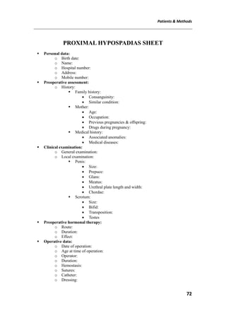

![Patients & Methods
76
Operative technique:
Under general anesthesia, the children were given caudal
anesthesia and third generation cephalosporin.
We adopt the Hayashi's modification of Koyanagi technique, done
in November 2000 [30]
.
A skin-incision line is drawn with a marking pen, as in the original
Koyanagi repair. An appropriate sized neleton catheter (usually 6 or 8 F)
is placed. A circumferential incision is made <5 mm proximal to the
corona, Fig [21]
The portion between the dartos and Buck's fascia is dissected on
the dorsal side. On the ventral side the urethral plate is incised as for
chordee repair, Fig [22]
A U-shaped skin incision is then made surrounding the meatus, to
extend the skin along the marked line. This second incision is extended
laterally and dorsally onto the dorsal prepuce, <8 mm parallel to the first
incision. The incised line of the dorsal prepuce is joined at the 12 o'clock
position, Fig [23]
This loop-shaped skin flap is used to create a new urethra.
Subsequently the portion between the prepuce and the dartos is dissected
on the dorsal side, to fix the prepuce as a new urethra to the dartos and to
maintain blood supply. The pedicle to the neourethra is suffciently
dissected down towards the penile base Fig [24]. On the ventral side,
bands of fibrous tissue, which can be seen passing proximal to the
hypospadiac meatus, should be excised until the corpus spongiosum
proximal to the meatus is completely exposed inside the scrotum.
In the original Koyanagi repair, the skin flap is divided into two
portions at the 12 o'clock position to form a Yshape, whereas in the
modified Koyanagi repair, a button-hole is made through the pedicle of
the dartos. While the glans is passed through this hole, the parameatal](https://image.slidesharecdn.com/0d76b122-16b8-4db3-a4a2-c2c7dd2f68b6-150812214802-lva1-app6892/85/02-All-Thesis-Dr-Aboubakr-Omar-FINAL1-76-320.jpg)
![Patients & Methods
77
skin flap and the vascular pedicle are mobilized to the ventral side,
keeping the loop shape, Fig [25]
The meatus is created by splitting the glans. A single midline
vertical incision is made extending to the tip of the glans, and sharp
dissection is carried out bilaterally to define the plane between the glans
cap and the corpora, mobilizing the wings, Fig [26]
The internal side of the loop is then closed from the front wall with
continuous full-thickness bites using 6/0 polyglactin sutures, Fig [27]
The external side is sutured as minutely as possible in a continuous
subcuticular manner with the same sutures from the back wall of the
neourethra Fig [27], although the distal sutures are placed interrupted
when excessive length is to be trimmed.
The neourethra is placed within the groove and anastomosed with
the tip. When the glans wings are approximated, a haemostat should be
placed between the neourethra and the glans, but not too close to the
glans. The edge of the neourethra and the glans is sutured as for
meatoplasty. Byar's flaps are created with the dorsal foreskin. The divided
dorsal flaps are turned towards the ventral side and sutured to cover the
ventral skin defect, Fig [27]](https://image.slidesharecdn.com/0d76b122-16b8-4db3-a4a2-c2c7dd2f68b6-150812214802-lva1-app6892/85/02-All-Thesis-Dr-Aboubakr-Omar-FINAL1-77-320.jpg)
![Patients & Methods
78
Fig [20] Penoscrotal hypospadias with adequate penile, prepuce, glanular size,
Urethral plate is short, causing severe chordae, scrotal size is adequate but bifid
and no transposition is present.
Fig [21] An appropriate sized neleton catheter (usually 6 or 8 F) is placed. A
circumferential incision is made <5 mm proximal to the corona.
Fig [22] The portion between the dartos and Buck's fascia is dissected on the
dorsal side. On the ventral side the urethral plate is incised as for chordae repair.](https://image.slidesharecdn.com/0d76b122-16b8-4db3-a4a2-c2c7dd2f68b6-150812214802-lva1-app6892/85/02-All-Thesis-Dr-Aboubakr-Omar-FINAL1-78-320.jpg)
![Patients & Methods
79
Fig [23] A U-shaped skin incision is then made surrounding the meatus, to extend the
skin along the marked line. This second incision is extended laterally and dorsally
onto the dorsal prepuce, <8 mm parallel to the first incision. The incised line of the
dorsal prepuce is joined at the 12 o'clock Position.
Fig [24] The portion between the prepuce and the dartos is dissected on the dorsal
side, to fix the prepuce as a new urethra to the dartos and to maintain blood supply.
The pedicle to the neourethra is sufficiently dissected down towards the penile base.
Fig [25] A button-hole is made through the pedicle of the dartos. While the glans is passed
through this hole, the parameatal skin flap and the vascular pedicle are mobilized to the
ventral side, keeping the loop shape.](https://image.slidesharecdn.com/0d76b122-16b8-4db3-a4a2-c2c7dd2f68b6-150812214802-lva1-app6892/85/02-All-Thesis-Dr-Aboubakr-Omar-FINAL1-79-320.jpg)
![Patients & Methods
80
Fig [26] The meatus is created by splitting the glans. A single midline vertical incision
is made extending to the tip of the glans, and sharp dissection is carried out bilaterally
to define the plane between the glans cap and the corpora, mobilizing the wings.
Fig [27] The internal side of the loop is then closed from the front wall with
continuous full-thickness bites using 6/0 polyglactin sutures. The external side is
sutured as minutely as possible in a continuous subcuticular manner with the same
sutures from the back wall of the neourethra, Byar's flaps are created with the dorsal
foreskin. The divided dorsal flaps are turned towards the ventral side and sutured to
cover the ventral skin defect.](https://image.slidesharecdn.com/0d76b122-16b8-4db3-a4a2-c2c7dd2f68b6-150812214802-lva1-app6892/85/02-All-Thesis-Dr-Aboubakr-Omar-FINAL1-80-320.jpg)
![RESULTS
82
RESULTS
This study was conducted in the General Pediatric Surgery Unit, in
Cairo University Specialized Pediatric hospital, during the period of
March 2014 to March 2015. Twenty cases with proximal hypospadias
were included in our prospective study. We performed the single stage
repair, modified Koyanagi technique to all of them.
Results were expressed as means ± standard deviation of the means,
minimum, maximum or number (%). Statistical Package for Social
Sciences (SPSS) computer program (version 19 windows) was used for
data analysis.
As regarding age, it ranged from 1 to 5 years. We performed the
operation to children with mean age of 2.83 ± 1.17. Five cases out of the
twenty were 2.5 years old (25%). Fig[28], table [5]
Fig [28 ] Age distribution](https://image.slidesharecdn.com/0d76b122-16b8-4db3-a4a2-c2c7dd2f68b6-150812214802-lva1-app6892/85/02-All-Thesis-Dr-Aboubakr-Omar-FINAL1-82-320.jpg)
![RESULTS
83
Table [5]: Mean age value
Characteristics Patients (n= 20)
Range (minimum-maximum) 1-5
Mean ±S.D 2.83 ± 1.17
As regarding the pathology, we included 20 males with proximal
hypospadias, all patients had severe chordae. 16 patients (80%) were
penoscrotal while 4 patients (20%) were perineal type. Fig[29], table [6]
14 patients (70%) were free from any associated local anomaly while 6
patients (30%) had associated local anomaly. 3 patients (15%) had
unilateral undescended testicle. 1 patient ( 5%) had bilateral undescended
testicle. 1 patient (5%) had unilateral oblique inguinal hernia. 1 patient
(5%) had on one side undescended testicle and the other side oblique
inguinal hernia. Fig[30], table [7]
Table [6]: Type of hypospadias
Characteristics Number Percent
Penoscrotal 16 80.0
Perineal 4 20.0
Fig. [29] Type of
hypospadias in the studied patients](https://image.slidesharecdn.com/0d76b122-16b8-4db3-a4a2-c2c7dd2f68b6-150812214802-lva1-app6892/85/02-All-Thesis-Dr-Aboubakr-Omar-FINAL1-83-320.jpg)
![RESULTS
84
Table [7]: Associated anomaly and its types in the studied patients
Characteristics Number Percent
Associated anomaly
No
Yes
14
6
70.0
30.0
Type of associated anomaly (n= 6)
Unilateral undescended testicle
Bilateral undescended testicle
Unilateral hernia
Rt. Hernia, Lt undescended testicle
3
1
1
1
15.0
5.0
5.0
5.0
Fig [30] Associated anomaly in the studied patients
As regarding etiology; positive consanguinity was proved in 5
patients (25%) while the rest 15 patients (75%) were negative for
consanguinity. Fig[31], table [8]
Drug usage whether to induce pregnancy or to fix the threatened
abortion or any other medical conditions were reported in 8 (40%)
patients while we found it negative in 12 patients (60%).](https://image.slidesharecdn.com/0d76b122-16b8-4db3-a4a2-c2c7dd2f68b6-150812214802-lva1-app6892/85/02-All-Thesis-Dr-Aboubakr-Omar-FINAL1-84-320.jpg)
![RESULTS
85
An interesting finding was reported is that 2 patients (10%) were a
twin to a female sister.
Table [8]: Incidence of drug usage during pregnancy, consanguinity and
twin sister in the studied patients.
Characteristics Number Percent
Drug usage during pregnancy
Negative
Positive
12
8
60.0
40.0
Consanguinity
Negative
Positive
15
5
75.0
25.0
Pregnancy
Single
Twin
18
2
90.0
10.0
Fig. [31] incidence of drug usage during pregnancy, consanguinity and twin
sister in the studied patients.](https://image.slidesharecdn.com/0d76b122-16b8-4db3-a4a2-c2c7dd2f68b6-150812214802-lva1-app6892/85/02-All-Thesis-Dr-Aboubakr-Omar-FINAL1-85-320.jpg)
![RESULTS
86
As regarding androgen used prior to surgery, we used topical
(androgen 5%), in 12 patients (60%). Fig[32], table [9] .The duration
ranged between 1 to three months prior to surgery. table [10] .
Table [9]: Percentage of patients using androgen prior to surgery in the
studied patients
Characteristics Number Percent
Negative 8 40.0
Positive 12 60.0
Fig. [32] Percentage of patients using androgen prior to surgery in the studied
patients.
Table [10]: Mean value of duration of androgen given prior to surgery
(months) in the studied patients
Characteristics Patients (n= 20)
Range (minimum-maximum) 1-3
Mean ± SD 1.79 ± 0.66](https://image.slidesharecdn.com/0d76b122-16b8-4db3-a4a2-c2c7dd2f68b6-150812214802-lva1-app6892/85/02-All-Thesis-Dr-Aboubakr-Omar-FINAL1-86-320.jpg)
![RESULTS
87
As regarding operation, in all the patients we used the modified
Koyanagi technique, the operative time ranged between 150 to 300
minutes. The mean operative time was 193.5 ± 41.84 minutes. table [11]
We used a second layer using the tunica vaginalis of the testes or scrotal
dartos flap in 7 patients (35%). Fig[33], table [12].
Table [11]: Operative time (minutes) in the studied patients
Characteristics Patients (n= 20)
Range (minimum-maximum) 150-300
Mean ± SD 193.5 ± 41.84
Table [12]: Second layer usage in the studied patients
Characteristics Number Percent
Negative 12 65.0
Positive 7 35.0
Fig [33] Second layer usage in the studied patients](https://image.slidesharecdn.com/0d76b122-16b8-4db3-a4a2-c2c7dd2f68b6-150812214802-lva1-app6892/85/02-All-Thesis-Dr-Aboubakr-Omar-FINAL1-87-320.jpg)
![RESULTS
88
As regarding postoperative follow up, the mean follow up time
in months was 8.15 ± 3.53, it ranged from 2-12 months. Fig[34],Fig [35],
table [13].
Table [13]: Follow up time (months) in the studied patients
Characteristics Patients (n= 20)
Range (minimum-maximum) 2-12
Mean ± SD 8.15 ± 3.53](https://image.slidesharecdn.com/0d76b122-16b8-4db3-a4a2-c2c7dd2f68b6-150812214802-lva1-app6892/85/02-All-Thesis-Dr-Aboubakr-Omar-FINAL1-88-320.jpg)
![RESULTS
89
Fig [34] Early outcome](https://image.slidesharecdn.com/0d76b122-16b8-4db3-a4a2-c2c7dd2f68b6-150812214802-lva1-app6892/85/02-All-Thesis-Dr-Aboubakr-Omar-FINAL1-89-320.jpg)
![RESULTS
90
Fig [35] Late outcome](https://image.slidesharecdn.com/0d76b122-16b8-4db3-a4a2-c2c7dd2f68b6-150812214802-lva1-app6892/85/02-All-Thesis-Dr-Aboubakr-Omar-FINAL1-90-320.jpg)
![RESULTS
91
As regarding early complications, we were able to detect
bleeding, retention, infection and stenosis, Fig [36] , table [14].
Immediate postoperative bleeding was reported in one patient (5%), to
which conservative measures were done and excellent healing was
achieved. Retention was reported in one patient (5%) , to which a supra
pubic cysto catheter was inserted, patient needed three sessions of
dilatation before removal of the supra pubic cysto catheter and developed
penoscrotal fistula , Fig [37] .Infection was reported in one patient (5%),
to which repeated dressings, wound swab was done for culture and
sensitivity, this patient developed recession of meatus . Stenosis was
reported in three patient (15%), to which three sessions of dilatations
were done each, Fig[38].
Table [14]: Early complications in the studied patients.
Characteristics Number Percent
Bleeding
Negative
Positive
19
1
95.0
5.0
Retention
Negative
Positive
19
1
95.0
5.0
Infection
Negative
Positive
19
1
95.0
5.0
Stenosis
Negative
Positive
17
3
85.0
15.0](https://image.slidesharecdn.com/0d76b122-16b8-4db3-a4a2-c2c7dd2f68b6-150812214802-lva1-app6892/85/02-All-Thesis-Dr-Aboubakr-Omar-FINAL1-91-320.jpg)
![RESULTS
92
Fig [36] Early complications in the studied patients
Fig [37] Early complications, urine retention, suprapubic cystocatheter inserted](https://image.slidesharecdn.com/0d76b122-16b8-4db3-a4a2-c2c7dd2f68b6-150812214802-lva1-app6892/85/02-All-Thesis-Dr-Aboubakr-Omar-FINAL1-92-320.jpg)
![RESULTS
93
Fig [38] Early complication, stenosis, small meatus and narrow stream
As regarding late complications, they were in the form of fistula,
meatal recession, glanular torsion, and diverticulum, Fig[39], table [15].
Fistula was reported in four patients (20%). All of them were penoscrotal
site , Fig[40]. Meatal recession was reported in 5 patients (25%). two
patients (15%) were mid penile. two patient (5%) were anterior penile,
one patient (5%) was penoscrotal , Fig[41]. Glanular torsion was reported
in one patient (5%) ,Fig[42]. Urethral diverticulum was reported in one
patient (5%) , Fig[43]. all of them were planned for a second stage repair
after six months' time.](https://image.slidesharecdn.com/0d76b122-16b8-4db3-a4a2-c2c7dd2f68b6-150812214802-lva1-app6892/85/02-All-Thesis-Dr-Aboubakr-Omar-FINAL1-93-320.jpg)
![RESULTS
94
Table [15]: Late complications in the studied patients
Characteristics Number Percent
Fistula
Negative
Positive (penoscrotal small)
Positive (penoscrotal large)
16
3
1
80.0
15.0
5.0
Site of meatal recession
Negative
Positive (anterior penile)
Positive (mid penile)
Positive (penoscrotal)
15
2
2
1
75.0
10.0
10.0
5.0
Torsion of the glands
Negative
Positive
19
1
95.0
5.0
Diverticulum
Negative
Positive
19
1
95.0
5.0](https://image.slidesharecdn.com/0d76b122-16b8-4db3-a4a2-c2c7dd2f68b6-150812214802-lva1-app6892/85/02-All-Thesis-Dr-Aboubakr-Omar-FINAL1-94-320.jpg)
![RESULTS
95
80
20
75
25
95
5
95
5
0
10
20
30
40
50
60
70
80
90
100
110
Percent
Fistula Site of
meatal
recession
Torsion of
the glands
Diverticulum
Negative Positive
Fig [39] Late complications in the studied patients
Fig [40] Late complication, penoscrotal fistula.](https://image.slidesharecdn.com/0d76b122-16b8-4db3-a4a2-c2c7dd2f68b6-150812214802-lva1-app6892/85/02-All-Thesis-Dr-Aboubakr-Omar-FINAL1-95-320.jpg)
![RESULTS
96
Fig [41] Late complication, recession of meatus , penoscrotal.
Fig [42] Late complication, torsion of the penis](https://image.slidesharecdn.com/0d76b122-16b8-4db3-a4a2-c2c7dd2f68b6-150812214802-lva1-app6892/85/02-All-Thesis-Dr-Aboubakr-Omar-FINAL1-96-320.jpg)
![RESULTS
97
Fig [43] Late complication, urethral diverticulum, filled on micturition
As regarding redo surgery, 9 patients (45%) needed redo
surgeries for the recession of meatus only (4 patients), for the fistula only
(2 patient), glanular torsion only (1 patient), for the recession of meatus
and fistula (1 patient), for the diverticulum and fistula (1 patient).
Fig[44],table [16]
Table [16]: Cases needed redo surgeries in the studied patients
Characteristics Number Percent
Negative 11 55.0
Positive 9 45.0
Positive, 45
Negative, 55
Fig [44] Cases needed redo surgeries in the studied patients.](https://image.slidesharecdn.com/0d76b122-16b8-4db3-a4a2-c2c7dd2f68b6-150812214802-lva1-app6892/85/02-All-Thesis-Dr-Aboubakr-Omar-FINAL1-97-320.jpg)
![DISCUSSION
98
DISCUSSION
Hypospadias repair is one of the commonest operations done in
pediatric surgery centers, with an incidence of 1 in 200 to 1 in 300[1]
.
Proximal hypospadias defects represent the most challenging and
complex manifestations of this entity and might be successfully treated
with one of several one- or two-stage repairs[5]
. Each technique, whether
two stage or one-stage, has its advantages and drawbacks and not one
technique has gained widespread popularity. Heralding one technique as
the gold standard for posterior hypospadias is probably unrealistic and
pediatric urologists have to master a variety of techniques because
various patient-related specifics can favor one or another technique[6]
.
Development of the technique:
Russell reported parallel circumferential incisions that he called a
stole procedure. His operation, published in 1900, was a two-stage
procedure where the part of the urethra that was taken from the dorsum
was divided and the blood supply was not preserved. Koyanagi et al.
developed similar incisions but divided the dorsal neourethra. Long
parameatal based flaps were created in a procedure called "wing flap-
flipping urethroplasty" . These are exceedingly long and narrow
perimeatal based flaps that did not maintain pedicle blood supply[32]
.
In the Koyanagi procedure, a long wide strip is harvested from the
penile shaft skin in continuity with the preputial hood. This is then
transferred ventrally and tubularized allowing a one-stage correction[28]
.
Koyanagi et al. developed a one-stage urethroplasty with a
parameatal foreskin flap. The advantages of the Koyanagi repair are that
only one anastomosis is necessary at the meatus, and neither torsion nor
bulking of the penile shaft are observed. It is highly suitable for severe
proximal hypospadias because it ensures that there will always be enough
skin for the neourethra, as the distance from the meatus around the shaft](https://image.slidesharecdn.com/0d76b122-16b8-4db3-a4a2-c2c7dd2f68b6-150812214802-lva1-app6892/85/02-All-Thesis-Dr-Aboubakr-Omar-FINAL1-98-320.jpg)
![DISCUSSION
99
to the dorsal preputial midline is always longer than the distance from the
meatus to the tip of the glans. The use of parameatal tissues and adjacent
skin flaps reduces the overall degree of tissue mobilization, and
eliminates the need for a circumferential anastomosis between the urethra
and neourethra. This procedure is applicable to any kind of proximal
hypospadias, even cases with bifid scrotum or penoscrotal
transposition[34]
.
The actual reported experience of the Koyanagi repair has been
discouraging because of high complication and reoperation rates, and
especially the occurrence of fistulae, although Koyanagi’s operation
theoretically appeared to provide an ideal treatment for severe proximal
hypospadias. In the largest and most recent series by Koyanagi et al. of
70 patients, the rate of complications requiring a second operation,
including urethral stricture, meatal stensis and urethrocutaneous fistula,
was 47%. In a recent series of patients who underwent this technique
reported by Glassberg et al. a secondary operation was required in 50%.
When we employed the Koyanagi repair we also encountered a 47%
complication rate[34]
.
Complication rates or rather reoperative rates vary from 20 to 50%.
This seemingly high number must be put into perspective in that if a
staged approach is used, 100% of patients will undergo at least two
operations with a substantial requiring a third for complications which
may develop after urethral tubularization. With the Koyanagi procedure,
the majority of patients only require one operation although a sizable
number may require a second procedure. Thus using this approach, the
total number of operations a cohort of patients with proximal hypospadias
may require will be much less than if a planned two-stage approach is
used[39]
.
A major advantage of the technique is that all major dissection is
performed in virgin, untouched tissue allowing neourethral reconstruction](https://image.slidesharecdn.com/0d76b122-16b8-4db3-a4a2-c2c7dd2f68b6-150812214802-lva1-app6892/85/02-All-Thesis-Dr-Aboubakr-Omar-FINAL1-99-320.jpg)
![DISCUSSION
100
without any scar tissue affecting vascularity. It has the added advantage
that the dissection needed permits simultaneous repair of associated
penoscrotal transposition, if present. Thus, all aspects of severe proximal
hypospadias can be corrected at one setting[39]
.
Belman commented that the vascularity is not as good as Koyanagi
et al. assumed it to be, because their high rate of complications was a
consequence of diminished blood supply. Rushton also mentioned that the
high complication rate associated with the Koyanagi repair was probably
caused by failure to preserve the axial blood supply to the long
parameatal ventral preputial skin flaps used to construct the neourethra.
Emir et al advocated that failure to main an adequate blood supply to the
flaps may result in ischemia with decreased tissue viability that in
hypospadias repair typically manifests as a urethrocutaneous fistula or
urethral stricture[34]
.
Modifications of the technique: table (17)
Modifications to this procedure have since been made in an attempt
to improve the blood supply. The proponents believe that this is in
essence a two-stage procedure completed in one stage. The published
complication rates for Koyanagi procedure ranges from 20 to 50%.
Jayanthi in a recent publication on modified Koyanagi repair for proximal
hypospadias acknowledged that a sizeable number of boys will need
reoperation following this procedure. His argument is that 100% of the
boys will have a second operation in a staged approach[28]
.
As a vascularized pedicle is not used and the perimeatally based
flaps are much too long and narrow to satisfy principles of reliable flap
survival, Snow and Cartwright modified the Koyanagi repair using an
island-flap technique, which preserved a reliable dual blood supply to a
long urethra. Their original series consisted of only four cases with a 50%
success rate, and they have not reported any additional results with their
modification[34]
.](https://image.slidesharecdn.com/0d76b122-16b8-4db3-a4a2-c2c7dd2f68b6-150812214802-lva1-app6892/85/02-All-Thesis-Dr-Aboubakr-Omar-FINAL1-100-320.jpg)
![DISCUSSION
101
Relatively large series of 20 cases each using a modified Koyanagi
repair for severe hypospadias were reported by Emir et al. and Hayashi et
al. They obtained acceptable success rates for the repair of severe
hypospadias of 80% and 70%, respectively. Emir et al.reported urethra-
cutaneous fistula as a complication of their modification of the Koyanagi
repair; we have encountered not only this but also meatal stenosis with
almost the same technique that they employed[34]
.
Sugita and his colleagues modified the technique by removal of the
subcutaneous tissue of the distal portion of the flap around the glans. The
authors believe that blood supply at this location is not reliable and this
tissue should act as a free graft rather a vascularized flap. A success rate
of 83% was achieved[33]
.
Another modification was added by Hayashi and his colleagues in
June 2006, they decided to preserve the distal portion of the urethral plate
after it is divided so that we could increase the caliber of the glandular
urethra, and provide a blood supply to the peripheral portion of the
neourethra from the preserved urethral plate and the spongy tissue
beneath the plate, as well as from an island vascular pedicle. There was
no meatal stenosis in this series of 12 cases with their latest modification
of the Koyanagi repair. The overall success rate was therefore 92 %[34]
.
Table [17]: Koyanagi and its modifications results
Year of Author
publication
Sample Country
size
Stenosis Fistula Complicate Success
1. Koyanagi et al. 1984, Japan 70 ptn 26% 21% 47% 53%
2. Catright et al 1994, USA 4 ptn 50% 50% 50%
3. Emir et al. 2000, Turkey 20 ptn 20% 20% 80%
4. Hayahi et al. 2001, Japan 20 ptn 15% 15% 30% 70%
5. Sugita et al. 2001, Japan 151 ptn 2% 13% 17% 83%
6. Hayshi et al. 2006, Japan 12 ptn 8% 8% 92%](https://image.slidesharecdn.com/0d76b122-16b8-4db3-a4a2-c2c7dd2f68b6-150812214802-lva1-app6892/85/02-All-Thesis-Dr-Aboubakr-Omar-FINAL1-101-320.jpg)
![DISCUSSION
103
Anand Alladi et al. published their study in 2013, it was done on
sample sized 24 patients with age ranging from 9 months to 11 years old
2.8 .The follow up period ranged from 6-42 months.
Our study was conducted in the general pediatric surgery unit, in
Cairo university specialized pediatric hospital, during the period of March
2014 to March 2015. 20 patients with proximal hypospadias were
included in our prospective study. We performed the single stage repair,
modified Koyanagi technique to all of them. The age ranged from 1-5
years and a mean of 2.8 years old. 30% of them had associated external
genitalia anomalies. 60% of them received preoperative topical androgen
for 1 to 3 months. They had a follow up ranged from 2- 12 months.
Table [18]: Studies done on the modified Koynagi
Follow
up
Androgen
preoperativ
e
Associate
d
anomalies
Mean of
age
Sampl
e size
Countr
y
Year of
publicatio
n
Author
6 months
(planned)
--2.5 yrs20 ptnJapan2001
Hayashi et
al
3-
36month
s (range)
100%-1.6 yrs11 ptnEgypt2006
Elhalaby et
al
34month
s
(mean)
80%16%2.5 yrs31 ptnFrance2009
P.
Mouriquan
d et al
3-
97monhs
(range)
-28%
3-9 yrs
(range)
14 ptnIndia2010
Rajendra
Nerli et al
3-
12month
s (range)
30%-1.3 yrs30 ptnEgypt2010
Adham
Elsaied et
al
4-8
months
(range)
-25%2.6 yrs20 ptnEgypt2013
M.
Elkassaby
et al
6-42
months
(range)
--
9m-
11yrs(range
)
24 ptnIndia2013
Anand
Alladi et al
2-12
months
(range)
60%30%2.8 yrs20 ptnEgypt2015
Present
study](https://image.slidesharecdn.com/0d76b122-16b8-4db3-a4a2-c2c7dd2f68b6-150812214802-lva1-app6892/85/02-All-Thesis-Dr-Aboubakr-Omar-FINAL1-103-320.jpg)
![DISCUSSION
106
Table [19]: Complication rates in studies on modified Koyanagi
SuccessComplicationDiverticulumTorsion
Meatal
recession
FistulaStenosisRetentionInfectionBleedingAuthor
70%30%---15%15%---
Hayashi et
al
82%18%9%-9%18%----
Elhalaby et
al
39%61%16%-19%39%16%---
P.
Mouriquand
et al
64%36%7%-7%21%----
Rajendra
Nerli et al
90%10%--3%7%----
Adham
Elsaied et al
60%40%-5%10%40%5%-20%5%
M.
Elkassaby et
al
54%46%-4%8%21%12%---
Anand
Alladi et al
55%45%5%5%25%20%15%5%5%5%
present
study
Proximal hypospadias remains a challenge for the paediatric
surgeon as the complication rate and the reoperation rate are high
whichever procedure is chosen. For severe forms of hypospadias with a
proximal division of corpus spongiosum, i.e. located proximal to the
penile midshaft, the choice of urethroplasty is very much surgeon
dependent. Literature data concerning outcome of the various techniques
commonly used to reconstruct proximal hypospadias are reviewed[37]
.
Table [20]: Comparison of the outcome of different techniques for
proximal hypospadias[37]
.](https://image.slidesharecdn.com/0d76b122-16b8-4db3-a4a2-c2c7dd2f68b6-150812214802-lva1-app6892/85/02-All-Thesis-Dr-Aboubakr-Omar-FINAL1-106-320.jpg)
![SUMMARY
107
SUMMARY
Hypospadias is the most common congenital anomaly of the penis,
affecting 0.4–8.2 of 1000 live male babies[17]
.
Hypospadias, a term derived from the Greek terms hypo (under)
and spadon (rent, fissure) In most cases, hypospadias in the male is
associated with three anomalies of the penis: (1) a ventral meatus that
may be located anywhere between the glans and the perineum, (2) ventral
deviation of the penis (chordee), and (3) the dorsal prepuce hood in
association with a ventral deficit of the prepuce. The second and third
abnormalities are not necessary for the diagnosis of hypospadias[4]
.
The position of the urethral meatus can be classified as anterior or
distal (glandular, coronal, or subcoronal; 60–65% of cases), middle
(midpenile; 20–30% of cases), or posterior or proximal (posterior penile,
penoscrotal, scrotal, or perineal; 10–15% of cases)[2]
.
In boys with more proximal hypospadias, cryptorchidism may
occur as frequently as 32%[16]
.
Hypospadias is considered to be a mild form of the 46, XY
disorders of sex development (DSD), In most cases, the degree of
hypospadias is relatively mild and a specific endocrine cause is not
sought or is not found. However, four main elements are involved in male
genital construction and may contribute to this malformation:
1. The genetic and endocrine background of the child, principally the
genes of phallic development, gonadal steroid synthesis (mainly
testosterone and its 5α reduced form, dihydrotestosterone, DHT),
and the responsiveness to these hormones. The genital tubercle thus
grows under the influence of androgens and any alteration in
androgen production or receptors may produce a hypospadiac penis.](https://image.slidesharecdn.com/0d76b122-16b8-4db3-a4a2-c2c7dd2f68b6-150812214802-lva1-app6892/85/02-All-Thesis-Dr-Aboubakr-Omar-FINAL1-107-320.jpg)
![SUMMARY
108
2. The placenta, which orchestrates the hormonal climate, especially
during the first part of gestation.
3. The mother, with her own hormonal production and possible
disorders.
4. The environment of mother and child, which may also interfere in
this fine balance[18]
.
Surgical techniques for hypospadias have evolved over the years
but the principles of any surgery remain the same; namely, to give the
child a straight penis with a terminal or near terminal meatus which is
cosmetically and functionally acceptable with minimal morbidity[3]
.
Hypospadias repair has a long and flat learning curve and requires
patience, experience, and great enthusiasm to achieve acceptable results.
The results published on the various operative techniques need to be
repeated and validated by other surgeons, and long-term results (up to
adulthood) are essential to justify operative methods and identify late
complications[4]
.
Proximal hypospadias defects represent the most challenging and
complex manifestations of this entity[5]
. The quality of the urethral plate
is the key to a successful hypospadias repair and the real difficulty is
deciding which urethral plate is of poor quality and needs to be
sacrificed[28]
.
No single technique is ideal, and pediatric surgeon has to master
many technique to deal with various anatomical variants of the proximal
hypospadias. A single staged repair can be safely and effectively
performed even in patients with the most severe proximal hypospadias.
Modified Koyanagi repair performed to severe hypospadias with chordee
gives a good cosmetic and functional result. Complications rate is low
once the learning curve is crossed.](https://image.slidesharecdn.com/0d76b122-16b8-4db3-a4a2-c2c7dd2f68b6-150812214802-lva1-app6892/85/02-All-Thesis-Dr-Aboubakr-Omar-FINAL1-108-320.jpg)
![REFERENCES
110
REFERENCES
[1] Raimund Stein. Hypospadias. European Urology Supplements 11
(2012) 33–45.
[2] Leung ARC, et al. Hypospadias: an update. Asian J Androl (2007);
9:16–22.
[3] Lisa Steven, et al. Current practice in paediatric hypospadias
surgery; A specialist survey. Journal of Pediatric Urology (2013)
1477-5131.
[4] Raimund Stein. Hypospadias. European Urology Supplements 11
(2012) 33–45
[5] Rajendra Nerli, et al. Modified Koyanagi’s procedure for
proximal hypospadias: Our experience. International Journal of
Urology (2010) 17, 294–296.
[6] Alexis Arnaud, et al. Choosing a technique for severe hypospadias.
African Journal of Paediatric Surgery.September-December (2011)
/ Vol 8 / Issue 3, (2011) , 286-290.
[7] MK Hassan, et al. Single Stage Urethroplasty for Proximal
Hypospadias with Chordee. Faridpur Med. Coll. J. (2011); 6(2):
66-69
[8] Adham Elsaied, Basem Saied, et al Modified Koyanagi
Technique in Management of Proximal Hypospadias. Annals of
Pediatric Surgery Vol. 6, No 1, January (2010), PP 22-26
[9] Bruce M. Carlson, Urogenital System Chapter 16, Human
Embryology and Development Biology, Fifth Edition, (2014), 376-
407.
[10] Sarah D. Blaschko, et al, Molecular mechanisms of external
genitalia development, International Society of Differentiation.
(2012) , 261-268.](https://image.slidesharecdn.com/0d76b122-16b8-4db3-a4a2-c2c7dd2f68b6-150812214802-lva1-app6892/85/02-All-Thesis-Dr-Aboubakr-Omar-FINAL1-110-320.jpg)
![REFERENCES
111
[11] T.W. Sadler, urogenital system chapter 16, Langman's Medical
Embryology Twelfth Edition, (2012), 232-260.
[12] Jenny H. Yiee et al, Penile Embryology and Anatomy, The
Scientific World Journal (2010) 10, 1174–1179.
[13] Klaus Steger , et al , Anatomy of the Male Reproductive System,
Practical Urology: Essential Principles and Practice, (2011), 54-69.
[14] Moira E. Dwyer, et al, Normal Penile, Scrotal, and Perineal
Anatomy with Reconstructive Considerations. . Semin Plast Surg
(2011); 25:179–188.
[15] Benjamin I. Chung, et al , Anatomy of the Lower Urinary Tract
and Male Genitalia, Campbell-Walsh Urology tenth edition,
(2012) 33-74.
[16] Kate H. Kraft, et al , Proximal Hypospadias, The Scientific World
Journal (2011) 11, 894–906.
[17] Antonella Giannantoni, Hypospadias Classification and Repair:
The Riddle of the Sphinx, European Urology 60 (2011) 1190 –
1192
[18] Kalfa, N. , et al , Hypospadias: interactions between environment
and genetics, Molecular and Cellular Endocrinology , (2010) 1-25.
[19] Livia Teresa Moreira Rios, et al, Prenatal Diagnosis of
Penoscrotal Hypospadia in Third Trimester by Two- and Three-
Dimensional Ultrasonography: A Case Report, Case Reports in
Urology, Volume (2012) 1-3.
[20] Snodgrass W, et al. Hypospadias dilemmas: A round table, Journal
of Pediatric Urology (2011) 1-13.
[21] Merriman LS, et al. The GMS hypospadias score: Assessment of
inter-observer reliability and correlation with post-operative
complications, Journal of Pediatric Urology (2013) 1-6.
[22] Bush NC, et al. Age does not impact risk for urethroplasty
complications after tubularized incised plate repair of hypospadias
in prepubertal boys, Journal of Pediatric Urology (2012) 1-5.](https://image.slidesharecdn.com/0d76b122-16b8-4db3-a4a2-c2c7dd2f68b6-150812214802-lva1-app6892/85/02-All-Thesis-Dr-Aboubakr-Omar-FINAL1-111-320.jpg)
![REFERENCES
112
[23] Brendan C. Jones, et al, Early Hypospadias Surgery May Lead to
a Better Long-Term Psychosexual Outcome, The Journal Of
Urology Vol. 182, (2009) 1744-1750.
[24] Netto JMB, et al. Hormone therapy in hypospadias surgery: A
systematic review, Journal of Pediatric Urology (2013) 1-9.
[25] Daniela B Gorduza, et al. Does androgen stimulation prior to
hypospadias surgery increase the rate of healing complications? A
preliminary report, J Pediatr Urol (2010) 1-4
[26] Ahmed T. Hadidi, et al , Chapter 94 Hypospadias, Paediatric
Surgery: A Comprehensive Text for Africa .(2011) 541-553.
[27] Michael Pfeil , et al , Hypospadias repair: an overview,
International Journal of Urological Nursing , Vol 4 No 1. (2010) 4
-12
[28] Ramnath Subramaniam, et al, Hypospadias Repair: An
Overview of the Actual Techniques. Semin Plast Surg (2011);
25:206–212.
[29] H. Emir, et al , Modification of The Koyanagi Technique for The
Single Stage Repair of Proximal Hypospadias ,The Journal of
Urology® Vol. 164, September (2000), 973–976.
[30] Y. Hayashi, et al , The modified Koyanagi repair for severe
proximal Hypospadias, BJU International (2001), 87, 235-238.
[31] Rajendra Nerli, et al, Modified Koyanagi’s procedure for
proximal hypospadias: Our experience. International Journal of
Urology (2010) 17, 294–296
[32] BrentLiv. Snow, The Yoke Hypospadias Repair , HADIDI •
AZMY Hypospadias Surgery, (2004) 203- 208 .
[33] Yoshifumi Sugita, et al , Severe Hypospadias Repair With Meatal
Based Paracoronal Skin Flap: The Modified Koyanagi Repair. The
Journal Of Urology® Vol. 166, September (2001) 1051–1053.](https://image.slidesharecdn.com/0d76b122-16b8-4db3-a4a2-c2c7dd2f68b6-150812214802-lva1-app6892/85/02-All-Thesis-Dr-Aboubakr-Omar-FINAL1-112-320.jpg)
![REFERENCES
113
[34] Yutaro Hayashi, et al , Neo-modified Koyanagi technique for the
single-stage repair of proximal hypospadias. Journal of Pediatric
Urology (2007) 3, 239-242
[35] Mamdouh Elkassaby, et al . Clinical Study, Comparative study
between modified Koyanagi and Snodgrass techniques in
management of proximal types of hypospadias . Journal of Taibah
University Medical Sciences (2013) 8(2), 97–104
[36] Elisangela de Mattos e Silva, et al, Outcome of severe
hypospadias repair using three different techniques,Journal of
Pediatric Urology (2009) 5, 205-211
[37] Massimo Catti, et al , Original Koyanagi urethroplasty versus
modified Hayashi technique: Outcome in 57 patients, Journal of
Pediatric Urology (2009) 5, 300-306
[38] Springer A, Assessment of outcome in hypospadias surgery – a
review, the journal Frontiers in Pediatrics. sJanuary 2014 | Volume
2 | Article 2 | 1-7 .
[39] Venkata R. Jayanthi. The modified Koyanagi hypospadias repair
for the one-stage repair of proximal hypospadias, Indian J Urol.
2008 Apr-Jun; 24(2): 206–209.
[40] Warren Snodgrass et al. Tubularized incised plate proximal
hypospadias repair: Continued evolution and extended
applications, J Pediatr Urol (2010), 1-8.
[41] Alexander Springer et al. Split Dorsal Dartos Flap Transposed
Ventrally as a Bed for Preputial SkinGraft in Primary Staged
Hypospadias Repair,UROLOGY (2012) , 79: 939–942.](https://image.slidesharecdn.com/0d76b122-16b8-4db3-a4a2-c2c7dd2f68b6-150812214802-lva1-app6892/85/02-All-Thesis-Dr-Aboubakr-Omar-FINAL1-113-320.jpg)

The document discusses the anatomy and embryology related to hypospadias. Regarding embryology, it describes the development of the genital system including gonadal differentiation, development of the male duct system, division of the cloaca, and sex differentiation of the external genitalia. It notes that hypospadias results from failure of fusion of the penile urethral folds during embryonic development. The document also reviews the anatomy of the penis, male urethra, and corpus spongiosum, noting the structures and their development are important for understanding hypospadias.


