7. Dega osteotomy extends through the outer
table of the ilium from the AIIS to the sciatic
notch.
A bicortical osteotomy is performed only at the
AIIS and with a kerison rongeur at the sciatic
notch.
The inner table of the ilium is not cut.
The lateral osteotomy made through the outer
table is extended with curved osteotomes to the
triradiate cartilage under fluroscopic guidance.
8. The osteotomy is then pried down laterally and
posteriorly with osteotomes and hinged on the
triaradiate cartilage, with the inner table of the
ilium being left intact.
Wedges of bone graft prop the osteotomy open,
and the direction of desired coverage is
addressed by where one places the bone graft.
The sponginess of the triradiate cartilage closes
the osteotomy around the bony wedges, so
fixation with pins is usually not necessary.
10. Study by mubarak and
colleagues
Dega osteotomy combined with
adductor, iliopsoas and proximal
hamstring release and a
shortening femoralVDRO
95% Of 104 hips
remained stable at 7
yrs follow up
AVN occurred in
8% of the hips.
Allowed excellent correction
of the superior and lateral
deficiency seen
preoperatively.
11. They advocated performing the osteotomy in
those with open triradiate cartilage , an
acetabular index greater than 25 degrees, MI
greater than 40%.
12. Open reduction if hip is 70% uncovered.
With open reduction , increased risk of AVN.
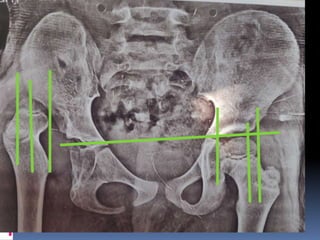
13. Severe subluxation of the right hip of a 7 year old boy
with spastic quadriplegia.The left hip is well contained.
14. A unilateral varus derotation osteotomy with a shelf
procedure and bilateral adductor releases were
performed.
15. The left hip subluxated 2.5 years after the right hip
reconstruction.
17. One year after contralateral reconstruction, both
hips were reduced and painless.
18. AP radiograph of the pelvis of a 10 yr old child with
cerebral palsy
19. A 3D CT scan of the right hip reveals global deficiency of
the acetabulum with anterior, superior and lateral lack of
coverage.
20. The hip was reconstructed by muscle release , femoral
VDRO with blade plate fixation and a dega pelvic
osteotomy.
21. The patient is positioned supine with the affected hip
raised on a bump. An anterior incision is made over
the iliac crest.The dega osteotomy is usually
performed during the same surgical setting as a
VDRO.
22. The iliac apophysis is split and the inner and outer
tables are exposed subperiosteally to the sciatic notch.
The direct head of the rectus femoris is detached.
23. Strip inner and outer surfaces of pelvis to
access sciatic notch.