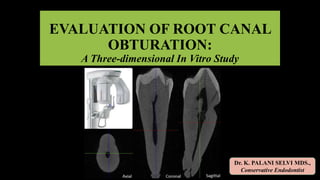
EVALUATION OF ROOT CANAL OBTURATION: A Three-dimensional In Vitro Study
•
1 like•204 views

CONE BEAM COMPUTED TOMOGRAPHIC EVALUATION OF 3 ROOT CANAL OBTURATION TECHNIQUES: AN IN-VITRO STUDY

Recommended
More Related Content
What's hot
What's hot (20)
Similar to EVALUATION OF ROOT CANAL OBTURATION: A Three-dimensional In Vitro Study
Similar to EVALUATION OF ROOT CANAL OBTURATION: A Three-dimensional In Vitro Study (20)
More from Palaniselvi Kamaraj
More from Palaniselvi Kamaraj (10)
Recently uploaded
Recently uploaded (20)
EVALUATION OF ROOT CANAL OBTURATION: A Three-dimensional In Vitro Study
- 1. EVALUATION OF ROOT CANAL OBTURATION: A Three-dimensional In Vitro Study Dr. K. PALANI SELVI MDS., Conservative Endodontist
- 3. CONTENTS
- 5. BIOMECHANICAL PREPARATION 3 DIMENSIONAL OBTURATION DISINFECTION OF THE CANAL SYSTEM INTRODUCTION
- 6. THE WASHINGTON STUDY • Study of endodontic success and failure • 60% of the failures is apparently caused by INCOMPLETE OBTURATION. • It is indispensable that the obturation should have a fluid tight hermetic seal.
- 7. OBTURATION MATERIALS • GUTTAPERCHA popularized by Bowman in 1867 is the most widely used root canal-filling material. • ROOT CANAL SEALERS enhance the adaptation of the core material to root dentin and are used in conjunction with GP.
- 8. COMPOSITION Polydimethyl Siloxane, Silicone Oil, Paraffin Base Oil, Hexachloroplatinic Acid, Zirconium Dioxide + GUTTAPERCHA POWDER • The excellent flow of this material made it the sealer of choice. • An excellent property of slight expansion after mixing which helps in better sealing. SEALER
- 9. OBTURATION TECHNIQUES 1. Cold Lateral Compaction 2. Warm Compaction (warm GP) A. Vertical B. Lateral 3. Continuous wave Compaction technique 4. Thermoplasticized GP injection 5. Carrier- based GP A. Thermafil thermoplasticized B. SimpliFill sectional obturation 6. McSpadden thermomechanical compaction 7. Chemically plasticized GP 8. Singe cone obturation 9. Custom cone
- 10. ASSESSMENT OF QUALITY OF ROOT FILLINGS SEMI-QUANTITATIVE ANALYSIS • Acid dissolution of roots • Electrochemical method • Fluid filtration • Dye penetration • Radiographs • Sections of the sample • SEM analysis. 3- DIMENSIONAL QUANTITATIVE ANALYSIS • CBCT • MICRO CT • SPIRAL CT
- 12. AIM • To compare the quality of 3 different root canal Obturation techniques: 1. Lateral compaction 2. Single cone method 3. Thermoplasticized GP - Calamus • using GUTTAFLOW 2 as sealer • Evaluation done by Cone Beam Computed Tomography.
- 16. ARMAMENTARIUM
- 17. M A T E R I A L S GROUP - A GROUP - C GROUP- B
- 18. M E T H O D O L O G Y 60 extracted lower premolars Pre –op radiograph Access cavity preparation Working length determination BMP WaveOne Gold NiTi rotary file system Size 35 - 6% ( medium) IRRIGATION 2.5% NaOCl 17% EDTA Normal Saline PRE- OBTURATION CBCT RANDOMISATION 3 groups(n= 20) A- CLC B- SINGLE CONE C- THERMOPLASTICIZED GP Post- Obturation CBCT
- 19. SAMPLE SELECTION
- 20. SAMPLE SELECTION • 60 human mandibular premolar teeth extracted for the orthodontic purpose were used for the study after ethical clearance. • Stored in 3% sodium hypochlorite solution for 1 week and later transferred to normal saline. • Single rooted teeth with mature apices without any defects were selected for the study after confirmation by taking radiographs.
- 22. RANDOMISATION GROUP --A COLD LATERAL CONDENSATION GROUP- B SINGLE CONE METHOD GROUP- C THERMOPLASTICIZED GP TECHNIQUE
- 23. PRE – OBTURATION CBCT
- 24. • The average height of the canal space(h) was measured from orifice to the root tip in the coronal and sagittal CBCT slices. • The D1 and D2 were the diagonals of the prepared canal space measured in the axial section. • The area of the canal space was then calculated using the formula, PRE – OBTURATION CBCT Volume of the prepared canal space (R) • The volume of the prepared canal space (R) was measured using diagonals method. R= Area of canal space x Height of the canal
- 25. Group A -Cold lateral condensation • A size 35 GP with 2% taper was coated with Guttaflow sealer and placed in the canal to the working length with tugback. • Lateral condensation was achieved, with additional accessory cones which were also coated with Guttaflow sealer, using a standardized finger spreader starting 1 mm short of working length. • When the points prevented the spreader penetration beyond the coronal third of the canal, the canal was considered to be adequately filled. • Excess GP was removed at CEJ using a heated condenser. • The GP at the CEJ was compacted using a cold plugger.
- 26. Group B -SINGLE-CONE OBTURATION • A size 35 GP point of 6% taper coated with Guttaflow sealer was used as a master cone and was placed in the canal up to the working length. • The excess cone was removed at CEJ using a heated condenser. • The GP at the CEJ was compacted using a cold plugger.
- 27. Group C - THERMOPLASTICIZED GUTTA-PERCHA TECHNIQUE • CALAMUS obturation unit was used for this technique. • A 35 size GP cone with 2% taper was coated with the sealer and placed in the canal up to the working length. • A medium-sized Calamus System -B insert tip which bound in the canal 3 mm short of working length was used at a temperature of 200°C and pressed against the cone so that the remaining cone in the canal was 3 mm and condensed using a plugger. • The 23-gauge cartridge’s needle tip was placed next to the master point to a depth at which the tip was neither forced nor bound to the canal wall. • The backfill was achieved by setting the temperature to 180°C and pressing the trigger so that the molten GP flowed in the tip was withdrawn slowly out of the canal. • The GP at the CEJ was compacted using a cold plugger.
- 28. Group C - THERMOPLASTICIZED GUTTA-PERCHA TECHNIQUE
- 29. POST- OBTURATION CBCT MODELS
- 31. MEASURING PARAMETERS • Volume percentage of the filling materials and Volume percentage of Voids were measured - Overall - Coronal 3rd - Middle 3rd - Apical 3rd
- 32. MEASURING PARAMETERS • At each levels the volume of filling materials was calculated using the formula d1 × d2 ×h - where d1 and d2 were the obturation material diagonals measured in the axial section - h - the height measured in coronal section. Volume percentage of the filling materials • The volume percentage of the obturated area in the 3 groups were calculated using formula: (Volume of filling /Total volume of prepared canal space ) × 100
- 33. MEASURING PARAMETERS Volume percentage of the Voids • The specimen where voids were seen, the inner area of the void was calculated using the linear measurements obtained using the Galileos viewer software and this value was multiplied by the slice thickness in order to calculate the Volume of the Void (V). • The Volume Percentage of the voids in the obturated root canal was calculated by using the formula, (R-V) ×100/R - where, R is the volume of the root canal space - V is the volume of the void space
- 35. STATISTICALANALYSIS • Statistical analysis was performed with nonparametric tests Kruskal-Wallis and Friedman tests. • The level of significance was set at P<0.05.
- 37. VOLUME PERCENTAGE OF FILLING MATERIALS A – cold lateral 83.2 81.2 83.4 85.2 B – single cone 81.5 82.5 80.5 81.5 C – thermoplastic GP 94.5 91.8 94.7 96.9 GROUPS OVERALL CORONAL MIDDLE APICAL A – Cold lateral 83.2 81.2 83.4 85.2 B – Single cone 81.5 82.5 80.5 81.5 C – Thermoplastic GP 94.5 91.8 94.7 96.9
- 38. VOLUME PERCENTAGE OF FILLING MATERIALS • The highest percentage of filling material in the apical third and the whole length of the root canal was observed in the THERMOPLASTIC GP OBTURATION ( Group- C) • In comparison, the percentage of filling material in this group was significantly higher than that of CLC and Single Cone obturation groups (P<0.05)
- 39. VOLUME PERCENTAGE OF VOIDS
- 40. VOLUME PERCENTAGE OF VOIDS • The voids which were detected in all samples, these results obtained were: OVERALL The highest VP (5.01%) was detected in single cone technique ( Group- B) The lowest VP (2.73%) was observed in (Group- C) THERMOPLASTICIZED GP method – showed lowest void % at all 3 levels. SINGLE CONE METHOD – showed highest void % at MIDDLE 3rd COLD LATERAL CONDENSATION- highest void % at APICAL 3rd .
- 41. • Since the p - value is between 0.010-0.050 there is significant difference between the methods/materials used for condensation. • Hence we reject the null hypothesis at 5%level. • Based on the mean rank, THERMOPLASTIC GP TECHNIQUE is better than Cold Lateral Condensation followed by the Single Cone method. INFERENCE
- 43. • Without sectioning the specimens and loss of material • Non-invasive technique • Specimens can be used for further research • Reconstruct overlapping structures at arbitrary intervals • Ability to resolve small objects. Why CBCT ???
- 44. • The only limitation of cone beam computed tomography (CBCT) method is that it couldn't distinguish between sealer and gutta percha. - Asheibi et al 2014 • On the other hand, found the same limitation when using other CT also. - Yigit et al 2014 Why CBCT ???
- 45. Cold Lateral Condensation Technique The final filling in CLC had the appearance of numerous GP cones tightly pressed together and joined by frictional grip and the cementing substance, while the spreader tracts can be devoid of sealer or the sealer can resorb later leading to voids . Schilder J Endod. 2006 • Our study showed that there were voids between the accessory cones throughout the length of the canal in CLC group
- 46. Single cone method • Among the previous studies which have compared the total volume percentage of various obturating techniques using different methods, very limited studies involve the Single-cone Technique which is the most common method of obturation in recent years. • Voids seen at the middle third of root canals in single-cone obturation can be attributed to the Root Canal Anatomy Of Mandibular Premolar where the single cone has failed to fill the canal space completely. The single-point technique is simple but its application must be limited to round canals that have assumed a precise shape given by the instrumentation procedure Daniele et al J Endod 2009 A matched taper single-cone obturation technique may be more effective in narrow round canals. Gordan et al Int Endod J 2005
- 47. • The 3D obturation was best with Calamus as compared to Single cone and lateral condensation. • Best results with Calamus could be explained on the basis of the maximum inert core material, minimum amount of sealer and a higher degree of homogeneity associated with the calamus Ruddle CJ. Filling root canal systems: the Calamus 3-D obturation technique. Dent Today. 2010;29(4):76, 78-81. Thermoplasticized Gutta-percha technique
- 48. Thermoplasticized Gutta-percha technique Overextension of GP Peng et al J Endod 2007 ,Ansari et al J Conserv Dent 2012 Overextension of GP reported in thermoplasticized GP technique in various studies therefore it was used as a backfill to obtain controlled placement in the present study. Voids seen with thermoplasticized GP Angerame D Ann Ist Super Sanita 2012 , Hammed M, J Endod 2009 • Internal voids probably created by air entrapment during the backfill. • Not in communication with the canal walls. • Can be regarded as less dangerous for the endodontic prognosis • Adaptation of GP in the root canal is almost complete.
- 51. SUMMARY Within the limitations of the present in vitro study, it is concluded that • None of the obturation technique is able to seal the root canal completely. • The Calamus may be a good alternative for perfect quality of 3D Obturation and it may be helpful in obtaining the good hermetic seal in endodontically treated teeth in future. • Further research is required in determining the amount of filling area and voids in other obturation techniques.
- 52. REFERENCES • Schilder H. Filling root canals in three dimensions. Dent Clin North Am 1967;11:723-44. • Keller RN, Kimbrough WF, Anderson RW. A comparison of thermoplastic obturation techniques: adaptation to the canal walls. J Endod. 1997;23:703-06 • Tasdemir T, Yesilyurt C, Ceyhanli TK, Celik D, Kursat Er. Evaluation of apical filling after root canal filling by 2 different techniques. J Can Dent Assoc. 2009;75:20 • Kandaswamy D, Venkateshbabu N, Reddy GK, Hannah R, Arathi G, Roohi R. Comparison of laterally condensed, vertically compacted thermoplasticized, cold free-flow GP obturations-A volumetric analysis using spiral CT. J Conserv Dent 2009;12:145-9 • Anbu R, Nandini S, Velmurugan N. Volumetric analysis of root fillings using spiral computed tomography: An in vitro study. Int Endod J 2010;43:64-8. • Chokkalingam M, Ramaprabha, Kandaswamy D. Three-dimensional helical computed tomographic evaluation of three obturation techniques: In vitro study. J Conserv Dent 2011;14:273-6. • Ansari B, Umer F, Khan FR. A clinical trial of cold lateral compaction with Obtura II technique in root canal obturation. J Conserv Dent 2012;15:156-60.
- 53. REFERENCES • FareaM, RaniA, HuseinA,MasudiS, PameijerCH. Evaluation of Gutta-Percha-Filled Areas in Root Canals after Filling by TwoDifferent Obturation Techniques. Aust J Basic ApplSci 2011; 5: 631-6. • Angerame D, De Biasi M, Pecci R, Bedini R, Tommasin E, Marigo L, et al. Analysis of single point and continuous wave of condensation root filling techniques by micro-computed tomography. Ann Ist Super Sanita 2012;48:35-41 • Naseri M, Kangarlou A, Khavid A, Goodini M. Evaluation of the quality of four root canal obturation techniques using micro-computed tomography. Iran Endod J 2013; 8: 89-93. • Asheibi F, Qualtrough AJE, Mellor A, Withers PJ, Lowe T. Micro-CT Evaluation of Voids in the Filling Material of Single-Rooted Teeth Obturated with Different Techniques. J Res Pract Dent 2014; 2014: 1-10 • Keles A, Alcin H, Kamalak A, Versiani MA. 3D Micro-CT evaluation of root flling quality in oval-shaped canal. Int Endod J. 2014;47:1177-84. • Schäfer E, Schrenker C, Zupanc J, Bürklein S. Percentage of Gutta-percha Filled Areas in Canals Obturated with Cross-linked Gutta-percha Corecarrier Systems, Single-Cone and Lateral Compaction Technique. J Endod 2016; 42: 294-8.