Call Girls In Nihal Vihar Delhi ❤️8860477959 Looking Escorts In 24/7 Delhi NCR
bacteria.ppt
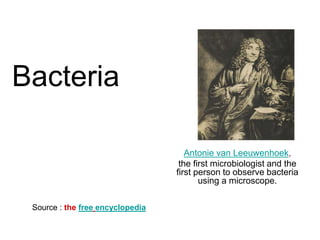
1. Bacteria
Antonie van Leeuwenhoek,
the first microbiologist and the
first person to observe bacteria
using a microscope.
Source : the free encyclopedia
2. Bacteria
• Bacteria were first observed by Antonie van Leeuwenhoek in 1676, using a
single-lens microscope of his own design.
• The name bacterium was introduced much later, by Christian Gottfried
Ehrenberg in 1838.
• Louis Pasteur demonstrated in 1859 that the fermentation process is
caused by the growth of microorganisms, and that this growth is not due to
spontaneous generation. (Yeasts and molds, commonly associated with
fermentation, are not bacteria, but rather fungi.) Along with his
contemporary, Robert Koch,
• Pasteur was an early advocate of the germ theory of disease.
• Robert Koch was a pioneer in medical microbiology and worked on cholera,
anthrax and tuberculosis. In his research into tuberculosis, Koch finally
proved the germ theory, for which he was awarded a Nobel Prize in 1905
• In Koch's postulates, he set out criteria to test if an organism is the cause of
a disease; these postulates are still used today.
3. Bacteria
• Though it was known in the nineteenth century that
bacteria are the cause of many diseases, no effective
antibacterial treatments were available.
• In 1910, Paul Ehrlich developed the first antibiotic, by
changing dyes that selectively stained Treponema
pallidum—the spirochaete that causes syphilis—into
compounds that selectively killed the pathogen.
• Ehrlich had been awarded a 1908 Nobel Prize for his
work on immunology, and pioneered the use of stains to
detect and identify bacteria, with his work being the basis
of the Gram stain and the Ziehl-Neelsen stain.
4. Bacteria
• A major step forward in the study of
bacteria was the recognition in 1977 by
Carl Woese that archaea have a separate
line of evolutionary descent from bacteria.
• This new phylogenetic taxonomy was
based on the sequencing of 16S ribosomal
RNA, and divided prokaryotes into two
evolutionary domains, as part of the three-
domain system.
6. Bacteria Morphology
A small number of species even have tetrahedral or
cuboidal shapes.
More recently, bacteria were discovered deep under the
Earth's crust that grow as long rods with a star-shaped
cross-section.
The large surface area to volume ratio of this morphology
may give these bacteria an advantage in nutrient-poor
environments.
This wide variety of shapes is determined by the bacterial
cell wall and cytoskeleton, and is important because it can
influence the ability of bacteria to acquire nutrients, attach
to surfaces, swim through liquids and escape predators.
7. Bacteria Morphology
• Most bacterial species are either spherical,
called cocci (sing. coccus, from Greek kókkos,
grain, seed)
• or rod-shaped, called bacilli (sing. bacillus,
from Latin baculus, stick). Elongation is
associated with swimming.
• Some rod-shaped bacteria, called vibrio, are
slightly curved or comma-shaped; others,
• It can be spiral-shaped, called spirilla, or tightly
coiled, called spirochaetes.
8. Bacteria Morphology
• Many bacterial species exist simply as single cells,
others associate in characteristic patterns: Neisseria
form diploids (pairs),
• Streptococcus form chains, and
• Staphylococcus group together in "bunch of grapes"
clusters.
• Bacteria can also be elongated to form filaments, for
example the Actinobacteria.
• Filamentous bacteria are often surrounded by a sheath
that contains many individual cells. Certain types, such
as species of the genus Nocardia, even form complex,
branched filaments, similar in appearance to fungal
mycelia.
10. Bacterial cell structure
Intracellular structures
• The bacterial cell is surrounded by a lipid membrane, or cell
membrane, which encloses the contents of the cell and acts as a
barrier to hold nutrients, proteins and other essential components of
the cytoplasm within the cell.
• As they are prokaryotes, bacteria do not tend to have membrane-
bound organelles in their cytoplasm and thus contain few large
intracellular structures.
• They consequently lack a nucleus, mitochondria, chloroplasts and
the other organelles present in eukaryotic cells, such as the Golgi
apparatus and endoplasmic reticulum.
• Bacteria were once seen as simple bags of cytoplasm, but elements
such as prokaryotic cytoskeleton, and the localization of proteins to
specific locations within the cytoplasm have been found to show
levels of complexity. These subcellular compartments have been
called "bacterial hyperstructures".
Intracellular structures
11. Bacterial cell structure
Intracellular structures
Many important biochemical reactions, such as energy generation, occur
by concentration gradients across membranes, a potential difference also
found in a battery.
The general lack of internal membranes in bacteria means reactions
such as electron transport occur across the cell membrane between the
cytoplasm and the periplasmic space.
However, in many photosynthetic bacteria the plasma membrane is
highly folded and fills most of the cell with layers of light-gathering
membrane.
These light-gathering complexs may even form lipid-enclosed structures
called chlorosomes in green sulfur bacteria.
Other proteins import nutrients across the cell membrane, or to expel
undesired molecules from the cytoplasm.
Intracellular structures
12. Bacterial cell structure
Extracellular structures
• Around the outside of the cell membrane is the bacterial
cell wall. Bacterial cell walls are made of peptidoglycan
(called murein in older sources), which is made from
polysaccharide chains cross-linked by unusual peptides
containing D-amino acids.
• Bacterial cell walls are different from the cell walls of
plants and fungi, which are made of cellulose and chitin,
respectively.
• The cell wall of bacteria is also distinct from that of
Archaea, which do not contain peptidoglycan. The cell
wall is essential to the survival of many bacteria, and the
antibiotic penicillin is able to kill bacteria by inhibiting a
step in the synthesis of peptidoglycan.
14. Bacterial Biofilm
• Bacteria often attach to surfaces and form dense
aggregations called biofilms or bacterial mats.
• These films can range from a few micrometers in
thickness to up to half a meter in depth, and may contain
multiple species of bacteria, protists and archaea.
• Bacteria living in biofilms display a complex
arrangement of cells and extracellular components,
forming secondary structures such as microcolonies,
through which there are networks of channels to enable
better diffusion of nutrients.
15. Bacterial Biofilm
• In natural environments, such as soil or the
surfaces of plants, the majority of bacteria are
bound to surfaces in biofilms.
• Biofilms are also important in medicine, as these
structures are often present during chronic
bacterial infections or in infections of implanted
medical devices, and bacteria protected within
biofilms are much harder to kill than individual
isolated bacteria.
16. Bacterial Biofilm
• A biofilm is an aggregate of microorganisms in which
cells adhere to each other and/or to a surface.
• These adherent cells are frequently embedded within a
self-produced matrix of extracellular polymeric substance
(EPS).
• Biofilm EPS, which is also referred to as slime, is a
polymeric conglomeration generally composed of
extracellular DNA, proteins, and polysaccharides in
various configurations.
• Biofilms may form on living or non-living surfaces, and
represent a prevalent mode of microbial life in natural,
industrial and hospital settings.
17. Bacterial Biofilm
• The microbial cells growing in a biofilm are
physiologically distinct from planktonic cells of the same
organism, which, by contrast, are single-cells that may
float or swim in a liquid medium.
• Microbes form a biofilm in response to many factors,
which may include cellular recognition of specific or non-
specific attachment sites on a surface, nutritional cues,
or in some cases, by exposure of planktonic cells to sub-
inhibitory concentrations of antibiotics.
• When a cell switches to the biofilm mode of growth, it
undergoes a phenotypic shift in behavior in which large
suites of genes are differentially regulated.
19. Bacterial Endospores
• Certain genera of Gram-positive bacteria, such as
Bacillus, Clostridium, Sporohalobacter, Anaerobacter
and Heliobacterium, can form highly resistant, dormant
structures called endospores.
• In almost all cases, one endospore is formed and this is
not a reproductive process, although Anaerobacter can
make up to seven endospores in a single cell.[81]
Endospores have a central core of cytoplasm containing
DNA and ribosomes surrounded by a cortex layer and
protected by an impermeable and rigid coat.
• Endospores show no detectable metabolism and can
survive extreme physical and chemical stresses, such as
high levels of UV light, gamma radiation, detergents,
disinfectants, heat, pressure and desiccation.
20. Bacterial Endospores
• In this dormant state, these organisms may
remain viable for millions of years, and
endospores even allow bacteria to survive
exposure to the vacuum and radiation in space.
• Endospore-forming bacteria can also cause
disease: for example, anthrax can be contracted
by the inhalation of Bacillus anthracis
endospores, and contamination of deep
puncture wounds with Clostridium tetani
endospores causes tetanus.