

Recommended
More Related Content
What's hot
What's hot (20)
Similar to Hair loss
Similar to Hair loss (20)
More from DR RML DELHI
More from DR RML DELHI (20)
Recently uploaded
Recently uploaded (20)
Hair loss
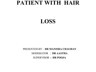
- 1. PATIENT WITH HAIR LOSS PRESENTED BY : DR MANISHA CHAUHAN MODERATOR : DR AASTHA SUPERVISOR : DR POOJA
- 2. History of hairloss •Complaints - Thinning or shedding or both •Duration •Onset- acute or chronic •Pattern of hairloss- Diffuse or localized •Associated symptoms- itching , pain , burning •Present & medical history
- 3. •Drug history •Nutritional history •Psychological history •History of hair care practices / use of hair cosmetics •Family history •Androgen excess ( in women )
- 4. Age related • Children - Alopecia areata , tinea capitis • Prepubertal to young adult- tractional alopecia , trichotillomania • Postpuberatal - Patterned hairloss ( PHL ) • Postmenopausal - frontal fibrosing alopecia
- 5. SCALP EXAMINATION OF NON SCARRING HAIR LOSS NORMAL LOOKING SCALP ABNORMAL LOOKING SCALP PSORIASIS DIFFUSE - ANAGEN HAIRLOSS TELOGEN HAIR LOSS SEBORRHOEIC DERMATITIS PATCHY - ALOPECIA AREATA, TRIANGULAR ALOPECIA DERMATOPHYTIC INFECTION TRICHOTILOMANIA, SECONDARY SYPHILIS TRACTION ALOPECIA PHL - MAGA & FAGA
- 6. Alopecia Non cicatricial alopecia Cicatricial alopecia Localized hairloss Diffuse hairloss Classification of alopecia
- 8. Evaluation technique Of Hair loss Methods Technique Merits Demerits DAILY COUNT Pt is ask to collect hairs for 7 days & count If >100/day hair m/s is performed Provide rough estimate of hairloss Daily counts are tedious, time consuming 60-s hair count Pt. collect hair before shampooing by combing for 60 s from back to front of scalp Repeat procedure for 3 consecutive shampooing Patient can self monitor their progress following therapy Estimate of daily hair shedding vary widely & age specific
- 9. Standardized and modified wash test Pt is refrain from shampoo for 5 days, then do & Collect hair shed & count and divide <3 cm & > 5cm Give a rough estimate about hairloss Very tedious Lack of correlation with vellus hair count AGA - <100 hairs/day with >10% telogen CTE- >100 hairs/day <10% telogen hair AGA+CTE - >100 hairs /day with >10% telogen hairs
- 10. HAIRPULL TEST ( traction test / sabouraud’s sign) Abstinence from shampoo for 2-5 days Approx. 50-60 hairs are grasp b/w thumb &index finger &tugged away Repeat in 4 section ( both parietal , frontal , occupital ) Merits Give estimate of severity of hairloss Simple & inexpensive test Demerits Difficult to standardized the pressure of hair pull Light microscopepy is essential to d/f in diff condition Test is positive - >10% hairs are grasped or >6 hairs are pulled away on scalp Positive test Seen in - Acute TE Active phase of CTE Early case of PHL Active edge of AA Loose anagen phase Active AGA - test is positive on top & negative on occiput
- 11. Trichogram / Hair pluck test/ Unit area trichogram Abstinence for shampoo for 5 days Use rubber forceps, 60-80 hairs pluck at 2 specifyic sites for AGA Hair grasp 0.5 cm from scalp & removed along direction of hair growth Examine under microscope Cut shaft of TG examined under polarized microscopy UAT >> TG Highly effective knowing hair density, diameter Painful for the pt Inappropriately perform cause dystrophic bulb & broken hair Method to evaluate hair root & hair cycle Results are expressed as A:T Normal - 14:1 CTE- 8:1 Merits Demerits
- 12. PHOTO TRICHOGRAM Day 0- hair in a selected area 1cm are cut closet scalp Vertex is selected in MAGA Midscalp is used in FAGA Hair is mounted & diameter of clipped hair measure under 40x microscope Marked area is photographed Non invasive test Measure hair growth cycle, hair density , linear hair growth rate, hair thickness Day 2- second photography after 48 hrs Hair variable on Day 0- density & length of hair Day 2- no. of hairs growing shows (anagen hair)
- 13. TRICHOSCAN Its a method of in vivo hair growth that combines epiluminescence microscopy with automated digital image analysis. Calculate a digital PTG within 15 min HPE Recommend only in cases with telogen loss lasting for 6 months 4mm biopsy taken from vertex & horizontally ATE- Absent follicular miniaturization & perillbulbar lymphocytic infillrate CTE- INCREASE in telogen hair A:T - 8:1 ( normal - 14:1) AGA - T:V - 1.9:1 (normal - 7:1)
- 14. Hair break with roots Hair break without root Telogen effluvium Tinea capitis Alopecia areata Trichotillomania PHL Improper hair care practices/ haircare cosmetics DRUGS Structural hairshaft disorders Loose anagen hairs Anagen effluvium
- 15. Localized hair loss Diffuse hair loss Alopecia areata Trichotillomania Loose anagen syndrome Tractional alopecia AG A Telogen effluvium Chronic diffuse telogen hairloss Chronic telogen effluvium Hairshaft disorders Syphilitic alopecia Tinea capitis On the Basis of Extent
- 16. Telogen effluvium • The term was coined by Kligman in 1961 • An increase shedding in the telogen club hairs due to premature termination of anagen phase of the cycle • Follicles is precipitating into catagen transform into resting stage that mimics telogen • Normally, Anagen - 86% , catagen - 1% , telogen -13% • In TE, Anagen- 70% and telogen become 30%, with daily shedding of unto 300 hairs
- 17. TELOGEN EFFLUVIUM ACUTE TE ( 2-4 months ) CHRONIC TE ( Idiopathic pattern ) CDTHL (Secondary to identifiable Cause )
- 18. Etiopathogenesis • The normal hair cycle consists of anagen( growth phase), catagen (involution), telogen ( dormant phase ), exogen ( release phase ) • Telogen hair remains in follicles for 4-6 weeks after onset of anagen • This cycle replace every hair on scalp every 3-5 year with Individual follicle undergoing 10-30 cycle • In humans , synchronous hair growth disappears in childhood
- 19. • Headington described five functional types of TE - IMMEDIATE ANAGEN RELEASE ( Stress, illness, drug induced) SHORT ANAGEN RELEASE (CTE & AGA) DELAYED ANAGEN RELEASE (Telogen gravidarum ) IMMEDIATE TELOGEN RELEASE Premature removal of Mature hair follicle ( therapy with minoxidil ) DELAYED TELOGEN RELEASE (Animals with synchronous hair cycle)
- 20. Physical & emotional stress Postpartum Shedding in newborn Post surgical , psychological, crash dieting, seasonal variation , UV induced Disease Severe infection High fever Endocrine causes Hyperthyroidism, Hypothyroidism , hypoPTH, Hypopituitarism , hyperandrenocortism, Diabetes, Cushing syndrome Acromegaly, hyperprolactenemia Dermatitis ACD to hair dye ( paraphenylenediamine) Etiology of TE
- 21. DRUGS RETINOIDS ( ETRETINATE > ACITRETIN ) CYTOTOXIC DRUGS, ANTIHYPERTENSIVE ( b blocker, ACE inhibitors ) , heavy metals ( thallium ,arsenic) Heavy metals ( thallium, arsenic, lead ) ANTICONVULSANT (PHENYTOIN, VALPROATE ) ANTICOAGULANTS, MINOXIDIL, Discontinuation of oral contraceptives Alopecia areata with slow progression Androgenetic alopecia Patient of CTE may overlap with FAGA Psychogenic pesodoeffluvium
- 22. Acute TE • TRIGGER FACTOR generally precedes excessive hairloss by 2-4 months • Sudden baldness over temples ( symm bitemporal thinning) • Hairloss is less than 50% • Factors - physiological stress , inadequate diet, hemorrhage ,shock, acute iron deficiency • Postpartum TE, persistent TE, seasonal hairloss
- 23. Chronic diffuse telogen hair loss •Refer to the hair shedding longer than 6 months, secondary to organic causes or coexist with FAGA. •When no triggering factor of chronic telogen loss is found labelled as CTE ( coexist with AGA ) • O/E- • Bitemporal thinning • Positive hair pull test ( vertex= occiput ) •No widening in central part ( commonly seen in AGA) •Mechanism - SYNCHRONIZATION of hair cycling, shortens anagen phase , premature teloptosis
- 24. • Causes - • Endocrine causes- • Hyperthyroidism, Hypothyroidism , hypoPTH, Hypopituitarism , hyperandrenocortism, Diabetes, Cushing syndrome , hyperprolactenemia • Metabolic & nutritional causes- • Vit D , FERRITIN , ZINC DEFICIENCY plays imp role • S . Ferritin - <30 microgram/dl precipitate telogen hairloss • Nutritional deficiencies - a/w dieting, malabsorption , parenteral nutrition
- 25. • Rare causes - • SLE - cause frontal , non scarring hair loss and thinning • Secondary syphilis - patchy alopecia ( bald patches over temporoparietal areas ) • External/ traumatic - frequent bushing, hairstyling • Psychological causes • Idiopathic (33%)
- 27. CHRONIC TE • It can result of an idiopathic change in hair cycle ( primary CTE ) OR secondary to causes including FPHL • Affecting middle age women 30-50 yrs • Sudden onset of increase in hair shedding persist for at least 6 months • No visible widening of central parting line, no miniaturization of hair follicle • Trichodynia or scalp dysaesthesia +/-
- 28. HAIR PULL TEST POSITIVE (10% of hairs are pulled from occiput & midscalp area ) Hair count test Daily hair count Standardized wash test 60 -s- hair test HAIR SHEDDING OF > 100hairs/day If <10% hairs are of 3cm - CTE If >10% hairs are of 3cm OR more - CTE with FPHL TRICHOSCOPY Decrease in hair density and empty follicle Numerous regrowing hairs + Trichogram/ hair pluck test ATE - >25% hairs are in TELOGEN phase Phototrichogram Helps in assess anagen to telogen hair ( 8:1) Scalp biopsy >25 % telogen follicles + = suggestive of TE
- 29. APPROACH TO INVESTIGATE HAIRLOSS INCREASE HAIR SHEDDING DIFFUSE HAIRLO SS S. FERRITIN - <30microgm/dl Vit B12 - <200 HYPOTHYROIDISM FREE ANDROGEN INDEX >5 PROLACTIN >100 AMH - 2-7 ( in case of hyperandrogegism )
- 30. • DIFFERENTIAL DIAGNOSIS- • AGA • DIFFUSE AA • ANAGEN EFFLUVIUM • LOOSE ANAGEN SYNDROME • STRUCTURE HAIR DISORDER • PSYCHOGENIC PSEUDOEFFLUVIUM
- 31. TREATMENT • Reassurance and counseling • Attempts at identifying specific cause • Potential therapeutic option like - • Inhibitition of catagen ( prolong anagen phase ) • Induction of anagen in telogen effluvium • Inhibition of exogen ( to reduce hair shaft shedding ) • Neither of the currently available FDA approved standard hair drug
- 32. • Correction of the catagen inducing endocrine disorders (thyroid disorders, hyperandrogenism, hyperprolactenemia) • Substitution therapy for catagen promoting deficiencies like iron, zinc, protein can also be initiated • Balanced diet and maintaining standard body weight is imp factors • It is suggested that maintaining S. Ferritin above 40ng/dl helps reversing hair loss
- 33. Management of TE TOPICAL MINOXIDIL ( 2% OR 5%) TOPICAL AGENTS DIETARY SUPPLEMENTS REASSURANCE AND COUNSELING OF THE PATIENT IRON - 300mg tds (60 mg elemental iron /day) For 3-6 months Millet extracts , pantothenic acid, Biotin, CYP COMPLEX
- 34. Androgenetic Alopecia • The term ‘pattern hair loss’ describe hairloss in distinctive pattern by progressive declining in hair fibre production & eventual miniaturization • Polygenic inheritance ( mutation in ANDROGEN RECEPTOR gene) • PHL IN MEN - AGA • PHL IN female - Female AGA/FPHL • It depends on genetic predisposition &interaction of endocrine factors
- 35. • Decrease in size & activity of hair follicle during successive hair cycle called miniaturization • Terminal thick , pigmented hair follicles turns to thinner ,shorter , less pigmented till they change to vellus hair • Anagen duration - decreased • Telogen- constant /prolonged • Testosterone DHT • Binding of DHT to its receptor in hair follicle lead to production of cytokines - TGF B1 & B2 promote senescence 5 alpha reductase
- 36. • Density of these receptors are more in frontoparietal scalp & vertex and less in occiput • PG-D2 shows inhibitory effect in hair growth( elevated in scalp of men with AGA )
- 37. Decrease in hair density Variation in hair thickness Decrease of hair per FHU
- 38. Norwood- Hamilton Classification ( MALE PATTERN CLASSIFICATION )
- 39. Investigation • Change of anagen to telogen ratio from 12:1 to 5:1 • Ratio of terminal to vellus hair( greater than 8:1 to less than 4:1) • Proportion of telogen hairs is increased from 5-10% to 15-20 % • No. Of hair follicle in FHU is reduced from 2-5 (normal) to 1-2 hair • On dermoscopy- diversity in hairshaft diameter>20% in frontoparietal area
- 40. Peripilar sign (Brown halo reflecting perifollicular inflammation) Yellow dot ( sebum & keratin accumulation with dilated follicular infundibulum) Increase in % of single hair follicular units
- 41. Female pattern hairloss • PHL in a women was first described by Ludwig in 1977 • Present with diffuse thinning ( loss of hair volume ) over mid-frontal scalp with minimum or no bitemporal recession • Ludwig graded FPHL into 3 grades • Genetic predisposition & polygenic inheritance • Shorter the length of CAG repeats in AR gene lead to significant risk of FPHL
- 42. • Stage I is characterized by a perceptible thinning of hair from the anterior part of the crown with minimal widening (rarefaction) of the part width . • The hair loss at this stage can easily be camouflaged. • This type may manifest in young women with SAHA syndrome (seborrhea, acne, hirsutism, and alopecia), generally of ovarian origin. • It may be accompanied by other manifestations of hyperandrogenism (seborrhoea, acne, hirsutism, seborrheic dermatitis, and slight menstrual alterations) • Biochemical levels are often normal
- 43. • Stage II is seen with advancing age when the rarefaction on the crown becomes more pronounced, and the number of thinner and shorter hairs increases • Camouflage of the denuded areas by special hair styles is no longer possible. • An excess of androstenedione, free T, and androstanediol glucuronide may be detectable
- 44. • Stage III is usually not encountered before menopause, and the crown may become literally bald • Contrary to what happens in men, a fringe of hair along the frontal hair line persists. • Adrenal diseases, tumoral or not, with very high levels of androstenedione, dehydroepiandrosterone sulfates (DHEA-S), free T, sometimes of prolactin, and always of androstanediol glucuronide
- 45. Normal Hair density Widening of central part Additional Thinning of hair At sides of part line Bald Patch distal To Hairline Advanced Hairloss Sinclair scale 5 visually distinct STAGES
- 46. Pathophysiology • FAGA can be related to excess of androstenedione serum level of ovaries or adrenal • androstenedione and DHEAS transformed into testosterone 7 convert into 5 alpha DHT mediated by 5alpha reductase • Conversion of testosterone to DHT requires free testosterone, highlight the importance of SHBG and free androgen • DHT metabolized into 3 a - androstanediol glucoronide(most sensitive marker of peripheral hyperdrogenemia • Hyperprolactenemia is associated with increase in DHEAS
- 47. Basic test Full blood count IRON PROFILE S. VIT B12 , VIT D Thyroid function test Fasting lipids/ fasting glucose Endocrine test S. Testosterone/ free androgen index DHEAS S. Prolactin , AMH 17- hydroxyprogesterone USG ABDOMEN INVESTIGATIONS
- 48. DIFFERENTIAL DIAGNOSIS OF FPHL CT E DIFFUSE AOPECIA AREATA ANAGEN EFFLUVIU M TRICHOTILLOMANI A Early acute onset Exclamation hairs + Yellow dots + Dystrophic Anagen Hairs Different length Of hairs Abrupt Diffuse Thinning Trigger + Others - hypothyroidism, SLE, Frontal fibrosing alopecia
- 49. FPHL CHRONIC TE DISTRIBUTION Central part of scalp Preserved frontal hairline Generalized ONSET Gradual Abrupt with trigger APPEARANCE Hair thinning with midline part Diffuse thinning HAIR SHEDDING Minimal Prominent HAIR PULL TEST Usually negative Positive OTHER HISTORY Family history + Stress , major illness SCALP BIOPSY T:V >4:1 T:V>8:1
- 52. Alopecia areata •Non scarring alopecia •Occur at any age ( peak incidence 2 to 4 decade ) •Involving any hair bearing areas •Seen over scalp , in a patchy distribution •Other sites- eyebrows , eyelashes, beard, thorax area •Nail changes(10-66%)- fine nail pitting, trachyonychia, onychorrhexis,onycholysis , onychomadesis
- 53. • It is a T cell mediated autoimmune disease occurs in genetically predisposed individuals & environmental factors • Genetic lineage focus to HLA class II ( HLA-D) on choromosome 6 • A receptor for stress induced proteins ( NKG2D) strongly associated • Rapid progression of hair follicle from anagen phase to catagen to telogen phase • Disorder affects anagen follicles with little or no inflammatory infiltrate around isthmus of hair follicle
- 54. IMMUNE RESPONSE TARG ET LOSS OF IMMUNE PRIVILEGE AUTOANTIG EN EPITOPE Early cortical differentiation in hair follicle matrix epithelium Vacuolar degeneration of affecting Anagen follicle Weakness of the hairshaft (EXCLAMATION HAIR) Reverts to telogen phase Renter anagen phase Not beyond iii/iv stage Cytotoxic subset ( CD8+ NKG2D+) Produce IFN-Y activates IL-15 sustains CD8+ T cell autoreactivity Involvement of JAK receptors Involvement of melanin, Melanin related protein Keratinocyte derived Ag Growing of white hair After AA
- 55. Clinical presentation of AA PATTER N DIFFUS E AA PATC HY OPHIASIS INVERSUS/SISAPHO ( band like loss of hair in Fronto Parietal) OPHIASIS ( band like loss of hair in tempero ocipital RETICULATE (Patchy, round to oval Hairloss) EXTEN T ALOPECIA UNIVERSALIS (Total loss of scalp & Body hair ) ALOPECIA TOTALIS (Total loss of scalp hair) ALOPECIA AREATA (Partial loss of scalp hair)
- 56. Poor prognostic of AA FOR WHOLE DISEASE FOR AN EPISODE ONSET IN CHILDHOOD DURATION OF EPISODE ASSOCIATED ENDOCRINE DISORDER ATOPY EXTENT OF AFFECTED AREA PRESENCE OF AUTOIMMUNE DISEASE (thyroiditis, LE, vitiligo, psoriasis ) C/L form- ophiasis, A totalis, A universalis POSITIVE HISTORY OF AA Nail dystrophy
- 57. Test Findings Hair pull test Positive >6 hair ( s/o disease is active & progressive ) Trichoscopy Exclamation hairs YELLOW DOT BLACK DOT Trichogram Increase no.of dystrophic anagen hair 4mm Punch biopsy Swarm of bees appearance (perifollicular & intrafollicular inflammatory infillrateof activated T cell ) Lab work up TSH , THYROID ANTIBODY, FERRITIN, VIT D, VIT B12 INVESTIGATIONS
- 61. TRICHOTILLOMANIA • Trichotillomania (TTM) is characterized by persistent hair pulling behavior, resulting in noticeable hair loss. • Patients report feeling anxious before pulling their hair out and pleasure, satisfaction, or relief after doing it • Asymmetrical patches of alopecia, particularly in the frontal and vertex regions.
- 62. • At dermoscopy, Fracture may occur at varying lengths, resulting in black dots, fraying hair, longitudinally split hair, coiled hair and stretching of the shaft. Hairs of varying lengths and short vellus hairs may be seen in these patches The hair pull test is negative along the edges. flame hair, empty hair folicular Ostia , follicular hemorrhages, decreased hair density , and no sign of exclamation signs
- 65. DIFFERENTIAL DIAGNOSIS • T. capitis • Alopecia areata • Monilethrix • Traction alopecia • Secondary syphilis
- 66. • Treatment - • Psychotherapy, behavioral therapy, and drugs – such as lithium salts, tricyclic antidepressants (TADs), selective serotonin reuptake inhibitors (SSRIs), and antipsychotics. • Recently, N-acetylcysteine has been proposed as an effective alter- native treatmen
- 67. Tinea capitis • Tinea capitis is a superficial fungal infection of the scalp. • The disease is primarily caused by dermatophytes in the Trichophyton and Microsporum genera that invade the hair shaft • A single or multiple patches of hair loss, sometimes with a “black dot” pattern, accompanied by inflammation, scaling, pustules, and itching
- 68. • Hair roots and skin scraping were mounted in 10% potassium hydroxide solution. • Slide was gently heated and microscopically examined for spores. • Trichoscopic findings- • Comma shape hair, zig zag hair, black dot hair, short broken hair, corkscrew hair • A therapeutic trial of anti- fungal treatment remains an acceptable alternative. • The treatment of choice is griseofulvin 10 - 20 mg/kg per day, with a meal, for 6 to 8
- 69. SYPHILITIC ALOPECIA •Syphilitic alopecia is considered an uncommon manifestation of secondary syphilis. •SA is further classified into three forms: •1) moth-eaten, or patchy alopecia characterized by small alopecia patches irregularly distributed over the scalp; •2) diffuse alopecia, characterized by a diffuse hair loss; resembling TE •3) mixed form (i.e., combination of diffuse hair loss and alopecia moth-eaten patches)
- 70. • Trichoscopic features—such as tapered bended hairs, erythematous background, diffuse scaling and perifollicular hyperkeratosis • Serology - VDRL + • Scalp biopsies - spirochetes in the peribulbar region and penetrating into the follicle matrix. • Current hypothesis supporting the pathogenesis of SA is a vasculitis of peribulbar capillaries causing a perifollicular lymphocytic infiltration with scattered plasma cells that stops the hair cell cycle • Treatment - • Inj benzathine penicillin G 2.4 million units intramuscularly Moth eaten type
- 71. Tapered Bent hairs Fine scaling Villus hair ( periphery) Perifollicular Hyperkeratosis Yellow dots + Trichoscopic finding in syphilitic alopecia
- 72. Tractional alopecia • Traction alopecia is a form of trauma-induced alopecia and results from continuous excessive pulling of hair shafts. • Hairstyling is the most common cause of longstanding traction and usually results in alopecia of scalp margins • TA most often affects the frontal and temporal scalp.
- 73. structure that encircles the hair shaft placed at the periphery of the alopecia plaque. • It is derived from the inner and/or outer hair root sheath cells and it develops because of persistent hair traction • Treatment - • loosen hairstyle is preferred • Avoid chemical treatment of hairs • I/L triamcinolone can be used at periphery • 2% minoxidil applecation promotes hair
- 74. Thank you