Model Call Girl in Subhash Nagar Delhi reach out to us at 🔝9953056974🔝
Head inj ury
1. INTRODUCTION: -
Any injury that results in trauma to the scalp, skull, or brain can be
classified as a head injury. The terms traumatic brain injury and head
injury are often used interchangeably in medical literature. Unlike a broken
bone where trauma to the body is obvious, head trauma can sometimes be
obvious or discrete. In the case of an open head injury, the skull is cracked
and broken by an object that makes contact with the brain, this leads to
bleeding. Other obvious symptoms can be neurological in nature. The
person may become sleepy, behave abnormally, lose consciousness, vomit,
develop a severe headache, have mismatched pupil sizes, and/or be unable
to move certain parts of the body. While these symptoms happen right after
head injury occurs, many problems can develop later in life. Alzheimer’s
disease, for example, is much more likely to develop in a person who has
experienced a head injury.
ANATOMY & PHYSIOLOGY:-
:BRAIN:
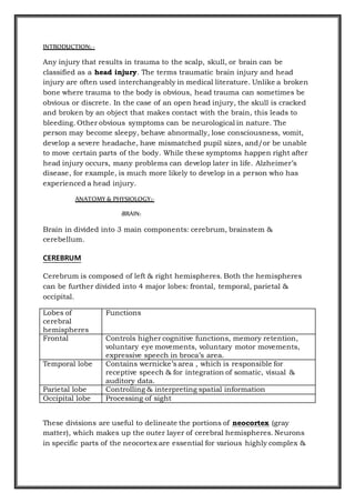
Brain in divided into 3 main components: cerebrum, brainstem &
cerebellum.
CEREBRUM
Cerebrum is composed of left & right hemispheres. Both the hemispheres
can be further divided into 4 major lobes: frontal, temporal, parietal &
occipital.
Lobes of
cerebral
hemispheres
Functions
Frontal Controls higher cognitive functions, memory retention,
voluntary eye movements, voluntary motor movements,
expressive speech in broca’s area.
Temporal lobe Contains wernicke’s area , which is responsible for
receptive speech & for integration of somatic, visual &
auditory data.
Parietal lobe Controlling & interpreting spatial information
Occipital lobe Processing of sight
These divisions are useful to delineate the portions of neocortex (gray
matter), which makes up the outer layer of cerebral hemispheres. Neurons
in specific parts of the neocortex are essential for various highly complex &
2. sophisticated aspects of mental functioning such as language, memory &
appreciation of visual spatial relationships.
Basal ganglia are a group of paired structures located centrally in the
cerebrum & midbrain, near lateral ventricles of both cerebral hemispheres.
It controls & facilitates learned & automatic movements.
Thalamus (part of diencephalon) is lying above the brain stem & below the
basal ganglia. It is the major relay center for sensory & other afferent (i.e.,
cerebellar) inputs to the cerebral cortex.
Hypothalamus is located just below the thalamus & slightly in front of the
midbrain. It regulates the ANS & endocrine system.
Limbic system located lateral to hypothalamus, near the inner surfaces of
the cerebral hemispheres. It is concerned with emotion, aggression, feeding
behaviour & sexual response.
BRAINSTEM
It includes midbrain, pons & medulla.
Ascending & descending fibers pass through the brainstem going to & from
the cerebrum & cerebellum. The cell bodies , or nuclei ,of cranial nerves iii
to xii are in the brainstem.
Reticular formation a diffusely arranged group of neurons & their axons that
extends from the medulla to the thalamus & hypothalamus, is also located
here. Its functions include relaying sensory information, influencing
excitatory & inhibitory control of spinal motor neurons, & controlling
vasomotor & respiratory activity.
RAS , a part of the reticular formation , is the regulatory system for arousal.
Medulla acts as respiratory, vasomotor & cardiac function regulatory center.
Brainstem also contains centers for sneezing, coughing, hiccupping,
vomiting, sucking & swallowing.
CEREBELLUM
Cerebellum located in the posterior part of the cranial fossa, along with the
brainstem under the occipital lobe of cerebrum.
Its function is maintain coordinate voluntary movement & to maintain trunk
stability & equilibrium.
3. :PROTECTIVE STUCTURES:
MENINGES
It consists of 3 protective layers: dura, arachnoid & pia mater.
The flax cerebri is a fold of the dura mater , that separates both
hemispheres & preventing expansion of brain tissue.
SKULL
The three bone layers of the skull.
The human skull is anatomically divided into two parts: the neurocranium,
formed by eight cranial bones that houses and protect the brain—and the
facial skeleton (viscerocranium) composed of fourteen bones, not including
the three ossicles of the inner ear.
The term skull fracture typically means fractures to the neurocranium,
while fractures of the facial portion of the skull are facial fractures, or if the
jaw is fractured, a mandibular fracture.
NEUROCRANIUM:
The eight cranial bones are separated by sutures : one frontal bone,
two parietal bones, two temporal bones, one occipital bone, one sphenoid
bone, and one ethmoid bone.
The bones of the skull are in three layers: the hard compact layer of the
external table (lamina externa), the dipole (a spongy layer of red bone
4. marrow in the middle, and the compact layer of the inner table (Lamina
interna).
SCALP
The scalp is the anatomical area bordered by the face anteriorly and
the neck to the sides and posteriorly.
Structure:
The scalp is usually described as having five layers, which can conveniently
be remembered as a mnemonic.
S: The skin on the head from which head hair grows. It contains numerous
sebaeceous glands and hair follicles.
C: Connective tissue. A dense subcutaneous layer of fat and fibrous tissue
that lies beneath the skin, containing the nerves and vessels of the scalp.
A: The aponeurosis called epicranial aponeurosis (or galea aponeurotica) is
the next layer. It is a tough layer of dense fibrous tissue which runs from
the frontalis muscle anteriorly to the occipitalis posteriorly.
L: The loose areolar connective tissue layer provides an easy plane of
separation between the upper three layers and the pericranium. This layer is
sometimes referred to as the "danger zone" because of the ease by which
infectious agents can spread through it to emissary veins which then drain
into the cranium. The loose areolar tissue in this layer is made up of
random collagen I bundles, collagen III. It will also be rich
in glycosaminoglycans (GAGs) and will be constituted of more matrix than
fibers.
P: The pericranium is the periosteum of the skull bones and provides
nutrition to the bone and the capacity for repair. It may be lifted from the
bone to allow removal of bone windows (craniotomy).
BLOOD SUPPLY OF BRAIN
CEREBRAL CIRCULATION
Cerebral circulation or blood supply of the brain arises from the internal
carotid arteries(anterior circulation) and the vertebral arteries(posterior
circulation). Each internal carotid artery supplies the ipsilateral hemisphere,
whereas the basilar artery form by the junction of the two vertebral arteries,
supplies structures within the posterior fossa (cerebellum and brain
5. stem).The circle of Wills arise from the basilar artery and the two internal
carotid arteries. This vascular circle may act as a safety valve when
differential pressures are present in these arteries. It also may function as
an anstomotic pathway when occlusion of a major artery on one side of the
brain occurs. In general, the two anterior cerebral arteries supply the medial
and anterior portion of the frontal lobes. The two middle cerebral arteries
supply the outer portions of the frontal, partial, and superior temporal
lobes. The two posterior cerebral arteries supply the medial portions of the
occipital and inferior temporal lobes. Venous blood drain from the brain
through the dural sinuses, which form channels that drain into the two
jugular veins.
SCALP CIRCULATION
The blood supply of the scalp is via five pairs of arteries, three from
the external carotid (superficial temporal artery, occipital artery &
posterior auricular artery) and two from the internal
carotid( supratrochlear artery & supraorbital artery ).
Note: The walls of the blood vessels are firmly attached to the fibrous
tissue of the superficial fascial layer, hence cut ends of vessels here do
not readily retract; Even a small scalp wound may bleed profusely.
INNERVATION OF SCALP
The innervation of scalp can be remembered using the mnemonic, "Z-
GLASS" for, Zygomaticotemporal nerve, Greater occipital nerve,Lesser
occipital nerve, Auriculotemporal nerve, Supratrochlear nerve
and Supraorbital nerve.
LYMPHATIC
Occipital and posterior auricular nodes, parotid nodes, submandibular and
deep cervical nodes .
EPIDEMIOLOGY IN INDIA:-
Traumatic brain injuries (TBIs) are a leading cause of morbidity, mortality,
disability and socioeconomic losses in India and other developing countries.
It is estimated that nearly 1.5 to 2 million persons are injured and 1 million
succumb to death every year in India. Road traffic injuries are the leading
cause (60%) of TBIs followed by falls (20%-25%) and violence (10%). Alcohol
involvement is known to be present among 15%-20% of TBIs at the time of
injury. The rehabilitation needs of brain injured persons are significantly
high and increasing from year to year. India and other developing countries
6. face the major challenges of prevention, pre-hospital care and rehabilitation
in their rapidly changing environments to reduce the burden of TBIs.
CAUSES:-
Motor vehicle traffic collisions ,Home and occupational accidents, falls,
Assaults.
CLASSIFICATION:-
There are many ways that head injuries can be classified. The three most
useful descriptions are:
Severity
Morphology
Mechanism of injury
Time of showing impact or consequences
Classification as per Severity
1. Severity based on Glassgow Coma Scale
Best Eye Response. (4)
No eye opening.
Eye opening to pain.
Eye opening to verbal command.
Eyes open spontaneously.
Best Verbal Response. (5)
No verbal response
Incomprehensible sounds.
Inappropriate words.
Confused
Orientated
Best Motor Response. (6)
No motor response.
Extension to pain.
Flexion to pain.
Withdrawal from pain.
Localising pain.
Obeys Commands.
7. Mild head inury (GCS 14 - 15)
Moderate head injury (GCS 9 - 13)
Severe head injury (GCS 3 - 8)
2. SEVERITY OF TBI BASED ON THE DURATION OF LOC
Severity of TBI Finding
Mild Mental status change or LOC < 30 min
Moderate Mental status change or LOC 30 min to 6 h
Severe Mental status change or LOC > 6 h
Morphological classification
Head injuries may also be classified by injury type. There are two broad
categories:
1. Focal injuries
2. Diffuse injuries
Focal injuries have an identifiable area of involvement. Examples include:
1. Cerebral contusion
2. Extradural haemorrhage:
3. Scalp lacerations
4. Skull fractures
5. Subarachnoid haemorrhage
6. Subdural haemorrhage
Diffuse injuries involve the entire brain. Examples include:
1. Concussion
2. Diffuse axonal injury
Classification as per time of showing impact:
PRIMARY INJURY:
8. Induced by mechanical force and occurs at the moment of injury; the 2 main
mechanisms that cause primary injury are contact (eg, an object striking the
head or the brain striking the inside of the skull) and acceleration-
deceleration.
SECONDARY INJURY:
Not mechanicallyinduced; it may be delayed from the moment of impact, and
it may superimpose injury on a brain already affected by a mechanical injury
Head injury as per Mechanism of injury:
Head injuries may also be classified by the mechanism of injury. The two
broad categories used are:
BLUNT HEAD INJURY:
Blunt force trauma is defined as trauma to tissue or organs without
penetration through the skin. It also can be called closed (non-missile) head
injury is where the dura mater remains intact. The skull can be fractured,
but not necessarily.
PENETRATING HEAD INJURY:
Penetrating trauma can be defined as injuries caused by foreign objects
penetrating the skin and disrupting underlying tissue and organs. The
penetrating object transfers it's kinetic energy to the body. This energy is
then dissipated into the surrounding tissue, often focussed in a small area.
DIFFERENT TYPES OF HEAD INJURY IN DETAILS:
SCALP LACERATIONS
WHAT IS SCALP LACERATION: The blunt force due to the direct effect of
scalp laceration (scalp laceration) for sharpening cutting or larger.
SYMPTOMS OF SCALP LACERATION: Scalp blood vessels is extremely rich,
dense and scalability small subcutaneous tissue, so once the scalp fracture.
Not easy to shrink blood vessels and bleeding are many and difficult to stop
on their own. Scalp laceration is large, and often in the short-term internal
massive blood loss caused by hemorrhagic shock.
CAUSES: For sharpening cutting or larger due to the direct effect of the
blunt force.
9. DIAGNOSIS: Sharp cuts wound neatly serrated edge cracked blunt injury to
the scalp and scalp contusions and abrasions.
TREATMENTS : Scalp laceration main emergency treatment to stop bleeding.
The most common method is bandaged, and then wound debridement in
areas where conditions permit.
SKULL FRACTURES
A head injury may cause skull fracture, which may or may not be associated
with injury to the brain.
CLASSIFICATION OF SKULL FRACTURES:
TYPE DESCRIPTION
Linea
r
Linear skull fractures are breaks in the bone that transverse the full
thickness of the skull from the outer to inner table. They are usually
fairly straight with no bone displacement. The common cause of injury
is blunt force trauma where the impact energy transferred over a wide
area of the skull.
Linear skull fractures are usually of little clinical significance unless
they parallel in close proximity or transverse a suture, or they involve a
venous sinus groove or vascular channel. The resulting complications
may include suture diastasis, venous sinus thrombosis, and epidural
hematoma. In young children, although rare, the possibility exists of
developing a growing skull fracture especially if the fracture occurs in
the parietal bone.
Depr
e
ssed
A depressed skull fracture is a type of fracture usually resulting from
blunt force trauma, such as getting struck with a hammer, rock or
getting kicked in the head. These types of fractures—which occur in
11% of severe head injuries—are comminuted fractures in which
broken bones displace inward. Depressed skull fractures present a
high risk of increased pressure on the brain, or a hemorrhage to the
brain that crushes the delicate tissue.
Compound depressed skull fractures occur when there is a laceration
over the fracture, putting the internal cranial cavity in contact with the
outside environment, increasing the risk of contamination and
infection. In complex depressed fractures, the dura mater is torn.
Depressed skull fractures may require surgery to lift the bones off the
brain if they are pressing on it.
Simp
le
Fracture without fragmentation or communicating lacerations.
Com
minu
ted
Multiple liner fracture with fragmentation of bone into many pieces.
10. Com
poun
d
A fracture in conjunction with an overlying laceration that tears the
epidermis and the meninges—or runs through the paranasal sinuses
and the middle ear structures, putting the outside environment in
contact with the cranial cavity—is a compound fracture.
Compound fractures may either be clean or contaminated. Intracranial
air (pneumocephalus) may occur in compound skull fractures. The
most serious complication of compound skull fractures is infection.
Increased risk factors for infection include visible contamination,
meningeal tear, loose bone fragments and presenting for treatment
more than eight hours after initial injury.
Com
poun
d
eleva
te
A compound elevated skull fracture is a rare type of skull fracture
where the fractured bone is elevated above the intact outer table of the
skull. This type of skull fracture is always compound in nature. It can
be caused during an assault with a weapon where the initial blow
penetrates the skull and the underlying meninges and, on withdrawal,
the weapon lifts the fractured portion of the skull outward. It can also
be caused the skull rotating while being struck in a case of blunt force
trauma, the skull rotating while striking an inanimate object as in a
fall, or it may occur during transfer of a patient after an initial
compound head injury.
Grow
ing
skull
fractu
re
A growing skull fracture (GSF) also known as a craniocerebral erosion or
leptomeningeal cyst due to the usual development of a cystic mass filled
with cerebrospinal fluid is a rare complication of head injury usually
associated with linear skull fractures of the parietal bone in children
under 3. It is characterized by a diastatic enlargement of the
fracture.The primary causative factor is a tear in the dura mater.
Crani
al
burst
skull
fract
ure
A cranial burst skull fracture usually occurring with severe injuries in
infants less than 1 year of age is a closed, diastatic skull fracture with
cerebral extrusion beyond the outer table of the skull under the
intact scalp.
Acute scalp swelling is associated with this type of fracture. In
equivocal cases without immediate scalp swelling the diagnosis may be
made via the use of magnetic resonance imaging thus insuring more
prompt treatment and avoiding the development of a "growing skull
fracture".
Diast
atic
fract
ure
Diastatic fractures occur when the fracture line transverses one or
more sutures of the skull causing a widening of the suture. While this
type of fracture is usually seen in infants and young children as the
sutures are not yet fused it can also occur in adults. When a diastatic
fracture occurs in adults it usually affects the lambdoidal suture as
this suture does not fully fuse in adults until about the age of 60.
11. CLINICAL MANIFESTATIONS OF SKULL FRACTURES DEPENDING ON
INVOLVED AREA
LOCATION SYNDROME / SEQUELE
Frontal fracture Exposure of brain to the contaminants through frontal air
sinus, possible association
Orbital fracture raccon eye
Temporal
fracture
battle’s sign
Parietal fracture deafness, otorreoa
Posterior fossa
fracture
occipital bruising, cortical blindness
Basilar skull
fracture
Basilar skull fractures are linear fractures that occur in the
floor of the cranial vault (skull base), which require more
force to cause than other areas of the neurocranium. Thus
they are rare, occurring as the only fracture in only 4% of
severe head injury patients.
Basilar fractures have characteristic signs: blood in
the sinuses; a clear fluid called cerebrospinal fluid (CSF)
leaking from the nose (rhinorrhea) or ears
(otorrhea); periorbital ecchymosis often called 'raccoon eyes'
(bruising of the orbits of the eyes that result from blood
collecting there as it leaks from the fracture site); and
retroauricular ecchymosis known as "Battle's sign"
(bruising over the mastoid process).
Dementia pugilistica , or "punch-drunk syndrome", caused by
repetitive head injuries, for example in boxing or other contact sports
A severe injury may lead to a coma or death
Shaken baby syndrome — a form of child abuse
Concussion
Concussion derives from the Latin term concutere ("to shake violently") or
concussus ("action of striking together"), is the most common type of
traumatic brain injury. The terms mild brain injury, mild traumaticbrain
injury (MTBI), mild head injury (MHI), minor head trauma, and concussion
may be use interchangeably.
Concussion defined as a head injury with a temporary loss of brain
function, concussion causes a variety of physical, cognitive, and emotional
symptoms, which may not be recognized if subtle.
12. Three grading systems have been most widely followed: by Robert Cantu, the
Colorado Medical Society, and the American Academy of Neurology. Each
employs three grades, as summarized in the following table:
SIGNS AND SYMPTOMS
Physical
1. Headache is the most common MTBI symptom.
2. Dizziness
3. Vomiting
4. Nausea
5. Lack of motor coordination
6. Difficulty balancing
7. Problems with movement or sensation
8. Visual symptoms include light sensitivity,seeing bright light, blurred
vision, and double vision.
9. Tinnitus, or a ringing in the ears, is also commonly reported.
10. Concussive convulsions are thought to result from temporary loss or
inhibition of motor function, and are not associated either with
epilepsy or with more serious structural damage.
Comparison of historic concussion grading scales – not currently
recommended for use by medical professionals
Guidelines Grade I Grade II Grade III
Cantu Post-traumatic
amnesia
<30 minutes, no
loss of
consciousness
Loss of
consciousness
<5 minutes or
amnesia lasting
30 minutes–
24 hours
Loss of
consciousness
>5 minutes or
amnesia
>24 hours
Colorado Medical
Society
Confusion, no
loss of
consciousness
Confusion, post-
traumatic
amnesia, no loss
of consciousness
Any loss of
consciousness
American
Academy of
Neurology
Confusion,
symptoms last
<15 minutes, no
loss of
consciousness
Symptoms last
>15 minutes, no
loss of
consciousness
Loss of
consciousness
(IIIa, coma lasts
seconds, IIIb for
minutes)
13. Cognitive and emotional
1. Confusion
2. disorientation
3. difficulty focusing attention
4. Loss of consciousness
5. Post-traumatic amnesia, in which events following the injury cannot
be recalled is a hallmark of concussion A person may repeat the same
questions, be slow to respond to questions or directions, have a
vacant stare, or have slurred or incoherent speech.
6. Changes in sleeping pattern
7. difficulty with reasoning, concentrating, and performing everyday
activities.
8. Concussion can result in changes in mood including crankiness, loss
of interest in favorite activities or items, tearfulness, and displays of
emotion that are inappropriate to the situation. Common symptoms in
concussed children include restlessness, lethargy, and irritability.
PATHOPHYSIOLOGY
The pathology of a concussion seems to start with the disruption of the cell
membrane of nerve cells.
This results in a migration of potassium from within the cell into the
extracellular space with subsequent release of glutamate which potentiates
further potassium shift, in turn resulting in depolarization and suppression
of nerve activity.
In an effort to restore ion balance, the sodium-potassium ion pumps
increase activity, which results in excessive ATP (adenosine triphosphate)
consumption and glucose utilization.
Lactate accumulates but, paradoxically, cerebral blood flow decreases,
which leads to a proposed "energy crisis."
After this increase in glucose metabolism, there is a subsequent lower
metabolic state which may persist for up to 4 weeks after injury.
14. A completely separate pathway involves a large amount of calcium
accumulating in cells, which may impair oxidative metabolism and begin
further biochemical pathways that result in cell death.
Again, both of these main pathways have been established from animal
studies and the extent to which they apply to humans is still somewhat
unclear.
Red flag criteria : warning features
1. Seizure
2. Worsening headache
3. Difficulty waking-up
4. Seeing double
5. Problem recognizing people or places
6. Repeated vomiting
7. Focal neurological problems
8. Not usual self
COMPLICATIONS OF CONCUSSION:
1. Post-concussion syndrome
In post-concussion syndrome, symptoms do not resolve for weeks, months,
or years after a concussion, and may occasionally be permanent. Symptoms
may include headaches, dizziness, fatigue, anxiety, memory and attention
problems, sleep problems, and irritability. Symptoms usually go away on
their own within months.
2. Dementia pugilistica
Chronic encephalopathy is an example of the cumulative damage that can
occur as the result of multiple concussions or less severe blows to the head.
The condition called dementia pugilistica, or "punch drunk" syndrome,
which is associated with boxers, can result in cognitive and physical deficits
such as parkinsonism, speech and memory problems, slowed mental
processing, tremor, and inappropriate behavior. It shares features with
Alzheimer's disease.
3. Second-impact syndrome
15. Second-impact syndrome, in which the brain swells dangerously after a
minor blow, may occur in very rare cases. The condition may develop in
people who receive a second blow days or weeks after an initial concussion,
before its symptoms have gone away
Intracranial hemorrhage
An intracranial hemorrhage (ICH) is a hemorrhage, or bleeding, within the
skull.
CAUSES
Blood vessel within the skull is ruptured or leaks. It can result from
physical trauma (as occurs in head injury) or nontraumatic causes (as
occurs in hemorrhagic stroke) such as a ruptured aneurysm.
Anticoagulant therapy, as well as disorders with blood clotting can heighten
the risk that an intracranial hemorrhage will occur.
DIAGNOSIS
CT scan (computed tomography) is the definitive tool for accurate diagnosis
of an intracranial hemorrhage.
CLASSIFICATION
1. Intra-axial hemorrhage:
Intra-axial hemorrhage is bleeding within the brain itself, or cerebral
hemorrhage. This category includes –
1.a. intraparenchymal hemorrhage, or bleeding within the brain tissue
1.b. intraventricular hemorrhage, bleeding within the
brain's ventricles (particularly of premature infants).
2. Extra-axial hemorrhage
Extra-axial hemorrhage, bleeding that occurs within the skull but outside of
the brain tissue, falls into three subtypes:
2.a. Epidural hemorrhage (extradural hemorrhage) :
It occur between the dura mater and the skull, is caused by trauma.
Cause- It may result from laceration of an artery, most commonly the
middle meningeal artery. This is a very dangerous type of injury because the
16. bleed is from a high-pressure system and deadly increases in intracranial
pressure can result rapidly.
Clinical presentations:
Patients have a loss of consciousness (LOC), then a lucid interval,
then sudden deterioration (vomiting, restlessness, LOC)
Head CT shows lenticular (convex) deformity.
Epidural hematoma:
Epidural hematoma (EDH) is a rapidly accumulating hematoma between the
dura mater and the cranium.
Clinical presentation:-
These patients have a history of head trauma with loss of
consciousness, then a lucid period, followed by loss of consciousness
Clinical onset occurs over minutes to hours. Many of these injuries
are associated with lacerations of the middle meningeal artery.
Diagnostic criteria: A "lenticular", or convex, lens-shaped extracerebral
hemorrhage that does not cross suture lines will likely be visible on a CT
scan of the head. Although death is a potential complication, the prognosis
is good when this injury is recognized and treated.
2.b. Subdural hemorrhage:
It results from tearing of the bridging veins in the subdural space between
the dura and arachnoid mater.
Head CT shows crescent-shaped deformity.
Subdural hematoma:
Subdural hematoma occurs when there is tearing of the bridging vein
between the cerebral cortex and a draining venous sinus.
Causes - 1. arterial lacerations on the brain surface 2. cerebral cortex
injury .
Clinical features-depend on the site of injury and severity of injury.
1. loss of consciousness 2. Clinical onset occurs over hours.
Diagnostic study: A crescent shaped hemorrhage compressing the brain
that does cross suture lines will be noted on CT of the head.
17. Treatment -Craniotomy and surgical evacuation is required if there is
significant pressure effect on the brain.
Complications-
focal neurologic deficits depending on the site of hematoma and brain
injury
increased intra cranial pressure leading to herniation of brain
ischemia due to reduced blood supply and seizures.
Subarachnoid hemorrhage: It occur between the arachnoid and pia
meningeal layers, like intraparenchymal hemorrhage.
Causes -Trauma or from ruptures of aneurysms or arteriovenous
malformations.
Classic presentation: sudden onset of a severe headache (a thunderclap
headache).
This can be a very dangerous entity, and requires emergent neurosurgical
evaluation, and sometimes urgent intervention.
Subarachnoid hematoma:
A subarachnoid hematoma is bleeding into the subarachnoid space—the
area between the arachnoid membrane and the pia mater surrounding the
brain.
Causes- 1. head injury 2. ruptured cerebral aneurysm.
Symptoms:
a severe headache with a rapid onset ("thunderclap headache"),
vomiting
confusion or a lowered level of consciousness
sometimes seizures
Diagnosis - CT scan of the head, occasionally by lumbar puncture.
Treatment is by prompt neurosurgery or radiologically guided interventions
with medications and other treatments to help prevent recurrence of the
bleeding and complications. Since the 1990s, many aneurysms are treated
by a minimal invasive procedure called "coiling", which is carried out by
instrumentation through large blood vessels. However, this procedure has
higher recurrence rates than the more invasive craniotomy with clipping.
18. CEREBRAL CONTUSION
Cerebral contusion, Latin contusio cerebri, a form of traumatic brain
injury, is a bruise of the brain tissue. Like bruises in other tissues, cerebral
contusion can be associated with multiple microhemorrhages, small blood
vessel leaks into brain tissue.
Signs and symptoms: headache; confusion; sleepiness; dizziness; loss of
consciousness; nausea and vomiting; seizures; and difficulty with
coordination and movement, difficulty with memory, vision, speech, hearing,
managing emotions, and thinking. Signs depend on the contusion's location
in the brain.
Causes:
Often caused by a blow to the head, contusions commonly occur in coup or
contre-coup injuries. In coup injuries, the brain is injured directly under the
area of impact, while in contrecoup injuries it is injured on the side opposite
the impact.
Contusions occur primarily in the cortical tissue, especially under the site of
impact or in areas of the brain located near sharp ridges on the inside of the
skull. The brain may be contused when it collides with bony protuberances
on the inside surface of the skull. The protuberances are located on the
inside of the skull under the frontal and temporal lobes and on the roof of
Hematoma type Epidural Subdural
Location Between the skull and
the outer endosteal layer of
the dura mater
Between the dura and
the arachnoid
Involved vessel Temperoparietal locus (most
likely) - Middle meningeal
artery
Frontal locus - anterior
ethmoidal artery
Occipital locus -
transverse or sigmoid
sinuses
Vertex locus - superior
sagittal sinus
Bridging veins
Symptoms(depen
d on severity)
Lucid interval followed
by unconsciousness
Gradually
increasing headache and
confusion
CT appearance Biconvex lens Crescent-shaped
19. the ocular orbit. Thus, the tips of the frontal and temporal lobes located
near the bony ridges in the skull are areas where contusions frequently
occur and are most severe. For this reason, attention, emotional and
memory problems, which are associated with damage to frontal and
temporal lobes, are much more common in head trauma survivors than are
syndromes associated with damage to other areas of the brain.
Treatment:
Since cerebral swelling presents a danger to the patient, treatment of
cerebral contusion aims to prevent swelling. Measures to avoid swelling
include prevention of hypotension (low blood pressure), hyponatremia
(insufficient sodium), and hypercapnia (increased carbon dioxide in the
blood).
Due to the danger of increased intracranial pressure, surgery may be
necessary to reduce it.
Diffuse axonal injury
Diffuse axonal injury (DAI) is a brain injury in which damage in the form of
extensive lesions in white matter tracts occurs over a widespread area. DAI
is one of the most common and devastating types of traumatic brain injury,
and is a major cause of unconsciousness and persistent vegetative state
after head trauma.It occurs in about half of all cases of severe head trauma.
DAI can occur in every degree of severity from very mild or moderate to very
severe. Concussion may be a milder type of diffuse axonal injury.
Mechanism
Unlike brain trauma that occurs due to direct impact and deformation of the
brain, DAI is the result of traumatic shearing forces that occur when the
head is rapidly accelerated or decelerated, as may occur in auto accidents,
falls, and assaults. It usually results from rotational forces or severe
deceleration. Vehicle accidents are the most frequent cause of DAI; it can
also occur as the result of child abuse such as in shaken baby syndrome.
The major cause of damage in DAI is the disruption of axons, the neural
processes that allow one neuron to communicate with another. Tracts of
axons, which appear white due to myelination, are referred to as white
matter. Acceleration causes shearing injury, which refers to damage inflicted
as tissue slides over other tissue. When the brain is accelerated, parts of
differing densities and distances from the axis of rotation slide over one
20. another, stretching axons that traverse junctions between areas of different
density, especially at junctions between white and grey matter.
Diagnosis:
1. Diffuse injury has more microscopic injury than macroscopic injury
and is difficult to detect with CT and MRI, but its presence can be
inferred when small bleeds are visible in the corpus callosum or the
cerebral cortex.
2. Newer studies such as Diffusion Tensor Imaging are able to
demonstrate the degree of white matter fiber tract injury even when
the standard MRI is negative.
Since axonal damage in DAI is largely a result of secondary biochemical
cascades, it has a delayed onset, so a person with DAI who initially appears
well may deteriorate later. Thus injury is frequently more severe than is
realized, and medical professionals should suspect DAI in any patients
whose CT scans appear normal but who have symptoms like
unconsciousness.
DAI IS CLASSIFIED INTO GRADES BASED ON SEVERITY OF THE INJURY:-
Grade I- widespread axonal damage is present but no focal abnormalities
are seen.
Grade II- damage found in Grade I is present in addition to focal
abnormalities, especially in the corpus callosum.
Grade III-damage encompasses both Grades I and II plus rostral (Rostral
(Latin: rostrum; beak or nose): situated toward the oral or nasal region)brain
stem injury and often tears in the tissue.
TREATMENT:
DAI currently lacks a specific treatment beyond what is done for any type of
head injury, including stabilizing the patient and trying to limit increases in
intracranial pressure (ICP).
Potential treatments
Polyethylene glycol acts as a membrane sealant, and may serve to prevent
the aforementioned devastating calcium influx. Rats treated with
polyethylene glycol immediately following DAI induction showed no cytotoxic
edema on diffusion weighted MRI 7 days later unlike controls.
21. Common Diagnostic studies
CT identifies and localizes lesions, cerebral edema, and bleeding.
Skull and cervical spine X-ray identify fracture and displacement.
Complete blood count, coagulation profile, electrolyte levels, serum
osmolarity, arterial blood gases, and other laboratory tests monitor for
complications.
Neuropsychological test during rehabilitation phase determine
cognitive deficits.
TREATMENT:-
The Brain Injury Association of America endorses the Brain Trauma
Foundation's Guidelines for the Management of Severe Brain Injury and
the Colorado Traumatic Brain Injury Medical Treatment Guidelines.
EMERGENCY MANAGEMENT:
Assessment Findings
1. Surface findings:-
Scalp lacerations
Fracture or depressions
Bruises or contusions on face, battle’s sign (bruising around eyes)
Raccon eyes
2. Respiratory –
Central neurogenic hyperventilation
22. Chyne- stokes respirations
Decresed oxygen saturation
Pulmonary edema
3. Central nervous system-
Unequal / dilated pupils
Asymmetric facial movements
Garbled speech, abusive speech
Confusion
Decreased level of consciousness
Combativeness
Involuntary movements
Seizures
Bowel & bladder incontinence
Flaccidity
Depressed or hyperactive reflexes
Decerebrate or decorticate posturing
GCS<12
CSF leaking from ears or nose
Interventions:
1. Initial—
Ensure patent airway
Stabilize cervical spine
Administer oxygen via non – rebreather mask.
Establish IV access with two large-bore catheters to infuse
normal saline or lacted ringer’s solution
Control external bleeding with sterile pressure dressing
2. Ongoing Monitoring-
Maintain patient warmth using blankets , warm IV fluids,
overhead warming lights, warm humidified oxygen
Monitor vital signs, level of consciousness, oxygen saturation,
cardiac rhythm, GCS score, pupil size & reactivity.
Anticipate need for intubation if gag reflex is impaired or absent
Administer fluids cautiously to prevent fluid overload &
increasing ICP
Assume neck injury with head injury.
Medications
Anti-Anxiety Agents may lesson feelingsof uncertainty, nervousness,
and fear.
Anti-Coagulants may be used to prevent blood clots.
23. Anti-Convulsants may be used to prevent seizures Phenytoin (Dilantin)
Anti-Depressants may be used to treat symptoms of depression.
Anti-Psychotics may be used to target psychotic symptoms of
combativeness, hostility, hallucinations, and sleep disorders.
Muscle Relaxants may be used to reduce muscle spasms or spasticity.
Sedative-Hypnotic Agents may be used to induce sleep or depress the
central nervous system in areasof mental and physical response,
awareness, sleep, and pain.
Stimulants may be used to increase levels of alertness and attention.
Analgesic; codein phosphate
Anesthetic; Lidocin (Xylocaine)
Anticonvulsant; Barbiturate; pentobarbital (Nembutal), if unable to
control ICP with diuresis
Diuretic; mannitol (Osmitrol), furosemide (Lasic) to combat cerebral
edema
Dopamine (Intropin) to maintain cerebral perfusion pressure above 50
mmHg (if blood pressure is low and ICP is elevated)
Glucocorticoid; dexamethasone (Decadron) to reduce cerebral edema
Histamin-2 (H2) receptor antagonist such as cimetidine (tagamet),
ranitidine (Zantag), famotidine (Pepcid), nizatidine (Axid)
Mucosal barriel fortifier; sucralfate (Carafate)
Posterior pituitary : vasopressin (Pitressin) if client develops diabetes
insipidus.
OTHER TREATMENTS:-
Cervical collar (until neck injury is ruled out)
Craniotomy; surgical incision into te cranium (may be necessary to
evacuate a hematoma or evacuate contents to make room for swelling to
prevent herniation)
Oxygen (O2) Therapy; intubation and mechanical ventilation (to provide
controlled hyperventilation to decrease elevate ICP)
Restricted oral intake for 24 to 48 hours
Ventriculostomy; insertion of a drain into the ventricles (to drain CSF in
the presence of hydrocephalus, which may occur as a result of head
injury; can also be used to monitor ICP).
Acute Rehabilitation
As early as possible in the recovery process, individualswho sustain brain
injuries will begin acute rehabilitation. The treatment is provided in a special
unit of the trauma hospital, a rehabilitation hospital or another inpatient
24. setting. During acute rehabilitation, a team of health professionalswith
experience and training in brain injury work with the patient to regain as
many activities of daily living as possible. Activities of daily living
including dressing, eating, toileting, walking, speaking and more.
Postacute Rehabilitation
When patients are well enough to participate in more intensive therapy, they
may be transferred to a postacute rehabilitation setting, such as a residential
rehabilitation facility. The goal of postacute rehabilitation is to help the patient
regain the most independent level of functioning possible. Rehabilitation
channelsthe body's natural healing abilitiesand the brain's relearning
processes so an individual may recover as quickly and efficiently aspossible.
Rehabilitation also involves learning new ways to compensate for abilities
that have permanently changed due to brain injury. There is much that is still
unknown about the brain and about brain injury rehabilitation. Treatment
methods and technologies are rapidly advancing asknowledge of the brain
and its function increases.
Subacute Rehabilitation
Patients who cannot tolerate intensive therapy may be transferred to a
subacute rehabilitation facility. Subacute rehabilitation programs are
designed for persons with brain injury who need a less intensive level of
rehabilitation services over a longer period of time. Subacute programs may
also be designed for persons who have made progress in the acute
rehabilitation setting and are still progressing but are not making rapid
functional gains. Subacute rehabilitation may be provided in a variety of
settings, often a skilled nursing facility or nursing home.
Day Treatment (Day Rehab or Day Hospital)
Day treatment provides rehabilitation in a structured group setting during the
day and allows the person with a brain injury to return home at night.
:NURSING MANAGEMENT:
NURSING ASSEment
Related Factors:
Hydrocephalus, Increased cerebral blood flow (hypercapnea, hyperemia),
Injury with cerebral edema, Intracranial mass, Systemic hypotension.
Defining Characteristics
25. 1.Decreased level of consciousness (LOC): confusion, disorientation,
somnolence, lethargy, and coma; 2.Headache; 3.Vomiting; 4.Papilledema;
Pupil asymmetry; 5.Decreased pupil reactivity;6.Impaired memory,
7.judgment, thought processes;8.Glasgow Coma Scale (GCS) score less than
13; 9.Unilateral or bilateral VI nerve palsy; 10.Repeated increases in ICP
greater than 10 mm Hg for more than 5 minutes;11.Elevated ICP waveforms;
12.Baseline ICP>10 mm Hg; 13.Wide amplitude ICP waveform; 14.Volume
pressure response test variation;15.Decreased cerebral blood flow (CBF);
16.Decreased cerebral perfusion pressure (CPP); 17.Hypertension;
18.Increased or decreased heart rate with arrhythmias; 19.Widening pulse
pressure; 20.vertigo, agitation, and restlessness; 21.Cerebrospinal fluid
leakageat ears and nose, which may indicate skull fracture; 22.Contusions
about eyes and ears indicating skull fractures; 23.Irregular respirations;
24.Cognitive deficit; 25.Pupillary abnormality; 26.Sudden onset of neurologic
deficits; 27.Otorrhea indicating posterior fossa skull fracture; 28.Rhinorrhea
indicating anterior fossa skul fracture.
ONGOING ASSESSMENT
1. Assess neurologic status as follows: LOC per Glasgow Coma Scale--
pupil size, symmetry, and reaction to light; extraocular movement
(EOM); gaze preference; speech and thought processes; memory; motor-
sensory signs and drift; increased tone; increased reflexes; Babinski
reflex.
Deteriorating neurological signs indicate increased cerebral ischemia.
2. Evaluate presence or absence of protective reflexes(e.g., swallowing,
gagging, blinking, coughing, and others).
(i) Monitor vital signs.
Continually increasing ICP results in life-threatening hemodynamic
changes; early recognition is essential to survival.
(c) Monitor arterial blood gases (ABGs) and/or pulse oximetry. Recommended
parameters of PaO2>80 mm Hg and PaCO2<35 mm Hg with normal ICP. If
patient's lungs are being hyperventilated to decrease ICP, PaCO2 should be
between 25 and 30 mm Hg.
26. A PaCO2<20 mm Hg may decrease CBF because of profound
vasoconstriction that produces hypoxia. PaCO2>45 mm Hg induces
vasodilation with increase in CBF, which may trigger increase in ICP.
(i) Monitor input and output with urine-specific gravity. Report urine-specific
gravity >1.025 or urine output <1.50 ml/kg/hr.
May indicate decreased renal perfusion and possible associated
decrease in CPP.
(i) Monitor ICP if measurement device is in place. Report ICP>15 mm Hg for 5
minutes.
(i) Calculate cerebral perfusion pressure (CPP).
Calculate CPP by subtracting ICP from the mean systemic arterial pressure
(MSAP):
CPP=MSAP-ICP
Determine MSAP using the following formula:
Systolic BP - Diastolic BP + Diastolic BP
3
Should be approximately 90 mm Hg to 100 mm Hg and not <50 mm Hg
to ensure blood flow to brain.
(c) Monitor serum electrolytes, blood urea nitrogen (BUN), creatinine, glucose,
osmolality, hemoglobin (HGB), and hematocrit (HCT) as indicated.
To detect treatment complications such as hypovolemia.
(c) Monitor closely when treatment of increased ICP begins to taper.
ICP may increase as treatment is tapered.
(c) Serially monitor ICP pressure and waveforms.
Sustained ICP>15 mm Hg causes transtentorial herniation and brain
stem compression/herniation with resultant compression of the
respiratory center, apnea, and cardiac arrest. Presence of A and B
waves indicates neurological deterioration; the physician should be
immediately informed.
27. Types of ICP waveforms:
Lundberg A waves (plateau waves) are increased ICP>50 mm Hg sustained
for more than 5 minutes.
These waves indicate a neurological emergency necessitating
immediate intervention to avoid brain damage.
B waves are increased ICP, usually between 20 mm Hg to 40 mm Hg and
may precede an A wave.
These can be seen with changes in respiratory pattern and must be
watched as a possible prelude to A waves.
C waves are nonpathological and often correlate with heart rate and
respiratory rate.
These waves are typically <20 mm Hg and occur every 4 to 8 minutes.
Nursing Diagnosis for Brain Injury
1. Ineffective airway clearance and impaired gas exchange related
to brain injury
Goal: Maintainsclear airway.
Plan of Intervention:
Positioning of the patient is to be done (head tilt, chin lift, extension of
the neck).
Secretions that obstruct the airway should be suctioned.
Blood gas analysisis to be done.
Endotracheal intubation can be done.
Tracheostomy can be done if there is upper airway obstruction.
Lung auscultation is to be done atleast three times per day.
If the patient is conscious, encourage him or her to cough out the
secretion.
Hyperoxygenate the patient before and after suctioning.
2. Ineffective cerebral tissue perfusion related to increased ICP,
decreased CPP, and possible seizures
Goal: Maintainscerebral perfusion with normal parameters
Plan of Intervention:
28. Monitor determinants of tissue oxygen delivery ( e.g PaCO2, SaO2, Hb,
Cardiac output) to ensure adequate oxygenation to support brain
function.
Calculate and monitor CPP to evaluate adequacy of cerebral blood
perfusion.
Monitor neurologic status to determine hemodynamic status.
Proper positioning ( head in neutral position or head end of the bed is
elevated 0-60 deg)
Extreme rotation of the neck and flexion of the neck is avoided to
prevent compression on jugular vein.
Valsalva maneuver is to be avoided.
Emotional stress and frequent arousal from sleep is to be avoided.
Increase blood pressure with volume expansion or inotropic or
vasoconstrictive agent as ordered to maintain hemodynamic parameters
and maintain cerebral perfusion pressure (CPP)
Monitor intake output to assess effects of diuretics and corticosteroid
therapy.
3. Deficient fluid volume related to decreased LOC and hormonal
dysfunction
Goal: Maintainsnormal fluid electrolyte balance.
Plan of intervention:
Assessment is to be done regarding fluid volume status including
edema, skin turgor, mucous membrane, tongue.
Intake output is to be monitored strictly.
Continuous hemodynamic monitoring is to be done.
Electrolytes values are to be monitored regularly.
Sign and symptoms of altered electrolyte level is to be identified early.
IV fluids or infusion is to be given through infusion pump in very slow
rate.
Sign of increased ICP and pulmonary edema is to be monitored.
4. Imbalanced nutrition, less than body requirements, related to
increased metabolic demands, fluid restriction, and inadequate
intake
Goal: To maintain balanced nutritional status.
Plan for intervention:
Nutritional status is to be assessed completely.
Oral intake is to be started within 3 days, if possible.
29. Enteral(through ryles tube) or parenteral feeding is to be started if oral
feeding can not be started according to the plan of nutritionist.
Strict intake output chart is to be maintained.
Daily weight is to be checked if possible.
5. Risk for injury (self-directed and directed at others) related to
seizures, disorientation, restlessness, or brain damage
Goal: Patient will have no injury.
Plan for intervention:
Risk and possible cause of injury is to be assessed.
Patient should be kept under constant observation.
Protective environment is to be provided.
If necessary, chemical or physical restraint is to be provided.
If the patient having seizure, anti-epileptic drug, airway, suction
apparatus should be kept ready at bedside.
Calm and reassuring approach is to be used always.
6. Risk for infection related to presence of various invasive lines,
prolonged hospitalization.
Goal: No sign and symptoms of infection.
Plan for intervention:
Sign and symptom of infection is to be assessed.
Strict aseptic technique is to be maintained during handling invasive
lines and performing invasive procedures.
Proper hand hygiene is to be maintained.
Personal hygiene of the patient is to be maintained.
If CSF drainage system is present, colour, character of the CSF is to be
monitored.
Broad spectrum antibiotics are to be administered.
Other nursing diagnosis--
7. Risk for imbalanced body temperature related to damaged
temperature-regulating mechanisms in the brain
8. Risk for impaired skin integrity related to bed rest, hemiparesis,
hemiplegia, immobility, or restlessness
9. Deficient knowledge about brain injury, recovery, and the
rehabilitation process
CONCLUSION:
Head injury can be from mild to severe, depeding on that treatment also
range from first aid to craniotomy with lifelong rehabilitation. So health
teaching to patient & family is very necessary to make the client able to
return in a normal life.
30. Bibliography:
1. Smeltzer SC, Bare BG, Hinkle JL, Cheever KH. Textbook of medical-
Surgical Nursing. 11th ed. New Delhi:Wolters kluwer;2008. p. 2180-85.
2. Lewis LS, Heitkmper MM, Dirksen SR, Brien PG, Bucher L. Medical
Surgical Nursing. 7th ed. Noida: Elsevier;2009. P. 1485-89.
3. Black JM, Hawks JH. Medical Surgical Nursing. 8th ed. Noida:
Elsevier;2009. P. 1933-39
PEER GROUP PRESENTATION ON
HEAD INJURY