RADIOGRAPHY OF SPINE .pptx
•
133 likes•673 views

This presentation will be helpful for Diploma, B.Sc. as well as M.Sc. students of radiology. I am sure they will grasp more information from this presentation and an explanation of pathologies related to this topic will also help you. This presentation will also help for making perfect position while taking radiography of Lumber spine, sacrum, and coccyx including specialized/functional views.

Recommended
More Related Content
What's hot
What's hot (20)
Similar to RADIOGRAPHY OF SPINE .pptx
Similar to RADIOGRAPHY OF SPINE .pptx (20)
More from SAMEER AHMAD GANAIE
Recently uploaded
Recently uploaded (20)
RADIOGRAPHY OF SPINE .pptx
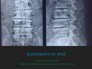
- 1. RADIOGRAPHY OF SPINE Mr. SAMEER AHMAD GANAIE ASSISTANT PROFESSOR COPMS ADESH UNIVERSITY, BATHINDA PUNJAB
- 2. CONTENTS Objective Purpose Landmarks Anatomy of Lumber Spine Anatomy of Sacrum Indications Positioning Basic and Special views Radiation Protection During Radiography References
- 3. OBJECTIVE X-ray of the Whole spine is performed to evaluate any area of the spine (Cervical, thoracic, Lumber, Sacral, or Coccygeal)
- 4. PURPOSE The main purpose of Radiography of spine is to diagnose or treat patients by imaging of the internal structures of the body to assess the presence or absence of disease, foreign objects, and structural damage or any congenital anomaly.
- 5. LANDMARKS
- 6. ANATOMY OF LUMBAR SPINE 5 vertebrae L1-L5 5 intervertebral discs Lumbar lordosis 30°–80° The apex of lumbar lordosis L3-L4
- 7. ANATOMY
- 9. INDICATIONS FRACTURES LOW BACK PAIN DISLOCATIONS
- 11. SCOLIOSIS
- 13. HERNIATED DISKS
- 15. AP VIEW
- 16. CENTRAL RAY Directed towards the midline at the level of the lower costal margin (L3). TECHNIQUES 70 – 80 KVP 55-65 MAS SID 100 CM
- 17. STRUCTURES SHOWN Lumbar vertebral bodies, disk spaces, spinous and transverse processes, lateral margin of psoas muscle, SI joints, the sacrum
- 18. EVALUATION CRITERIA Lumbar vertebral bodies, disk spaces, spinous and transverse processes, lateral margin of psoas muscle, SI joints, and the sacrum should be clearly demonstrated. There should be no rotation of the vertebral column. Spinous processes should be in the midline of the vertebral bodies. Right and left transverse processes equal in length. Sacroiliac joints demonstrate equal distance from the spine. Optimal exposure should clearly demonstrate soft tissues as well as margins of psoas muscle and bony vertebrae.
- 19. LATERAL VIEW
- 20. CENTRAL RAY Directed perpendicular to a point 7.5 cm anterior to the third lumbar spinous process at the level of the lower costal margin. TECHNIQUES 70 – 80 KVP 100-150 MAS SID 100 CM
- 21. STRUCTURES SHOWN Lumbar vertebral bodies, intervertebral foramina, disk spaces, spinous processes, LS joint, sacrum
- 22. EVALUATION CRITERIA Lumbar vertebral bodies, intervertebral foramina, disk spaces, spinous and transverse processes, SI joints, and sacrum should be clearly demonstrated. There should be no rotation of the vertebral column Nearly superimposed iliac crests Superimposed posterior margins of each vertebral body. Open intervertebral disc spaces. The vertebrae should be aligned down in the middle of the radiograph. Optimal exposure should demonstrate clearly soft tissues as well as joint spaces and bony vertebrae.
- 24. CENTRAL RAY Directed horizontally a point 7.5 cm anterior to the third lumbar spinous process at the level of the lower costal margin.(parallel to a line joining the anterior superior iliac spines) TECHNIQUES 70 – 80 KVP 100-150 MAS SID 100 CM
- 25. STRUCTURES SHOWN Lumbar vertebral bodies, intervertebral foramina, disk spaces, spinous processes, LS joint, sacrum
- 26. EVALUATION CRITERIA Lumbar vertebral bodies, intervertebral foramina, disk spaces, spinous processes, and sacrum should be clearly demonstrated. There should be no rotation of the vertebral column Nearly superimposed iliac crests Superimposed posterior margins of each vertebral body. Open intervertebral disc spaces. The vertebrae should be aligned down in the middle of the radiograph. Optimal exposure should demonstrate clearly soft tissues as well as joint spaces and bony vertebrae.
- 27. LATERAL FLEXION AND EXTENSION
- 28. CENTRAL RAY Directed perpendicular to a point 7.5 cm anterior to the third lumbar spinous process at the level of the lower costal margin. TECHNIQUES 70 – 80 KVP 100-150 MAS SID 100 CM
- 29. STRUCTURES SHOWN Lumbar vertebral bodies, intervertebral foramina, disk spaces, spinous processes joint, sacrum
- 30. EVALUATION CRITERIA Lumbar vertebral bodies, intervertebral foramina, disk spaces, spinous processes and sacrum should be clearly demonstrated. There should be no rotation of the vertebral column Nearly superimposed iliac crests Superimposed posterior margins of each vertebral body. Open intervertebral disc spaces. The vertebrae should be aligned down in the middle of the radiograph. Optimal exposure should demonstrate clearly soft tissues as well as joint spaces and bony vertebrae.
- 31. OBLIQUE VIEW
- 32. CENTRAL RAY Directed perpendicular to midclavicular line on the raised side at the level of the lower costal margin TECHNIQUES 70 – 80 KVP 55-65 MAS SID 100 CM
- 33. STRUCTURES SHOWN
- 34. EVALUATION CRITERIA Demonstrate the articular process and facet joints of the side closest to the cassette. They should be open and uniformly visible through the vertebral bodies. Adequate rotation of the spine is evidenced by the position of the pedicles. If the pedicle is anterior on the vertebral body, the patient is not rotated enough, if the pedicle is posterior on the vertebral body, the patient is rotated too much When the patient has been properly positioned in a 30°-45° oblique position, the articular process and facet joints have the appearance of "Scottie dogs."
- 36. AP VIEW
- 37. Directed to the midline at the level of the anterior superior iliac spines with 10–20 degrees cranially angulation TECHNIQUES 70 – 80 KVP 55-65 MAS SID 100 CM CENTRAL RAY
- 38. STRUCTURES SHOWN 4th and 5th Lumbar vertebral bodies, intervertebral foramina, disk spaces, spinous processes joint, sacrum
- 39. EVALUATION CRITERIA There should be no rotation of the vertebral column. Spinous processes in the midline of the vertebral bodies. Right and left transverse processes equal in length. Symmetric vertebrae. Sacroiliac joints demonstrate equal distance from the spine. Optimal exposure should clearly demonstrate soft tissues as well as margins of psoas muscle and bony vertebrae
- 40. LATERAL VIEW
- 41. CENTRAL RAY Directed perpendicular to level of the tubercle of the iliac crest or midway between the level of the upper border of the iliac crest and the anterior superior iliac spine. TECHNIQUES 70 – 80 KVP 100-150 MAS SID 100 CM
- 42. STRUCTURES SHOWN
- 43. EVALUATION CRITERIA There should be no rotation of the vertebral column. Nearly superimposed iliac crests Superimposed posterior margins of each vertebral body. Open intervertebral disc spaces. The L5-S1 lumbosacral junction lateral projection should demonstrate the lower one or two lumbar vertebrae and the upper sacrum with lumbosacral joint in the center of the radiograph. Optimal exposure should demonstrate clearly soft tissues as well as joint spaces and bony vertebrae.
- 44. OBLIQUE VIEW
- 45. CENTRAL RAY Directed perpendicular to the midline at the level of the anterior superior iliac spines. TECHNIQUES 70 – 80 KVP 55-65 MAS SID 100 CM
- 46. STRUCTURES SHOWN articular process facet joints , 4th and 5thlumbar vertebrae, Proximal sacrum
- 47. EVALUATION CRITERIA Demonstrate the articular process and facet joints of the side closest to the cassette. They should be open and uniformly visible through the vertebral bodies. Adequate rotation of the spine is evidenced by the position of the pedicles. If the pedicle is anterior on the vertebral body, the patient is not rotated enough, if the pedicle is posterior on the vertebral body, the patient is rotated too much.
- 48. SACRUM
- 49. AP VIEW
- 50. CENTRAL RAY Directed perpendicular to a point midway between the level of the anterior superior iliac spines and the superior border of the symphysis pubis with tube angulation of 10–25 degrees cranially TECHNIQUES 70 – 80 KVP 55-65 MAS SID 100 CM
- 52. EVALUATION CRITERIA No motion since blurring can occur Optimum contrast and density to demonstrate vertebral bodies and soft tissue No rotation
- 53. LATERAL
- 54. CENTRAL RAY Directed perpendicular to the long axis of the sacrum and to a point at a level midway between the posterior superior iliac spines and the sacro- coccygeal junction. TECHNIQUES 70 – 80 KVP 100-150 MAS SID 100 CM
- 55. STRUCTURES SHOWN
- 56. EVALUATION CRITERIA No motion since blurring can occur Optimum contrast and density to demonstrate vertebral bodies and soft tissue No rotation
- 57. COCCYX
- 58. AP VIEW
- 59. CENTRAL RAY Directed to a point in the midline 2.5 cm superior to the symphysis pubis with tube angulation of 15 degrees caudally TECHNIQUES 70 – 80 KVP 55-65 MAS SID 100 CM
- 61. EVALUATION CRITERIA No motion since blurring can occur Optimum contrast and density to demonstrate vertebral bodies and soft tissue No rotation
- 62. LATERAL VIEW
- 63. CENTRAL RAY Directed perpendicular to long axis of the sacrum and towards the palpable coccyx. TECHNIQUES 70 – 80 KVP 100-150 MAS SID 100 CM
- 64. STRUCTURES SHOWN
- 65. EVALUATION CRITERIA No motion since blurring can occur Optimum contrast and density to demonstrate vertebral bodies and soft tissue No rotation
- 67. CENTRAL RAY Directed perpendicular to ischial tuberosities TECHNIQUES 70 – 80 KVP 55-65 MAS SID 100 CM
- 68. STRUCTURES SHOWN
- 69. EVALUATION CRITERIA No motion since blurring can occur Optimum contrast and density to demonstrate soft tissue No rotation
- 70. RADIATION PROTECTION Collimate the beam to region of interest Follow 10 day rule
- 71. References Clark’s positioning. Bhargava (For Residents and Technicians)
Editor's Notes
- ANY RADIOLOGICAL PROCEDURES OF PELVIS OF WOMEN OF CHILD BEARING AGE,SHOULD BE CARRIED OUT WITHIN 10 DAYS FROM ONSET OF MENSTRUAL CYCLE