
Recommended
More Related Content
What's hot
What's hot (20)
Similar to bones of Upper limbs and anatomy of upper limbs
Similar to bones of Upper limbs and anatomy of upper limbs (20)
More from sadhamhussain52
Recently uploaded
Recently uploaded (20)
bones of Upper limbs and anatomy of upper limbs
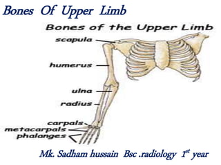
- 1. Bones Of Upper Limb Mk. Sadham hussain Bsc .radiology 1st year
- 2. Clavicle(collar bone) Paculiarities of clavicle • Only long bone placed horizontally. • Subcutaneous and can easily palpated. • 1st bone to ossify in the body. • The only long bone with two primary center of ossification for shaft. • No madullary cavity.
- 3. Applied Anatomy. • Common sit of fractured in clavicle is the junction of medial 2/3 and lateral 1/3.
- 4. Scapula(shoulder blade) 2 surfaces • Costal surface is also called subscapular fossa • Dorsal surface is divided into an upper supraspinous and infraspinous fossa by spinous process 3 broder • Superior border • Lateral border • Medial border
- 5. 3 processes • Acromion process – it articulates with the lateral end of the clavicle at the acromioclavicular joint. • Coracoid process • Spinous process Applied anatomy Paralysis of a muscle called serratus anterior causes winging of the scapula .
- 7. Applied Anatomy of Scapula • Paralysis of serratus anterior muscle causes winging of the scapula .
- 8. Humerus • It has upper end , lower end , shaft . Upper end • Head – it articulates with glenoid cavity of the scapula to form shoulder joint . • Greater tubercle • Lesser tubercle • Bicipital groove (intertubercular sulcus) – it is an area between GT and LT
- 12. (radial fossa , coronoid fossa , olecranon fossa)
- 13. Applied Anatomy Humerus. • The fracture affecting the surgical neck can damage axillary nerve leads to paralysis of deltoid muscle. • Fracture affecting the shaft can cause damage to radial nerve which result wrist drop. • Supracondylar fracture can damage median nerve and brachial artery. Damage of brachial artery cause volkman’s ischemci contracture.
- 14. • Fracture affecting the medial epicondyle can damage ulnar nerve which leads to ‘claw hand’ sensory loss in the medial side of the palm and medial one and a half finger .
- 15. Radius (arm) •The shorter of the two long bones of the forearm, extending from the elbow to the wrist; •it is the bone on the thumb side of the arm. •The radius rotates around the ulna, permitting the hand to rotate and be flexible. •A projection just above the thumb side of the wrist marks the end of the radius.
- 19. Applied Anatomy of Radius • Colles’ fracture : Fracture of distal end of the radius , the distal fractured fragment is displaced upwards and posteriorly by brachio radialis muscle . This produces ‘dinner fork deformity’. • smith’s fracture : it is reverse of colles’ fracture . It produced by a fall on the back of the hand .
- 22. Applied Anatomy Of Ulna. • Fracture of shaft of ulna may be associated with fracture of radius . • Fracture of olecranon process occurs if one falls on the point of elbow .
- 23. Bones of the Hand
- 32. Applied anatomy of carpal bone . • Scaphoid fracture : scaphoid is the most common carpal bone fractured due to fall on the outstretched hand. • Lunate dislocation : though it is uncommon , its forward dislocation can cause carpal tunnel syndrome .
- 33. Applied Anatomy of phalanges • Bennett’s fracture : fracture involving the base of the 1st metacarpal bone . • Mallet finger : the distal phalanx undergoes extreme flexion due to detachment of extensor tendon from the distal phalanx .