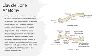
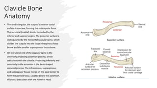
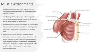
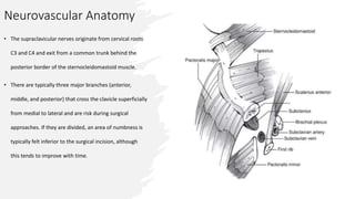
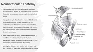
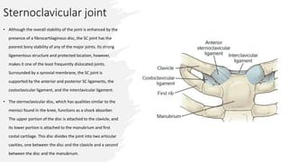
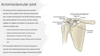
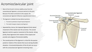
SlideShare a Scribd company logo
1 of 16
ORTHOFIXAR.COM
• Clavicle anatomy consists of medial end,
middle and lateral end. There are many
ligaments and muscles connecting to these
parts of clavicle.
• Clavicle bone is the First bone in the body to
ossify (at 5 weeks’ gestation) and last to fuse
(medial epiphysis at 25 years of age).
Clavicle Bone
Anatomy
• Serving as a strut between the sternum and scapula,
the clavicle bone elevates and rotates to maintain
the alignment of the scapula, allowing for additional
motion when the arm is raised and preventing
excessive anterior displacement of the scapula.
• The proximal two-thirds of the clavicle bone is
characterized by an anteriorly convex bend. The
distal one-third begins to flatten while curving
concavely to meet with the scapula. The point at
which the clavicle begins to transition from a convex
to a concave bend, approximately two-thirds of the
way along its shaft, is relatively weak and is a
common site for fractures.
Clavicle Bone
Anatomy
• Thin and triangular, the scapula’s anterior costal
surface is concave, forming the subscapular fossa.
The vertebral (medial) border is marked by the
inferior and superior angles. The posterior surface is
distinguished by the horizontal scapular spine, which
divides the scapula into the large infraspinous fossa
below and the smaller supraspinous fossa above.
• On the lateral end of the scapular spine is the
anteriorly projecting acromion process, which
articulates with the clavicle. Projecting inferiorly and
anteriorly to the acromion is the beak-shaped
coracoid process. The infraspinous, supraspinous,
and subscapular fossae merge on the axial border to
form the glenoid fossa. Located below the acromion,
this fossa articulates with the humeral head.
Clavicle Ligament
• Medial Clavicle articulates with sternal bone forming the
sternoclavicular joint, there is relatively little motion at the
sternoclavicular joint. Medially the clavicle is secured to the
sternum by the sternoclavicular capsule. The thickening of the
posterior capsule has been determined to be the single most
important soft tissue constraint to anterior or posterior
translation of the medial clavicle.
• There is also an interclavicular ligament which runs from the
medial end of one clavicle, gains purchase from the superior
aspect of the sternum at the sternal notch and attaches to the
medial end of the contralateral clavicle. Acting as a tension
wire at the base of the clavicle, this ligament helps prevent
inferior angulation or translation of the clavicle.
• There are extremely stout ligaments that originate on the first
rib and insert on the undersurface or the inferior aspect of the
clavicle.
Clavicle Ligament
• Laterally the clavicle articulates with the acromion bone.
• The coracoclavicular ligaments are stout ligaments that arise from
the base of the coracoid:
1. The trapezoid (more lateral): inserts onto the small osseous ridge of
the inferior clavicle.
2. Conoid (more medial): inserts onto the clavicular conoid tubercle.
• These ligaments are very strong and provide the primary resistance
to superior displacement of the clavicle. Their integrity, or lack
thereof, plays an important role in the decision making and fixation
selection in the treatment of displaced lateral third clavicle fractures.
• Clavicle fractures in this location will often have an avulsed inferior
fragment to which these ligaments are attached, especially in
younger individuals.
• The capsule of the AC joint is thickened superiorly and is primarily
responsible for resisting AP displacement of the joint.
• If one is inserting a hook plate for fixation of a very distal fracture, a
small defect can be made in the posterolateral aspect.
Muscle Attachments
• Medially: the pectoralis major muscle originates from the
clavicular shaft anteroinferiorly, and the sternocleidomastoid
originates superiorly.
• Laterally: the pectoralis origin merges with the origin of the
anterior deltoid, while the trapezius insertion blends superiorly
with the deltoid origin at the lateral margin.
• The medial clavicular fragment is elevated by the unopposed pull
of the sternocleidomastoid muscle, while the distal fragment is
held inferiorly by the deltoid and medially by the pectoralis
major.
• The platysma or “shaving muscle” is variable in terms of
thickness and extent, but usually envelopes the anterior and
superior aspects of the clavicle and runs in the subcutaneous
tissues, extending superiorly to the mandible and the deeper
facial muscles. It is divided during the surgical approach, and is
typically included in the closure of the superficial, or
skin/subcutaneous layer.
Neurovascular Anatomy
• The supraclavicular nerves originate from cervical roots
C3 and C4 and exit from a common trunk behind the
posterior border of the sternocleidomastoid muscle.
• There are typically three major branches (anterior,
middle, and posterior) that cross the clavicle superficially
from medial to lateral and are risk during surgical
approaches. If they are divided, an area of numbness is
typically felt inferior to the surgical incision, although
this tends to improve with time.
Neurovascular Anatomy
• The subclavian vein runs directly below the subclavius
muscle and above the first rib, where it is readily accessible
(for central venous access) and vulnerable (to inadvertent
injury).
• More posteriorly lie the subclavian artery and the brachial
plexus, separated from the vein and clavicle by the
additional layer of the scalenus anterior muscle medially.
subclavian vessels were closest at the medial end, with the
vein directly apposed to the posterior cortex of the medial
clavicle in some cases.
• In the middle third, the artery and vein were a mean of 17
and 13 mm from the clavicle, respectively, at an
approximate angle of 60 degrees to the horizontal (i.e., the
vessels were posterior-inferior to the clavicle).
• Laterally, the distances were greater, with the artery and
vein a mean of 63 and 76 mm, respectively form the clavicle.
Sternoclavicular joint
• The manubrium of the sternum serves as the site of
attachment for each clavicle. Projecting above the body
of the sternum, the superior surface of the manubrium
is indented by the jugular (suprasternal) notch.
Projecting off each side of the jugular notch is the
clavicular notch, which accepts the medial head of the
clavicle
• At the Sternoclavicular joint, the proximal portion of the
clavicle meets the manubrium of the sternum and a
portion of the first costal cartilage to form a gliding joint
that allows three degrees of freedom of motion:
• Elevation and depression,
• Protraction and retraction,
• Anterior and posterior rotation.
Sternoclavicular joint
• Although the overall stability of the joint is enhanced by the
presence of a fibrocartilaginous disc, the SC joint has the
poorest bony stability of any of the major joints. Its strong
ligamentous structure and protected location, however,
makes it one of the least frequently dislocated joints.
Surrounded by a synovial membrane, the SC joint is
supported by the anterior and posterior SC ligaments, the
costoclavicular ligament, and the interclavicular ligament.
• The sternoclavicular disc, which has qualities similar to the
menisci found in the knee, functions as a shock absorber.
The upper portion of the disc is attached to the clavicle, and
its lower portion is attached to the manubrium and first
costal cartilage. This disc divides the joint into two articular
cavities, one between the disc and the clavicle and a second
between the disc and the manubrium.
Acromioclavicular joint
• The distal end of the clavicle meets the acromion
process of the scapula to form the Acromioclavicular
joint. A plane synovial joint, the AC joint allows a gliding
articulation between the acromion and the clavicle,
capable of 3 degrees of freedom of movement, each
around an oblique axis:
• Internal and external rotation around a vertical axis,
• Upward and downward rotation around an axis
perpendicular to the plane of the scapula,
• Anterior and posterior scapular tilting around a horizontal
axis.
• This articulation allows for the motion necessary to
maintain the relationship between the scapula and the
clavicle in the early and late stages of the GH joint’s ROM
Acromioclavicular joint
• Surrounded by a synovial membrane, the AC joint is
supported by the AC ligament and the coracoclavicular
ligament, which suspend the scapula from the clavicle.
• A synovial disc is present between the clavicle and the
acromion that disappears by the fourth decade of life.
• Divided into two separate bands, the superior and
inferior portions of the AC ligament function to maintain
continuity between the articulating surfaces of the
acromion and clavicle.
• With much of its restraint in the horizontal plane, this
ligament maintains stability by preventing the clavicle
from riding up and over the acromion process.
Acromioclavicular joint
• Most of the AC joint’s intrinsic stability arises from the
coracoclavicular ligament, a structure extrinsic to the joint.
Because of its direct connection to the scapula, the
coracoclavicular ligament influences scapulohumeral motion.
• This ligament is divided into two distinct portions:
1. The lateral quadrilateral-shaped trapezoid ligament.
2. The medial triangular-shaped conoid ligament.
• Separated by a bursa, the trapezoid ligament limits lateral
movement of the clavicle over the acromion. The conoid
ligament restricts superior movement of the clavicle. Acting
jointly, these ligaments limit rotation of the scapula and
provide some degree of horizontal stability.
• The conoid portion of the ligament is critical for the passive
posterior rotation of the clavicle that occurs during shoulder
elevation. A horizontal dislocation of the AC joint can occur
with the coracoclavicular ligament remaining intact.
References:
1. Orthofixar | Orthopedic Surgery Learning
2.Millers Review of Orthopaedics -7th Edition Book.
3.Rockwood and Green's Fractures in Adults 8th Edition book.
4.Hyland S, Charlick M, Varacallo M. Anatomy, Shoulder and Upper
Limb, Clavicle. [Updated 2021 Jul 26]. In: StatPearls [Internet].
Treasure Island (FL): StatPearls Publishing; 2021 Jan-. Available from:
NBK525990.

More Related Content

What's hot (20)

The femur
The femurThe femur
The femur
 
Ankle joint Anatomy
Ankle joint AnatomyAnkle joint Anatomy
Ankle joint Anatomy
 
Slideshow: Scapula
Slideshow: ScapulaSlideshow: Scapula
Slideshow: Scapula
 
Upper limb bones
Upper limb bonesUpper limb bones
Upper limb bones
 
Development,structure and organization of bone
Development,structure and organization of boneDevelopment,structure and organization of bone
Development,structure and organization of bone
 
Elbow joint anatomy
Elbow joint anatomyElbow joint anatomy
Elbow joint anatomy
 
Cartilage
Cartilage Cartilage
Cartilage
 
Joints of lower limb
Joints of lower limbJoints of lower limb
Joints of lower limb
 
urinary Bladder.ppt
urinary Bladder.ppturinary Bladder.ppt
urinary Bladder.ppt
 
Anatomy of shoulder joint
Anatomy of shoulder jointAnatomy of shoulder joint
Anatomy of shoulder joint
 
Vertebral column
Vertebral columnVertebral column
Vertebral column
 
Lower limb complete
Lower limb completeLower limb complete
Lower limb complete
 
Anatomy of vertebral column
Anatomy of vertebral columnAnatomy of vertebral column
Anatomy of vertebral column
 
upper limb joints.
upper limb joints.upper limb joints.
upper limb joints.
 
Anatomy of Humerus
Anatomy of HumerusAnatomy of Humerus
Anatomy of Humerus
 
Shoulder joint
Shoulder jointShoulder joint
Shoulder joint
 
Lower limb bones
Lower limb bonesLower limb bones
Lower limb bones
 
Hip bone (Gross Anatomy)
Hip bone (Gross Anatomy)Hip bone (Gross Anatomy)
Hip bone (Gross Anatomy)
 
Bones and joint of foot
Bones and joint of footBones and joint of foot
Bones and joint of foot
 
The clavicle
The clavicleThe clavicle
The clavicle
 

Similar to clavicle Anatomy.pptx

Clavicle anatomy.pdf
Clavicle anatomy.pdfClavicle anatomy.pdf
Clavicle anatomy.pdfmhmad farooq
 
Osteology upper limb by Dr G Kamau
Osteology upper limb by Dr G KamauOsteology upper limb by Dr G Kamau
Osteology upper limb by Dr G KamauMathewJude
 
Biomechanics of shoulder
Biomechanics of shoulderBiomechanics of shoulder
Biomechanics of shoulderSayali Gujjewar
 
applied aspects of shoulder joint
applied aspects of shoulder jointapplied aspects of shoulder joint
applied aspects of shoulder jointmrinal joshi
 
Anatomy and xrays of shoulder joint
Anatomy and xrays of shoulder jointAnatomy and xrays of shoulder joint
Anatomy and xrays of shoulder jointAkshay Mylar
 
Introduction to upper limb
Introduction to upper limbIntroduction to upper limb
Introduction to upper limbLucidante1
 
introductiontoupperlimb-140210053432-phpapp01.pdf
introductiontoupperlimb-140210053432-phpapp01.pdfintroductiontoupperlimb-140210053432-phpapp01.pdf
introductiontoupperlimb-140210053432-phpapp01.pdfRCGaur1
 
anatomy of vertebral column.pptx
anatomy of vertebral column.pptxanatomy of vertebral column.pptx
anatomy of vertebral column.pptxJaseerAk1
 
OSTEOLOGY OF THE VERTEBRAE
OSTEOLOGY OF THE VERTEBRAE OSTEOLOGY OF THE VERTEBRAE
OSTEOLOGY OF THE VERTEBRAE Kamal Deen
 
The vertebral column.pptx
The vertebral column.pptxThe vertebral column.pptx
The vertebral column.pptxmeharkinan
 
Clavicle Anatomy and its Articulation - ANAND GURMAITA (BSc. RADIOLOGY)
Clavicle Anatomy and its Articulation - ANAND GURMAITA (BSc. RADIOLOGY)Clavicle Anatomy and its Articulation - ANAND GURMAITA (BSc. RADIOLOGY)
Clavicle Anatomy and its Articulation - ANAND GURMAITA (BSc. RADIOLOGY)ANAND GURMAITA
 
Shoulder joint_nursing.pptx
Shoulder joint_nursing.pptxShoulder joint_nursing.pptx
Shoulder joint_nursing.pptxABHIJIT BHOYAR
 
Anatomy and Examination of the Knee
Anatomy and Examination of the KneeAnatomy and Examination of the Knee
Anatomy and Examination of the KneeSri Harsha Gutta
 
Joints of-upper-limb
Joints of-upper-limbJoints of-upper-limb
Joints of-upper-limbCIMS
 
sterno acromioclavicular.pptx
sterno acromioclavicular.pptxsterno acromioclavicular.pptx
sterno acromioclavicular.pptxminel5
 

Similar to clavicle Anatomy.pptx (20)

Clavicle anatomy.pdf
Clavicle anatomy.pdfClavicle anatomy.pdf
Clavicle anatomy.pdf
 
Osteology upper limb by Dr G Kamau
Osteology upper limb by Dr G KamauOsteology upper limb by Dr G Kamau
Osteology upper limb by Dr G Kamau
 
Biomechanics of shoulder
Biomechanics of shoulderBiomechanics of shoulder
Biomechanics of shoulder
 
applied aspects of shoulder joint
applied aspects of shoulder jointapplied aspects of shoulder joint
applied aspects of shoulder joint
 
Anatomy and xrays of shoulder joint
Anatomy and xrays of shoulder jointAnatomy and xrays of shoulder joint
Anatomy and xrays of shoulder joint
 
anatomy of spine
anatomy of spineanatomy of spine
anatomy of spine
 
Biomechanich of the spine ppt (2)
Biomechanich of the spine ppt (2)Biomechanich of the spine ppt (2)
Biomechanich of the spine ppt (2)
 
Introduction to upper limb
Introduction to upper limbIntroduction to upper limb
Introduction to upper limb
 
introductiontoupperlimb-140210053432-phpapp01.pdf
introductiontoupperlimb-140210053432-phpapp01.pdfintroductiontoupperlimb-140210053432-phpapp01.pdf
introductiontoupperlimb-140210053432-phpapp01.pdf
 
Sc joint
Sc joint Sc joint
Sc joint
 
anatomy of vertebral column.pptx
anatomy of vertebral column.pptxanatomy of vertebral column.pptx
anatomy of vertebral column.pptx
 
OSTEOLOGY OF THE VERTEBRAE
OSTEOLOGY OF THE VERTEBRAE OSTEOLOGY OF THE VERTEBRAE
OSTEOLOGY OF THE VERTEBRAE
 
The vertebral column.pptx
The vertebral column.pptxThe vertebral column.pptx
The vertebral column.pptx
 
Clavicle Anatomy and its Articulation - ANAND GURMAITA (BSc. RADIOLOGY)
Clavicle Anatomy and its Articulation - ANAND GURMAITA (BSc. RADIOLOGY)Clavicle Anatomy and its Articulation - ANAND GURMAITA (BSc. RADIOLOGY)
Clavicle Anatomy and its Articulation - ANAND GURMAITA (BSc. RADIOLOGY)
 
Shoulder joint_nursing.pptx
Shoulder joint_nursing.pptxShoulder joint_nursing.pptx
Shoulder joint_nursing.pptx
 
Appendicular Skeleton.pptx
Appendicular Skeleton.pptxAppendicular Skeleton.pptx
Appendicular Skeleton.pptx
 
The hip complex
The hip complexThe hip complex
The hip complex
 
Anatomy and Examination of the Knee
Anatomy and Examination of the KneeAnatomy and Examination of the Knee
Anatomy and Examination of the Knee
 
Joints of-upper-limb
Joints of-upper-limbJoints of-upper-limb
Joints of-upper-limb
 
sterno acromioclavicular.pptx
sterno acromioclavicular.pptxsterno acromioclavicular.pptx
sterno acromioclavicular.pptx
 

More from mhmad farooq

Hand Flexor tendon injury.pdf
Hand Flexor tendon injury.pdfHand Flexor tendon injury.pdf
Hand Flexor tendon injury.pdfmhmad farooq
 
oswestry disability index pdf.pdf
oswestry disability index pdf.pdfoswestry disability index pdf.pdf
oswestry disability index pdf.pdfmhmad farooq
 
Neck Disability Index.pdf
Neck Disability Index.pdfNeck Disability Index.pdf
Neck Disability Index.pdfmhmad farooq
 
Homan’s Sign for DVT.pptx
Homan’s Sign for DVT.pptxHoman’s Sign for DVT.pptx
Homan’s Sign for DVT.pptxmhmad farooq
 
Hip muscles anatomy
Hip muscles anatomyHip muscles anatomy
Hip muscles anatomymhmad farooq
 
Thomas Test | Iliopsoas Tightness
Thomas Test | Iliopsoas TightnessThomas Test | Iliopsoas Tightness
Thomas Test | Iliopsoas Tightnessmhmad farooq
 
Extensor tendon injuries
Extensor tendon injuriesExtensor tendon injuries
Extensor tendon injuriesmhmad farooq
 

More from mhmad farooq (10)

Hand Flexor tendon injury.pdf
Hand Flexor tendon injury.pdfHand Flexor tendon injury.pdf
Hand Flexor tendon injury.pdf
 
oswestry disability index pdf.pdf
oswestry disability index pdf.pdfoswestry disability index pdf.pdf
oswestry disability index pdf.pdf
 
Neck Disability Index.pdf
Neck Disability Index.pdfNeck Disability Index.pdf
Neck Disability Index.pdf
 
Thomas Test.pdf
Thomas Test.pdfThomas Test.pdf
Thomas Test.pdf
 
Homan’s Sign for DVT.pptx
Homan’s Sign for DVT.pptxHoman’s Sign for DVT.pptx
Homan’s Sign for DVT.pptx
 
Hip muscles anatomy
Hip muscles anatomyHip muscles anatomy
Hip muscles anatomy
 
Mortons neuroma
Mortons neuromaMortons neuroma
Mortons neuroma
 
Mortons neuroma
Mortons neuromaMortons neuroma
Mortons neuroma
 
Thomas Test | Iliopsoas Tightness
Thomas Test | Iliopsoas TightnessThomas Test | Iliopsoas Tightness
Thomas Test | Iliopsoas Tightness
 
Extensor tendon injuries
Extensor tendon injuriesExtensor tendon injuries
Extensor tendon injuries
 

Recently uploaded

Organic Name Reactions for the students and aspirants of Chemistry12th.pptx
Organic Name Reactions  for the students and aspirants of Chemistry12th.pptxOrganic Name Reactions  for the students and aspirants of Chemistry12th.pptx
Organic Name Reactions for the students and aspirants of Chemistry12th.pptxVS Mahajan Coaching Centre
 
Framing an Appropriate Research Question 6b9b26d93da94caf993c038d9efcdedb.pdf
Framing an Appropriate Research Question 6b9b26d93da94caf993c038d9efcdedb.pdfFraming an Appropriate Research Question 6b9b26d93da94caf993c038d9efcdedb.pdf
Framing an Appropriate Research Question 6b9b26d93da94caf993c038d9efcdedb.pdfUjwalaBharambe
 
Full Stack Web Development Course for Beginners
Full Stack Web Development Course  for BeginnersFull Stack Web Development Course  for Beginners
Full Stack Web Development Course for BeginnersSabitha Banu
 
POINT- BIOCHEMISTRY SEM 2 ENZYMES UNIT 5.pptx
POINT- BIOCHEMISTRY SEM 2 ENZYMES UNIT 5.pptxPOINT- BIOCHEMISTRY SEM 2 ENZYMES UNIT 5.pptx
POINT- BIOCHEMISTRY SEM 2 ENZYMES UNIT 5.pptxSayali Powar
 
“Oh GOSH! Reflecting on Hackteria's Collaborative Practices in a Global Do-It...
“Oh GOSH! Reflecting on Hackteria's Collaborative Practices in a Global Do-It...“Oh GOSH! Reflecting on Hackteria's Collaborative Practices in a Global Do-It...
“Oh GOSH! Reflecting on Hackteria's Collaborative Practices in a Global Do-It...Marc Dusseiller Dusjagr
 
Painted Grey Ware.pptx, PGW Culture of India
Painted Grey Ware.pptx, PGW Culture of IndiaPainted Grey Ware.pptx, PGW Culture of India
Painted Grey Ware.pptx, PGW Culture of IndiaVirag Sontakke
 
MARGINALIZATION (Different learners in Marginalized Group
MARGINALIZATION (Different learners in Marginalized GroupMARGINALIZATION (Different learners in Marginalized Group
MARGINALIZATION (Different learners in Marginalized GroupJonathanParaisoCruz
 
How to Make a Pirate ship Primary Education.pptx
How to Make a Pirate ship Primary Education.pptxHow to Make a Pirate ship Primary Education.pptx
How to Make a Pirate ship Primary Education.pptxmanuelaromero2013
 
Enzyme, Pharmaceutical Aids, Miscellaneous Last Part of Chapter no 5th.pdf
Enzyme, Pharmaceutical Aids, Miscellaneous Last Part of Chapter no 5th.pdfEnzyme, Pharmaceutical Aids, Miscellaneous Last Part of Chapter no 5th.pdf
Enzyme, Pharmaceutical Aids, Miscellaneous Last Part of Chapter no 5th.pdfSumit Tiwari
 
Proudly South Africa powerpoint Thorisha.pptx
Proudly South Africa powerpoint Thorisha.pptxProudly South Africa powerpoint Thorisha.pptx
Proudly South Africa powerpoint Thorisha.pptxthorishapillay1
 
Employee wellbeing at the workplace.pptx
Employee wellbeing at the workplace.pptxEmployee wellbeing at the workplace.pptx
Employee wellbeing at the workplace.pptxNirmalaLoungPoorunde1
 
Software Engineering Methodologies (overview)
Software Engineering Methodologies (overview)Software Engineering Methodologies (overview)
Software Engineering Methodologies (overview)eniolaolutunde
 
Biting mechanism of poisonous snakes.pdf
Biting mechanism of poisonous snakes.pdfBiting mechanism of poisonous snakes.pdf
Biting mechanism of poisonous snakes.pdfadityarao40181
 
CARE OF CHILD IN INCUBATOR..........pptx
CARE OF CHILD IN INCUBATOR..........pptxCARE OF CHILD IN INCUBATOR..........pptx
CARE OF CHILD IN INCUBATOR..........pptxGaneshChakor2
 
Historical philosophical, theoretical, and legal foundations of special and i...
Historical philosophical, theoretical, and legal foundations of special and i...Historical philosophical, theoretical, and legal foundations of special and i...
Historical philosophical, theoretical, and legal foundations of special and i...jaredbarbolino94
 
EPANDING THE CONTENT OF AN OUTLINE using notes.pptx
EPANDING THE CONTENT OF AN OUTLINE using notes.pptxEPANDING THE CONTENT OF AN OUTLINE using notes.pptx
EPANDING THE CONTENT OF AN OUTLINE using notes.pptxRaymartEstabillo3
 
History Class XII Ch. 3 Kinship, Caste and Class (1).pptx
History Class XII Ch. 3 Kinship, Caste and Class (1).pptxHistory Class XII Ch. 3 Kinship, Caste and Class (1).pptx
History Class XII Ch. 3 Kinship, Caste and Class (1).pptxsocialsciencegdgrohi
 
KSHARA STURA .pptx---KSHARA KARMA THERAPY (CAUSTIC THERAPY)————IMP.OF KSHARA ...
KSHARA STURA .pptx---KSHARA KARMA THERAPY (CAUSTIC THERAPY)————IMP.OF KSHARA ...KSHARA STURA .pptx---KSHARA KARMA THERAPY (CAUSTIC THERAPY)————IMP.OF KSHARA ...
KSHARA STURA .pptx---KSHARA KARMA THERAPY (CAUSTIC THERAPY)————IMP.OF KSHARA ...M56BOOKSTORE PRODUCT/SERVICE
 

Recently uploaded (20)

Organic Name Reactions for the students and aspirants of Chemistry12th.pptx
Organic Name Reactions  for the students and aspirants of Chemistry12th.pptxOrganic Name Reactions  for the students and aspirants of Chemistry12th.pptx
Organic Name Reactions for the students and aspirants of Chemistry12th.pptx
 
Model Call Girl in Bikash Puri Delhi reach out to us at 🔝9953056974🔝
Model Call Girl in Bikash Puri  Delhi reach out to us at 🔝9953056974🔝Model Call Girl in Bikash Puri  Delhi reach out to us at 🔝9953056974🔝
Model Call Girl in Bikash Puri Delhi reach out to us at 🔝9953056974🔝
 
Framing an Appropriate Research Question 6b9b26d93da94caf993c038d9efcdedb.pdf
Framing an Appropriate Research Question 6b9b26d93da94caf993c038d9efcdedb.pdfFraming an Appropriate Research Question 6b9b26d93da94caf993c038d9efcdedb.pdf
Framing an Appropriate Research Question 6b9b26d93da94caf993c038d9efcdedb.pdf
 
Full Stack Web Development Course for Beginners
Full Stack Web Development Course  for BeginnersFull Stack Web Development Course  for Beginners
Full Stack Web Development Course for Beginners
 
POINT- BIOCHEMISTRY SEM 2 ENZYMES UNIT 5.pptx
POINT- BIOCHEMISTRY SEM 2 ENZYMES UNIT 5.pptxPOINT- BIOCHEMISTRY SEM 2 ENZYMES UNIT 5.pptx
POINT- BIOCHEMISTRY SEM 2 ENZYMES UNIT 5.pptx
 
“Oh GOSH! Reflecting on Hackteria's Collaborative Practices in a Global Do-It...
“Oh GOSH! Reflecting on Hackteria's Collaborative Practices in a Global Do-It...“Oh GOSH! Reflecting on Hackteria's Collaborative Practices in a Global Do-It...
“Oh GOSH! Reflecting on Hackteria's Collaborative Practices in a Global Do-It...
 
Painted Grey Ware.pptx, PGW Culture of India
Painted Grey Ware.pptx, PGW Culture of IndiaPainted Grey Ware.pptx, PGW Culture of India
Painted Grey Ware.pptx, PGW Culture of India
 
MARGINALIZATION (Different learners in Marginalized Group
MARGINALIZATION (Different learners in Marginalized GroupMARGINALIZATION (Different learners in Marginalized Group
MARGINALIZATION (Different learners in Marginalized Group
 
How to Make a Pirate ship Primary Education.pptx
How to Make a Pirate ship Primary Education.pptxHow to Make a Pirate ship Primary Education.pptx
How to Make a Pirate ship Primary Education.pptx
 
Enzyme, Pharmaceutical Aids, Miscellaneous Last Part of Chapter no 5th.pdf
Enzyme, Pharmaceutical Aids, Miscellaneous Last Part of Chapter no 5th.pdfEnzyme, Pharmaceutical Aids, Miscellaneous Last Part of Chapter no 5th.pdf
Enzyme, Pharmaceutical Aids, Miscellaneous Last Part of Chapter no 5th.pdf
 
9953330565 Low Rate Call Girls In Rohini Delhi NCR
9953330565 Low Rate Call Girls In Rohini  Delhi NCR9953330565 Low Rate Call Girls In Rohini  Delhi NCR
9953330565 Low Rate Call Girls In Rohini Delhi NCR
 
Proudly South Africa powerpoint Thorisha.pptx
Proudly South Africa powerpoint Thorisha.pptxProudly South Africa powerpoint Thorisha.pptx
Proudly South Africa powerpoint Thorisha.pptx
 
Employee wellbeing at the workplace.pptx
Employee wellbeing at the workplace.pptxEmployee wellbeing at the workplace.pptx
Employee wellbeing at the workplace.pptx
 
Software Engineering Methodologies (overview)
Software Engineering Methodologies (overview)Software Engineering Methodologies (overview)
Software Engineering Methodologies (overview)
 
Biting mechanism of poisonous snakes.pdf
Biting mechanism of poisonous snakes.pdfBiting mechanism of poisonous snakes.pdf
Biting mechanism of poisonous snakes.pdf
 
CARE OF CHILD IN INCUBATOR..........pptx
CARE OF CHILD IN INCUBATOR..........pptxCARE OF CHILD IN INCUBATOR..........pptx
CARE OF CHILD IN INCUBATOR..........pptx
 
Historical philosophical, theoretical, and legal foundations of special and i...
Historical philosophical, theoretical, and legal foundations of special and i...Historical philosophical, theoretical, and legal foundations of special and i...
Historical philosophical, theoretical, and legal foundations of special and i...
 
EPANDING THE CONTENT OF AN OUTLINE using notes.pptx
EPANDING THE CONTENT OF AN OUTLINE using notes.pptxEPANDING THE CONTENT OF AN OUTLINE using notes.pptx
EPANDING THE CONTENT OF AN OUTLINE using notes.pptx
 
History Class XII Ch. 3 Kinship, Caste and Class (1).pptx
History Class XII Ch. 3 Kinship, Caste and Class (1).pptxHistory Class XII Ch. 3 Kinship, Caste and Class (1).pptx
History Class XII Ch. 3 Kinship, Caste and Class (1).pptx
 
KSHARA STURA .pptx---KSHARA KARMA THERAPY (CAUSTIC THERAPY)————IMP.OF KSHARA ...
KSHARA STURA .pptx---KSHARA KARMA THERAPY (CAUSTIC THERAPY)————IMP.OF KSHARA ...KSHARA STURA .pptx---KSHARA KARMA THERAPY (CAUSTIC THERAPY)————IMP.OF KSHARA ...
KSHARA STURA .pptx---KSHARA KARMA THERAPY (CAUSTIC THERAPY)————IMP.OF KSHARA ...
 

clavicle Anatomy.pptx

  • 2. • Clavicle anatomy consists of medial end, middle and lateral end. There are many ligaments and muscles connecting to these parts of clavicle. • Clavicle bone is the First bone in the body to ossify (at 5 weeks’ gestation) and last to fuse (medial epiphysis at 25 years of age).
  • 3. Clavicle Bone Anatomy • Serving as a strut between the sternum and scapula, the clavicle bone elevates and rotates to maintain the alignment of the scapula, allowing for additional motion when the arm is raised and preventing excessive anterior displacement of the scapula. • The proximal two-thirds of the clavicle bone is characterized by an anteriorly convex bend. The distal one-third begins to flatten while curving concavely to meet with the scapula. The point at which the clavicle begins to transition from a convex to a concave bend, approximately two-thirds of the way along its shaft, is relatively weak and is a common site for fractures.
  • 4. Clavicle Bone Anatomy • Thin and triangular, the scapula’s anterior costal surface is concave, forming the subscapular fossa. The vertebral (medial) border is marked by the inferior and superior angles. The posterior surface is distinguished by the horizontal scapular spine, which divides the scapula into the large infraspinous fossa below and the smaller supraspinous fossa above. • On the lateral end of the scapular spine is the anteriorly projecting acromion process, which articulates with the clavicle. Projecting inferiorly and anteriorly to the acromion is the beak-shaped coracoid process. The infraspinous, supraspinous, and subscapular fossae merge on the axial border to form the glenoid fossa. Located below the acromion, this fossa articulates with the humeral head.
  • 5. Clavicle Ligament • Medial Clavicle articulates with sternal bone forming the sternoclavicular joint, there is relatively little motion at the sternoclavicular joint. Medially the clavicle is secured to the sternum by the sternoclavicular capsule. The thickening of the posterior capsule has been determined to be the single most important soft tissue constraint to anterior or posterior translation of the medial clavicle. • There is also an interclavicular ligament which runs from the medial end of one clavicle, gains purchase from the superior aspect of the sternum at the sternal notch and attaches to the medial end of the contralateral clavicle. Acting as a tension wire at the base of the clavicle, this ligament helps prevent inferior angulation or translation of the clavicle. • There are extremely stout ligaments that originate on the first rib and insert on the undersurface or the inferior aspect of the clavicle.
  • 6. Clavicle Ligament • Laterally the clavicle articulates with the acromion bone. • The coracoclavicular ligaments are stout ligaments that arise from the base of the coracoid: 1. The trapezoid (more lateral): inserts onto the small osseous ridge of the inferior clavicle. 2. Conoid (more medial): inserts onto the clavicular conoid tubercle. • These ligaments are very strong and provide the primary resistance to superior displacement of the clavicle. Their integrity, or lack thereof, plays an important role in the decision making and fixation selection in the treatment of displaced lateral third clavicle fractures. • Clavicle fractures in this location will often have an avulsed inferior fragment to which these ligaments are attached, especially in younger individuals. • The capsule of the AC joint is thickened superiorly and is primarily responsible for resisting AP displacement of the joint. • If one is inserting a hook plate for fixation of a very distal fracture, a small defect can be made in the posterolateral aspect.
  • 7. Muscle Attachments • Medially: the pectoralis major muscle originates from the clavicular shaft anteroinferiorly, and the sternocleidomastoid originates superiorly. • Laterally: the pectoralis origin merges with the origin of the anterior deltoid, while the trapezius insertion blends superiorly with the deltoid origin at the lateral margin. • The medial clavicular fragment is elevated by the unopposed pull of the sternocleidomastoid muscle, while the distal fragment is held inferiorly by the deltoid and medially by the pectoralis major. • The platysma or “shaving muscle” is variable in terms of thickness and extent, but usually envelopes the anterior and superior aspects of the clavicle and runs in the subcutaneous tissues, extending superiorly to the mandible and the deeper facial muscles. It is divided during the surgical approach, and is typically included in the closure of the superficial, or skin/subcutaneous layer.
  • 8. Neurovascular Anatomy • The supraclavicular nerves originate from cervical roots C3 and C4 and exit from a common trunk behind the posterior border of the sternocleidomastoid muscle. • There are typically three major branches (anterior, middle, and posterior) that cross the clavicle superficially from medial to lateral and are risk during surgical approaches. If they are divided, an area of numbness is typically felt inferior to the surgical incision, although this tends to improve with time.
  • 9. Neurovascular Anatomy • The subclavian vein runs directly below the subclavius muscle and above the first rib, where it is readily accessible (for central venous access) and vulnerable (to inadvertent injury). • More posteriorly lie the subclavian artery and the brachial plexus, separated from the vein and clavicle by the additional layer of the scalenus anterior muscle medially. subclavian vessels were closest at the medial end, with the vein directly apposed to the posterior cortex of the medial clavicle in some cases. • In the middle third, the artery and vein were a mean of 17 and 13 mm from the clavicle, respectively, at an approximate angle of 60 degrees to the horizontal (i.e., the vessels were posterior-inferior to the clavicle). • Laterally, the distances were greater, with the artery and vein a mean of 63 and 76 mm, respectively form the clavicle.
  • 10. Sternoclavicular joint • The manubrium of the sternum serves as the site of attachment for each clavicle. Projecting above the body of the sternum, the superior surface of the manubrium is indented by the jugular (suprasternal) notch. Projecting off each side of the jugular notch is the clavicular notch, which accepts the medial head of the clavicle • At the Sternoclavicular joint, the proximal portion of the clavicle meets the manubrium of the sternum and a portion of the first costal cartilage to form a gliding joint that allows three degrees of freedom of motion: • Elevation and depression, • Protraction and retraction, • Anterior and posterior rotation.
  • 11. Sternoclavicular joint • Although the overall stability of the joint is enhanced by the presence of a fibrocartilaginous disc, the SC joint has the poorest bony stability of any of the major joints. Its strong ligamentous structure and protected location, however, makes it one of the least frequently dislocated joints. Surrounded by a synovial membrane, the SC joint is supported by the anterior and posterior SC ligaments, the costoclavicular ligament, and the interclavicular ligament. • The sternoclavicular disc, which has qualities similar to the menisci found in the knee, functions as a shock absorber. The upper portion of the disc is attached to the clavicle, and its lower portion is attached to the manubrium and first costal cartilage. This disc divides the joint into two articular cavities, one between the disc and the clavicle and a second between the disc and the manubrium.
  • 12. Sternoclavicular joint • Although the overall stability of the joint is enhanced by the presence of a fibrocartilaginous disc, the SC joint has the poorest bony stability of any of the major joints. Its strong ligamentous structure and protected location, however, makes it one of the least frequently dislocated joints. Surrounded by a synovial membrane, the SC joint is supported by the anterior and posterior SC ligaments, the costoclavicular ligament, and the interclavicular ligament. • The sternoclavicular disc, which has qualities similar to the menisci found in the knee, functions as a shock absorber. The upper portion of the disc is attached to the clavicle, and its lower portion is attached to the manubrium and first costal cartilage. This disc divides the joint into two articular cavities, one between the disc and the clavicle and a second between the disc and the manubrium.
  • 13. Acromioclavicular joint • The distal end of the clavicle meets the acromion process of the scapula to form the Acromioclavicular joint. A plane synovial joint, the AC joint allows a gliding articulation between the acromion and the clavicle, capable of 3 degrees of freedom of movement, each around an oblique axis: • Internal and external rotation around a vertical axis, • Upward and downward rotation around an axis perpendicular to the plane of the scapula, • Anterior and posterior scapular tilting around a horizontal axis. • This articulation allows for the motion necessary to maintain the relationship between the scapula and the clavicle in the early and late stages of the GH joint’s ROM
  • 14. Acromioclavicular joint • Surrounded by a synovial membrane, the AC joint is supported by the AC ligament and the coracoclavicular ligament, which suspend the scapula from the clavicle. • A synovial disc is present between the clavicle and the acromion that disappears by the fourth decade of life. • Divided into two separate bands, the superior and inferior portions of the AC ligament function to maintain continuity between the articulating surfaces of the acromion and clavicle. • With much of its restraint in the horizontal plane, this ligament maintains stability by preventing the clavicle from riding up and over the acromion process.
  • 15. Acromioclavicular joint • Most of the AC joint’s intrinsic stability arises from the coracoclavicular ligament, a structure extrinsic to the joint. Because of its direct connection to the scapula, the coracoclavicular ligament influences scapulohumeral motion. • This ligament is divided into two distinct portions: 1. The lateral quadrilateral-shaped trapezoid ligament. 2. The medial triangular-shaped conoid ligament. • Separated by a bursa, the trapezoid ligament limits lateral movement of the clavicle over the acromion. The conoid ligament restricts superior movement of the clavicle. Acting jointly, these ligaments limit rotation of the scapula and provide some degree of horizontal stability. • The conoid portion of the ligament is critical for the passive posterior rotation of the clavicle that occurs during shoulder elevation. A horizontal dislocation of the AC joint can occur with the coracoclavicular ligament remaining intact.
  • 16. References: 1. Orthofixar | Orthopedic Surgery Learning 2.Millers Review of Orthopaedics -7th Edition Book. 3.Rockwood and Green's Fractures in Adults 8th Edition book. 4.Hyland S, Charlick M, Varacallo M. Anatomy, Shoulder and Upper Limb, Clavicle. [Updated 2021 Jul 26]. In: StatPearls [Internet]. Treasure Island (FL): StatPearls Publishing; 2021 Jan-. Available from: NBK525990.