IMMUNOTECHNIQUES (immunoprecipitation)
•Download as PPTX, PDF•
16 likes•7,246 views

IMMUNOTECHNIQUES (immunoprecipitation) for M.Sc. students

Recommended
More Related Content
What's hot
What's hot (20)
Similar to IMMUNOTECHNIQUES (immunoprecipitation)
Similar to IMMUNOTECHNIQUES (immunoprecipitation) (20)
More from Richa Tiwari I.T. College (Lucknow university)
More from Richa Tiwari I.T. College (Lucknow university) (10)
Recently uploaded
Recently uploaded (20)
IMMUNOTECHNIQUES (immunoprecipitation)
- 2. INTRODUCTION “Immunoprecipitation is a technique used to enrich or purify a specific protein from a complex mixture using an antibody” • Also provides a sensitive assay for the presence of a particular antigen, in a cell or tissue • It can be used for a wide variety of applications such as protein function and studying protein- protein complexes
- 3. TYPES OF IMMUNOPRECIPITATION INDIVIDUAL PROTEIN IMMUNOPRECIPITATION (IP) PULL DOWN ASSAY PROTEIN COMPLX IMMUNOPRECIPITATION (CO-IP)
- 4. INDIVIDUAL PROTEIN IMMUNOPRECIPITATION It is an assay designed to purify a single antigen from a complex mixture using a specific antibody attached to a beaded support (Immobilized protein complex). • A common sequential method to incubate antibody and sample (cell lysate), followed by addition of affinity beads (directly or indirectly) A common sequential
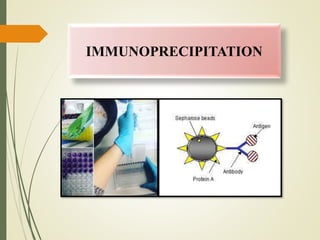
- 5. Antibody may be incubate first with beads (it become binds with IgG binding proteins, protein A, G or A/G) Antigen containing sample added Antigen, antibody and support are binding out Beads are washed extensively Antigen eluted from support using appropriate elution buffer
- 6. PROTEIN COMPLEX IMMUNOPRECIPITTATION It is very similar to IP because it uses an immobilization antibody specific to an antigen of interest. A Co-IP is designed to isolate the antigen along with any protein or ligands that are bound to it. Such instances the known antigen is termed as Bait protein and the proteins interacts with , are called Prey proteins. Number of factors which affect the Co-IP- • Optimization of binding and wash conditions must include consideration of effect on Bait- Prey interaction
- 7. PULL DOWN ASSAY It is similar in concept to a Co-IP, performed in order to investigate protein or ligands that bound to a bait protein. It proves a suspected interaction between two proteins and investigate unknown proteins it is not based on Ab- Ag interaction. The bait protein capture to solid support (beads) by a non- antibody affinity system ( by covalent attachment and affinity tag) Example- immobilized metal affinity chromatography (IMAC) can be used to perform pull down assay with histidine-tagged bait proteins
- 9. INDIRECT METHOD Protein A, G or A/G binding proteins Beaded support Forms Ab- binding platform Antibodies binds with protein Protein A, G or A/G binding proteins Beaded support Antibody Beaded support removed out and desired protein immunoprecipitated
- 10. DIRECT METHOD
- 12. PROCEDURE OF IMMUNO- PRECIPITATION 2. Preparation of immune complex 4. Elution of immune complex 3. Capture of immune complex 1. Material preparation
- 13. MATERIAL PREAPRATION 1. IP Lysis / Wash Buffer 0.025M Tris, 0.15 M NaCl 0.001 M EDTA 1% NP-40 5% Glycerol 2. Saline solution- 0.15 M NaCl 3. SDS PAGE Sample Buffer Lane marker reducing sample buffer dilution Use 100mM Tris pH 6.8 40mM DTT, 2%SDS, 20% Glycerol, 0.2% brmophenol blue 4. ELUTION BUFFER IgG Elution Buffer or 0.1-0.2 M Glycine. HCl at pH 2.5-3.0
- 14. PREPARATION OF IMMUNE COMPLEX Prepare protein sample, use the IP Lysis / wash buffer Use 500ul of IP Lysis / wash buffer per 50mg of wet cell pellet Combine 2-10ug of affinity purified antibody with the cell lysate Total protein/IP REACTION 500-1000ug Dilute the antibody/ lysate solution to 300-600ul with IP Lysis/ wash buffer Incubate for 1 hours to overnight at 4°C Immune complex formed
- 15. CAPTURE OF IMMUNE COMPLEX Gently shake the water of protein A/G Agarose to obtain an even suspension Add 10 ul of resin into spin column Place the column into micro centrifuge (1000 x g) for 1 min Wash the resin twice with 100 ul of cold IP Lysis/wash buffer. Gently tap the bottom of spin column to remove excess liquid and insert the bottom plug into the spin column Add the antibody/lysate sample to protein A/G plus agarose in spin column Remove the bottom plug Centrifuge column and save the flow through
- 16. ELUTION OF THE IMMUNE COMPLEX Sample Buffer Elution • Ideal for Western Blot analysis Place the spin column containing the resin into new collection tube and add 50 ul 2 x SDS PAGE • Incubate at 100 C for 5-10 mins • Centrifuge to collect elute, cool at room temperature before apply an SDS PAGE Gel Low Buffer Elution • Ideal for enzymatic and functional assay • Place the spin column containing the resin into the new collection tube and add 50 ul 2 X SDS PAGE • Incubate for 10 min at room temperature • Centrifuge the tube and collect the flow through
- 17. APPLICATIONS • It is used to estimate the molecular weight, identity or quantity of a protein of interest. • Study protein- protein interaction • Determine the concentration of protein • Analyse the expression level of protein of interest • Studying Cancer development, cell signalling pathway and diseases • Post translational modification • Verify protein expression in a specific tissue.
- 18. THANK YOU…..