
Recommended
Recommended
More Related Content
Similar to CORNYBACTERIUM.pptx
Similar to CORNYBACTERIUM.pptx (20)
Recently uploaded
Recently uploaded (20)
CORNYBACTERIUM.pptx
- 1. BY : Dr GAURAV MATHUR
- 2. Slender Gram-positive rods, pleomorphic; easily decolousised; 0.6-0.8μ diameter and 3-6 μ length; Irregular swelling at one or both ends (‘club shaped’); Non-capsulate, Non-sporing and nonmotile Granules containing polymetaphosphate are seen in the cells; Take up bluish purple color against lightly stained cytoplasm, when stained with Loeffler’s Methylene Blue, and hence called ‘Metachromatic granules’; • Also called, ‘volutin granules’ or ‘Babes Ernst granules’; • They are often situated at poles- ‘polar bodies
- 3. Special stains for demonstrating the granules : – Albert’s stain – Neisser’s stain – Ponder’s stain • The bacilli are arranged in pairs, palisades or small groups; the bacilli lie at various angles to each other, resembling the letters, V or L; • This is called, “Chinese letter pattern” or “cuneiform pattern”;
- 4. • Blood agar • Loeffler’s serum slope • Tellurite blood agar • Tinsdale’s medium (cystine added to tellurite containing agar)
- 5. • Blood agar : small, granular and gray with irregular edges; Hemolysis may or may not present; Loeffler’s serum slope: – Very rapid growth; – Colonies in 6-8 hrs – Initially circular white opaque colonies and acquire yellowish tint on incubation
- 6. Tinsdale’s medium (also contain cystine in addition to tellurite): – Grey black colonies with dark brown haloes indicate C.diphtheriae
- 7. • Ferment- glucose, galactose, maltose and dextrose; but not lactose, sucrose, mannitol; • Proteolytic activity is absent; • Do not hydrolyse urea; • Do not form phosphatase; • Produce cystinase (halo on Tinsdale’s medium)
- 8. The pathognomonic effects are due to the Toxin Toxin is a protein Two fragments, A and B; Fragment B : binds to a cell surface receptor and helps in transport of toxin into the cell; • After entering the cell, A subunit is released ; • A subunit catalyses the transfer of ‘adenosine diphosphate ribose (ADPR)’ • ADPR binds with the elongation factor EF 2 • “ADPR-EF2” complex is inactive protein synthesis stops abruptly necrotising and neurotoxic effects of the toxin;
- 9. • Commonest site of infection: Upper respiratory tract (fauces, larynx,nose) • Ocassionally, other cutaneous or mucocutaneous areas ( otitic/conjunctival/ genitovulval/vaginal/ prepucial/skin) • Faucial diphtheria is the commonest type; • Sore throat is frequently the presenting symptom;
- 10. • The toxin causes local necrotic changes; • The resulting fibrinous exudate, together with the epithelial cells, leucocytes, erythrocytes and bacteria constitute : “pseudomembrane” • Any effort to remove it will tear off capillaries beneath it and cause bleeding; • Mechanical complications are due to pseudomembrane and systemic effects are due to the toxin
- 11. • The toxin is absorbed systemically and damages heart muscle, liver, adrenals etc. Nontoxigenic strains can also produce local disease but systemic effects are absent • Incubation period : usually 3-4 days; • Acute infection : in the form of – – Membranous tonsillitis – Nasal infection – Laryngeal infection – Skin infection –uncommon
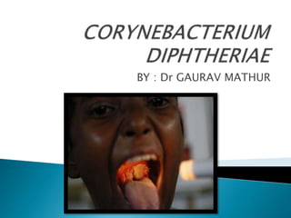
- 12. Characteristic feature is : ‘wash –leather’ eleveted greyish greeen membrane in the tonsils with a well defined Edge surrounded by a zone of inflammation;
- 13. CLASSIFICATION BASED ON CLINICAL SEVERITY • Malignant or hypertoxic: – ‘Bull neck’ due to marked adenitis in neck; – Severe toxemia – Circulatory failure – Death – Paralytic squealae in survivors • Septic : ulceration, cellulitis and gangrene around pseudomembrane; • Hemorrhagic: bleeding from the edge of pseudomembrane, epistaxis, purpura etc.
- 14. • Asphyxia : due to mechanical obstruction Emergency tracheostomy may be necessary; • Acute circulatory failure • Myocarditis • Postdiphtheritic paralysis- palatine(soft palate) and ciliary ( eye muscles) nerves Recovery – spontaneous and complete • Septic : pneumonia and otitis media
- 15. • Specimens : – Swabs from – nose, throat or other suspected lesions; • Smear examination: Gram stain – shows beaded rods in typical arrangement; corynebacteria normally found in throat; – Albert’s stain or Neisser’s stain is useful for demonstrating the granules
- 16. • Inoculate on : – Loeffler’s serum slope – Tellurite blood agar or Tinsdale medium – Blood agar • In vivo methods: – Subcutaneous test – Intracutaneous test • In vitro methods: – Elek’s gel precipitation test – Tissue culture test
- 17. • In vitro test; • A rectangular strip of filter paper is saturated with the diphtheria antitoxin(1000 units/ml); • This strip is placed on : agar plate with 20% horse serum, while the medium is setting; • The cultures to be tested are streaked at right angles to the filter paper strip
- 21. Mainly a disease of childhood(pediatrics) in endemicareas – uncommon below 1st year; peaks at 5. • In nature, C.diphtheriae occurs in the respiratory tract, in the wounds or in the skin of the infected persons or carriers; • Transmission is by- – Droplet dissemination from cases or carriers – Direct contact – Occasionally, fomites
- 22. ACTIVE IMMUNISATION Two preparations of toxoid are available: – Formol toxoid – Adsorbed toxoid • Formal toxoid : prepared by incubating the toxin with formalin for 3-4 weeks; • Adsorbed toxoid : purified toxoid is adsorbed onto an adjuvant, either aluminium phosphate or aluminium hydroxide; this is more immunogenic;
- 23. • Adsorbed toxoid –given as IM injections • Recommended vaccines: – As a trivalent preperation : DPT (adsorbed Diphtheria/Pertusis/Tetanus) – Adsorbed Diphtheria/Tetanus (DT) – Adsorbed low dose diphtheria vaccine for adults (d) – Adsorbed Tetanus/low dose diphtheria vaccine(Td) for adults; – A quadraple vaccine containing DPT+inactivated polio vaccine is also available;
- 24. 3 DOSE of DPT at 6 , 10 and 14 week AND 2 BOOSTER dose at 16-24 month and 5-6 years of age TOTAL 5 DOSE– 0.5ml ROUTE Intramuscular PASSIVE : Antidiptheric serum As an emergency measure when susceptibles are exposed to infection Subcutaneous administration of500-1000 units of antitoxin orADS(Antidiphtheritic serum);
- 25. • Specific measure : prompt administration of antitoxin to neutralize the circulating toxin; • Antibiotics : Penicillin or Erythromycin for 14 days; • Complete bed rest; • Supportive therapy and treatment of complications • Erythromycin: for treatment of carriers
- 26. THANK YOU SEE YOU IN NEXT CLASS