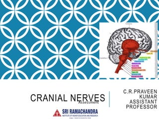
Carinal nerve assessment.ppt
•Download as PPT, PDF•
0 likes•15 views

Cranial nerves I through XII were assessed using various tests. CN I (olfactory) was tested by having the patient smell different odors. CN II (optic) assessment included visual acuity, visual fields, and ophthalmoscopy. CN III, IV, and VI were evaluated using light reflexes, accommodation, and eye movements. Sensory and motor functions of CN V (trigeminal) were assessed using touch, temperature, and jaw movement tests. Facial expression was used to examine CN VII (facial) motor skills. Hearing was roughly tested for CN VIII (acoustic). Swallowing and palate movement checked CN IX and X. Shoulder shrugging assessed CN XI (

Recommended
More Related Content
Similar to Carinal nerve assessment.ppt
Similar to Carinal nerve assessment.ppt (20)
More from praveen Kumar
More from praveen Kumar (11)
Recently uploaded
Recently uploaded (20)
Carinal nerve assessment.ppt
- 2. CN I – OLFACTORY (SENSORY) Not often checked – impairment usually due to other causes (allergies, colds) Impairment will also lead to decreased taste Check each nostril independently with patient’s eyes closed Use non-toxic substances such as Coffee Tobacco Mild soap (Ivory) Cloves
- 3. CN II – OPTIC (SENSORY) Three tests 1. Visual acuity (sharpness or keenness) 2. Visual fields (peripheral vision) 3. Ophthalmoscope (internal eye inspection of optic fundus, where CNII joins the eye)
- 4. CN II – OPTIC: CHECKING VISUAL ACUITY Snellen chart may be used for greater accuracy Simple acuity test Patient covers one eye at a time Hold up fingers and ask how many he/she sees Or simply read a newspaper at arm’s length
- 5. CN II – OPTIC: CHECKING VISUAL FIELDS Method called confrontation Sit 2-3 feet from patient, your left eye aligned with patient’s right Your eye acts as control, so you need good peripheral vision! You close or cover your eye aligned with patient’s eye Holding up your index finger, mid- distance between the patient and yourself, just beyond your own peripheral field, wiggle your finger as you slowly bring it into the visual field. Ask the patient to tell you when he first sees your finger. It should be at about the same time that you see it. Repeat the test to cover the entire visual field for each eye, which is to test at each of the six even-numbered positions on a clock's face.
- 6. CN II – OPTIC: OPHTHALMOSCOPE •Expensive piece of equipment •Use appropriately
- 7. ALL TOGETHER NOW … Cranial Nerves III, IV, and VI III Oculomotor IV Trochlear VI Abducens Observation Examine the upper and lower lids by observation. Look to see that the opening between the eyelids, or the "palpebral fissures", are equal on both sides, and that each lid relates symmetrically to the cornea. Make sure to observe both upper and lower lids. Next, observe the pupils in normal room light to see if they are symmetric.
- 8. CRANIAL NERVES III, IV, AND VI (MOTOR) Four tests 1. Direct Light Reflex 2. Consensual Light Reflex 3. Accommodation 4. Six Cardinal Fields of Gaze
- 9. CRANIAL NERVES III, IV, AND VI Direct Light Reflex Each pupil should constrict briskly when the light strikes the pupil. Move the light in from the temporal side. Document the reaction in mm (e.g., 6mm 4mm).
- 10. CRANIAL NERVES III, IV, AND VI Consensual Light Reflex Perform the procedure again, exactly as before for Direct Light Reflex… only this time watch the opposite pupil. It should react the same as the pupil in which the light is shined.
- 11. CRANIAL NERVES III, IV, AND VI Accommodation - adaptation of the eyes for near vision Ask the patient to focus on a distant light (e.g., a wall or door) at their eye level and maintain that gaze until directed otherwise. Hold an object (pencil, penlight, finger) about 18" from the patient's nose. Ask him to change his focus from the distant object to the closer one. As he does so, observe his eyes as they converge (turn inward) and the pupils constrict.
- 12. CRANIAL NERVES III, IV, AND VI Document normal reactions as PERLA! Pupils Equally Reactive to Light and Accommodation
- 13. CRANIAL NERVES III, IV, AND VI Document abnormal eye movements Some key terms to know 1. Nystagmus: constant, involuntary, cyclical movement 2. Saccadic: jerky, rapid, intermittent movements 3. Tracking: lagging, catching up movement 4. Vergence: turning of one eye without reference to the other, which may indicate weakness of oblique muscles.
- 14. CRANIAL NERVES III, IV, AND VI Six Cardinal (primary) Fields of Gaze: tests for extraocular movement Six fields correspond roughly to 12, 2, 4, 6, 8, 10 on a clock face Hold an object (pen, penlight, finger) about 12" from the patient's nose. Instruct the patient to keep his head still and to follow the object's movement to the six cardinal fields with his eyes only. Slowly move the object through each vision field separately, observing both eyes simultaneously.
- 15. CN V – TRIGEMINAL (SENSORY AND MOTOR) As the name suggests, the trigeminal nerve innervates 3 sections of the face: 1.ophthalmic 2.maxillary 3.mandibular
- 16. CN V – TRIGEMINAL Sensory Assessment Begin by assessing ability to sense light touch to the face. Ask the patient to close his/her eyes and to tell you what he/she feels, and when and where he/she feels it. Using a fine wisp of cotton or your fingertip, gently test the forehead, cheeks, and jaw, randomly and bilaterally. The patient should be able to identify the same sensation bilaterally, and tell when and where he/she feels the touch. If not, repeat the test using the sharp and blunt ends of a sterile pin to check sensitivity to pain. Sensitivity to temperature may also be tested using test tubes with warm and cool water.
- 17. CN V – TRIGEMINAL Corneal Reflex Test Usually not done if light touch is intact Instruct the patient to look up and away from you. Approaching the patient laterally, out of his line of vision, and avoiding the eyelashes, touch the cornea lightly with a fine wisp of cotton. Look for blinking of the eyes, the normal reaction to this stimulus. Be aware that use of contact lenses frequently diminishes, or may even eliminate, the corneal reflex response. The corneal reflex tests the afferent (sensory) arc of CN V, and the efferent (motor) arc of CN VII.
- 18. CN V – TRIGEMINAL Motor Assessment Ask the patient to clench his/her teeth. While he/she is clinching, palpate the temporal muscles. You should note symmetrical strength. Move your hands to the area of the masseter muscles and ask the patient to clench again. Bilateral contraction should be equally strong. To assess chewing ability, ask the patient to clench and unclench his/her jaws several times while you observe for distorted movements or asymmetry.
- 19. CN VII – FACIAL (SENSORY AND MOTOR) Motor assessment Observation during conversation Facial symmetry during spontaneous expression Wrinkling the nose Smiling and frowning Closing eyes Grimacing Intentional expression; ask the patient to Raise and lower eyebrows Squeeze eyes shut tightly Smile showing teeth Puff out cheeks
- 20. CN VII – FACIAL Sensory assessment Usually not done unless problems are noted during motor assessment CNVII responsible for taste on anterior 2/3 of the tongue With patient’s eyes closed, check for recognition of common, easily distinguishable tastes such as chocolate or lemon
- 21. CN VIII – ACOUSTIC (SENSORY AND MOTOR) aka Vestibulocochlear nerve Two branches Vestibular nerve branch controls balance and equilibrium Cochlear nerve branch controls hearing Vestibular branch not usually checked unless several symptoms of abnormality exist Vertigo N/V Nystagmus Postural deviation Pallor Sweating Hypotension
- 22. CN VIII – ACOUSTIC Sensory assessment To make a gross assessment of the cochlear division, begin by instructing the patient to close his eyes and tell you what he hears and in which ear. Gently rub your fingers together about 6" away from first one ear, then the other, then both simultaneously. If deficit is suggested, a more precise assessment may be done with a tuning fork.
- 23. CN IX & X – SENSORY AND MOTOR CN IX Glossopharyngeal CN X Vagus Cranial nerves nine and ten are tested together, because they are closely associated and similar in function. The motor aspect of the glossopharyngeal nerve innervates the muscle used to swallow. Its sensory component supplies sensation to the pharynx and is responsible for taste perception on the posterior 1/3 of the tongue, and for salivation.
- 24. CN X – VAGUS! The vagus nerve controls: swallowing phonation (the process of uttering vocal sounds) movement of the uvula and soft palate. CN X also innervates the thoracic and abdominal visceral organs! Carries sensory impulses from the GI tract, the heart, and the lungs
- 25. CN IX & X – ASSESSMENT Begin assessment of these 2 nerves by inspecting the soft palate. When the patient says "ah", the palate should rise promptly and symmetrically. The uvula should NOT be used to assess symmetry, because there are many normally odd-shaped structures.
- 26. CN IX & X – ASSESSMENT The gag reflex assesses the sensory component of CN IX, and the motor response of CN X. Touch the posterior pharynx lightly with a cotton- tipped applicator. Test the palatal reflex, stroke the posterior portion of the palate on each side with the applicator. In both instances, the palate should elevate and a gag response should be induced. However, remember that normal patients frequently manifest bilateral loss of the gag reflex, especially patients with a history of smoking or tobacco use.
- 27. CN XI – SPINAL ACCESSORY (MOTOR) The spinal accessory nerve supplies the sternocleidomastoid muscles and the upper portion of the trapezius muscles.
- 28. CN XI – SPINAL ACCESSORY To assess sternocleidomastoid strength, apply resistance to the jaw and have the patient try to turn his head to the side against your pressure. To evaluate the trapezius, watch the patient shrug his shoulders, which should move at the same speed and with roughly the same extent of movement. Next, ask the patient to shrug his shoulders upward while you try to hold them down.
- 29. CN XII – HYPOGLOSSAL (MOTOR) Responsible for normal tongue movements. First observe the tongue at rest on the floor of the mouth. Look for: asymmetry deviation to one side loss of bulk on one side fasciculations (involuntary contractions, twitching) Next, ask the patient to stick out his tongue. It should protrude along the midline [the "median raphe" (midline crease) lines up with the notch between the "medial incisors" (two front teeth)].
- 30. CN XII – HYPOGLOSSAL Finally, have the patient push his/her tongue as hard as he/she can against the inside of the cheek, while you push using your thumb against the outside of the cheek. Compare right and left sides. Remember that weakness pushing the tongue into the right cheek indicates an abnormality in the left hypoglossal nerve, and vice versa.