2 Carbohydrate metabolism_mai_2564.pptx
•Download as PPTX, PDF•
0 likes•202 views

introduction of carbohydrate metabolism

Recommended
More Related Content
What's hot
What's hot (20)
Similar to 2 Carbohydrate metabolism_mai_2564.pptx
Similar to 2 Carbohydrate metabolism_mai_2564.pptx (6)
2 Carbohydrate metabolism_mai_2564.pptx
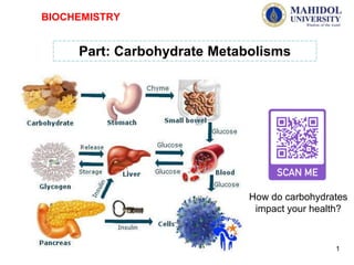
- 1. Part: Carbohydrate Metabolisms BIOCHEMISTRY 1 How do carbohydrates impact your health?
- 2. Why do we first learn carbo metabolism? 2
- 3. 3
- 4. 1. Glucose metabolism 1.1 Glycolysis (glucose degradation), TCA cycle and oxidative phosphorylation 1.2 Gluconeogenesis (glucose synthesis) 2. Glycogen metabolism (Glycogenolysis/Glycogenesis) 3. Pentose phosphate pathway (Glucose oxidation) Carbohydrate Metabolisms: 4
- 5. Cell break down food molecules gradually, capturing a little bit of chemical energy at key steps. This enables cells to use the energy stored in the chemical bonds of foods like glucose to produce compounds such as ATP that directly power the activities of the cell. 6O2 + C6H12O6 6CO2 + 6H2O + Energy Cellular respiration is the process that releases energy from food in the presence of oxygen. What kind of EN 5
- 6. Glucose เป็นศูนย์กลางของเมแทบอลิซึมในร่างกาย เนื่องจากเป็นแหล่งพลังงานที่ร่างการนาใช้ง่ายที่สุดและทุกเซลล์ใน ร่างกายใช้ Glc เป็นแหล่งพลังงานได้ และเป็นสารตั้งต้นการสังเคราะห์สารเคมีหลายชนิดในเซลล์ อีกทั้งยังเป็นแหล่ง พลังงานเดียวที่ของสมองและสาหรับเซลล์ที่ไม่มี mitochondria (RBC) ดังนั้น ร่างกายจึงจาเป็นต้องรักษาระดับ น้าตาลในเลือด --แหล่งของ Glucose ในเลือด มาจาก 3 แหล่ง: 1) Diet 2) Degradation of Glycogen 3) Gluconeogenesis Part: Carbohydrate Metabolisms 6
- 7. Different organs and tissues handle fuels differently At rest, the brain uses approximately 20% of all oxygen consumed by the body. Glucose is normally its only fuel: during starvation, however, the brain adapts to the use of ketone bodies as an alternative energy source. The two pathways that provide glucose are glycogenolysis and gluconeogenesis. When glucose concentration in the extracellular fluid decreases, it is first replenished by degrading liver glycogen. Part: Carbohydrate Metabolisms 7 However, when the fasting period extends, gluconeogenesis is initiated. Gluconeogenesis takes place mostly in the liver, and the kidneys also contribute during prolonged fast. Its main substrates are lactate (from anaerobic glycolysis), alanine (from the amino acids released during breakdown of muscle protein) and glycerol (from the breakdown of triacylglycerols in the adipose tissue).
- 8. Muscle uses both glucose and fatty acids as energy sources. During short- term exercise, glucose is the preferred substrate, however, at rest and during prolonged exercise, fatty acids are the main energy source. Part: Carbohydrate Metabolisms 8 Glucose concentration in plasma reflects the balance between, on the one hand, its intake (absorption from the gut) or its endogenous production (glycogenolysis and gluconeogenesis), and on the other, its tissue utilization in glycolysis, pentose phosphate pathway, tricarboxylic acid (TCA) cycle and in glycogen synthesis
- 9. Transport of glucose into cells -Glucose ไม่สามารถเข้าสู่เซลล์ได้โดยตรง จึงต้องการ glucose transporter เรียกว่า GLUT มีหลายชนิด (GLUT1-14) -GLUT is tissue specific. For example GLUT2 in liver GLUT3 in neuron GLUT1 in erythrocyte and blood brain barrie r จะเกิดอะไรขึ้นถ้า GLUT4 ดื้อหรือไม่ตอบสนองต่อ INSULIN!!!!!!? GLUT 1 - 3 are insulin-independent transporters GLUT4 in adipose tissue and skeleton muscle (GLUT4 activity is dependent on INSULIN) 9
- 10. • Insulin and the Regulation of Glucose in the Blood Transport of glucose into cells The Role of Insulin in the Human Body
- 11. 11
- 12. - Glycolysis (Glykys “sweet or sugar” and Lysis “splitting”) ─ degradation of one glucose to yield pyruvate Glucose (6-carbon compound) Glycolysis (Exergonic process) Pyruvate (2x) (3-carbon compound) Glucose metabolism: 1. Glycolysis Occur in Cytosol 12
- 13. Glycolysis is composed of 3 stages I. Use energy: เอนไซม์Hexokinase เปลี่ยน Glucose in the Cell เพื่อผลิต Fructose 1,6-bisphosphate II. Cleavage: C6 Sugar ถูกตัดเป็น 2x(C3) Fragments III. Generate energy: เกิด Oxidation ของ Aldehyde ไปเป็น Acid และ ผลิต High energy compounds. Glucose metabolism: 2. Glycolysis 13
- 14. Glucose metabolism: Glycolysis Stage I Stage II Stage I = การลงทุนด้วยพลังงาน Control point of Glycolysis Arsenic 14
- 15. Stage III = ได้พลังงาน (Pay off ATP) Glucose metabolism: Glycolysis Stage I Stage II Stage III Substrate-level phosphorylation Substrate-level phosphorylation Control point of Glycolysis 15
- 16. Glucose metabolism: Glycolysis Hexokinase Vs Glucokinase Hexokinase/Glucokinase เร่งปฏิกิริยาเดียวกัน แต่พบใน tissue ต่างกัน และถูกควบคุมการทางานต่างกัน Hexokinase Glucokinase -พบใน tissue ทั่วไป สามารถเร่งปฏิกิริยากับ Hexose ทุกชนิด -ถูกยับยั้งโดย G-6-P (Product) -Low Km = ทางานมีประสิทธิภาพสูงแม้ภาวะที่มี Glc ต่า -พบใน เซลล์ตับ เป็น Glc sensor มีความสาคัญต่อการหลั่ง Insulin ควบคุม Glc homeostasis -จาเพาะ only Glc -High Km = จะทางานก็ต่อเมื่อภาวะที่มี Glc ในเซลล์ตับสูง เช่น หลังการกินอาหาร Carbo สูง -ถูก activate โดย Glc แต่ถูกยับยั้งโดย F-6-P -Mutation of Glucokinase ทาให้เกิดโรคเบาหวาน (Diabete). 16
- 17. ATP and NADH Formation (exergonic process) (endergonic process) Overall DG’o drives the glycolysis exergonically Glucose metabolism: Glycolysis 17
- 18. Glucose metabolism: Glycolysis Feeder Pathways for Glycolysis -Sugar แต่ละตัวเข้าสู่ glycolysis ที่ตาแหน่ง แตกต่างกัน -Glycogen เข้าสู่ glycolysis ในรูป Glc-1-phosphate 18
- 19. Glucose metabolism: Glycolysis ภาวะ Aerobic: Pyruvate จะเข้าสู่ TCA Cycle ส่วน NADH ก็จะถูกนาไปสร้าง ATP ใน Oxidative phosphorylation ภาวะ Anaerobic: Pyruvate จะถูกเปลี่ยนไปเป็น Lactate โดยใช้ NADH เพื่อ recycle NAD+ กลับไป 19
- 20. Metabolic Fates of Pyruvate Glucose metabolism: Glycolysis 1 2 3 4 Oxaloacetate 20
- 21. Metabolic Fates of Pyruvate 1. Reduction of pyruvate to lactate (Anaerobic condition ในเซลล์ eukaryote ) -Lactate คือผลิตภัณฑ์สุดท้ายในวิถี glycolysis ในภาวะ anaerobic ในเซลล์ eukaryote -แต่การผลิต lactate จาก pyruvate คือ วิถีหลักใน lens and cornea of the eye, kidney medulla, leukocytes and red blood cells เพราะว่า tissue เหล่านี้poorly vascularized and/or lack mitochondria. Glucose metabolism: Glycolysis - NADH/NAD+ ratio (NADH production มาจากปฏิกิริยา glyceraldehyde 3-phosphate dehydrogenase และจาก TCA cycle), ทาให้การผลิต lactate จาก pyruvateเกิดได้ดีขึ้น -ในระหว่างการออกกาลังกายอย่างหนัก (intense exercise) จะเกิดการสะสม lactate ในกล้ามเนื้อ, causing a drop in the intracellular pH, ทาให้กล้ามเนื้อล้า - จากนั้น lactate ที่มีมากก็จะ diffuses into the bloodstream, และจะถูกใช้โดย 1. liver เพื่อผลิต glucose โดยวิถี gluconeogenesis หรือ 2. ที่หัวใจก็จะใช้ lactate เป็นแหล่งพลังงาน โดยผ่าน TCA cycle. Lactate formation in muscle: In exercising skeletal muscle 21
- 22. Metabolic Fates of Pyruvate Glucose metabolism: Glycolysis 2. Reduction of pyruvate to Ethanol (Anaerobic Microorganism) -The conversion of pyruvate to ethanol occurs by the two reactions. -The decarboxylation of pyruvate by pyruvate decarboxylase occurs in yeast and certain other microorganisms, but not in humans. 3. Oxidative decarboxylation of pyruvate to Acetyl CoA -is an important pathway in tissues with a high oxidative capacity, เช่น กล้ามเนื้อหัวใจ - Pyruvate dehydrogenase irreversibly converts pyruvate, into acetyl CoA, a major fuel for the TCA cycle. 22
- 23. Metabolic Fates of Pyruvate Glucose metabolism: Glycolysis 4. Carboxylation of pyruvate to oxaloacetate -This reaction is important because เป็น pathway ที่ช่วยสร้าง the citric acid cycle intermediates, and ช่วยสร้างตัวกลางสาหรับวิถี gluconeogenesis. 23
- 24. 24
- 25. 25
- 26. Glucose metabolism: 1.2 การสร้าง Acetyl CoA Pyruvate dehydrogenase (PDH) Glucose Pyruvate Glycolysis Pyruvate Cytosol Mitochrondia -Pyruvate จะต้องถูก transport เข้าสู่ mitochondrion ก่อนเข้าสู่ the TCA cycle -ซึ่งจะมีตัว transporter จาเพาะสาหรับขนส่ง pyruvate ผ่าน inner mitochondrial membrane - เมื่อเข้าสู่ matrix ของ mitochondria, pyruvate จะถูกเปลี่ยนเป็น acetyl CoA โดยเอนไซม์ pyruvate dehydrogenase ซึ่งเป็น multienzyme complex 26
- 27. -- Thiamin deficiency = PDH complex is inactive = brain cells ไม่สามารถผลิต ATP (via the TCA cycle) ได้อย่างพอเพียง - เรียกว่าโรค กลุ่มอาการเวอร์นิคเค-คอร์ซาคอฟ (Wernicke- Korsakoff Syndrome) เกิดจาก thiamine deficiency อาจจะพบในคนที่ติดเหล้า Glucose metabolism: 1.2 การสร้าง Acetyl CoA การขาด Thiamin ส่งผลต่อระบบประสาทส่วนกลางอย่างไร? PDH complex Pyruvate Acetyl CoA - Thiamin is a PDH co-enzyme arsenic PDH complex Pyruvate Acetyl CoA Lactic acid Brain Acidosis 27
- 28. Glucose metabolism: 1.2 การสร้าง Acetyl CoA - When it binds to lipoic acid, the PDH complex is inactive, pyruvate (and consequently lactate) accumulates. affects the brain, causing neurologic disturbances and death. PDH complex Pyruvate Acetyl CoA arsenic PDH complex Pyruvate Acetyl CoA Lactic acid Brain Acidosis ทาไมสารหนู ทาให้คนตายได้ และพิษของสารหนูเกิดขึ้นได้อย่างไร? Mechanism of arsenic poisoning: - Arsenic can interfere with glycolysis at the glyceraldehyde 3-phosphate step, thereby decreasing ATP production. -Arsenic poisoning can inhibition of PDH enzyme complex by binding to lipoic acid which is a one of coenzyme of PHD. 28
- 29. -The TCA cycle (Krebs cycle or the citric acid cycle) มีบทบาทสาคัญหลายอย่างใน metabolism -เกิดใน mitochondria 1.3 Tricarboxylic acid cycle (TCA cycle) 1. เป็นวิถีสุดท้ายในการเกิดออกซิเดชันของ carbohydrates, amino acids, และ fatty acids, โดย การเปลี่ยน carbon skeletons ของสารเหล่านี้เป็น CO2 29
- 30. 2. The TCA cycle เป็นแหล่ง intermediates สาหรับใช้ใน การสังเคราะห์สาร เช่น สาร intermediates Oxaloacetate ที่มาจาก amino acids สามารถนาไปสังเคราะห์ glucose โดยวิถี Gluconeogenesis 1.3 Tricarboxylic acid cycle (TCA cycle) 30
- 31. TCA cycle and Energy Produced 3. ในกระบวนการเกิดออกซิเดชันของ สารต่างๆ ผ่านวิถี TCA cycle จะ ทาให้ได้ NADH, FADH ซึ่ง สามารถนาไปผลิตสารพลังงานสูง หรือ ATP ในกระบวนการ oxidative phosphorylation 31
- 32. 1.4 Oxidative Phosphorylation -NADH and FADH2 เกิดการ oxidation โดยการส่ง e- ผ่าน “e- transport chain” เพื่อผลิตเป็นสารพลังงานสูง ATP (เปลี่ยน ADP to ATP) -เกิดที่ “inner membrane of mitochondria” -e-transport chain ประกอบด้วย: (1) โปรตีนตัวพา e- ที่มีโคเอนไซม์ เช่น NAD+, FMN, Co enzyme Q, Cytochrom b c a a3 (Fe2+/Fe3+) (2) เอนไซม์ ATP synthase https://www.youtube.com/watch?v=39HTpUG1MwQ How Mitochondria Create Energy 32
- 33. 1.4 Oxidative Phosphorylation NADH NAD+ TCA cycle NADH เข้าที่ complex I (3 ATP/NADH) FADH2 เข้าที่ complex II (2 ATP/NADH) มาจาก Glycolysis and TCA cycle 33
- 34. 1.4 Oxidative Phosphorylation การส่ง e- ผ่าน “e- transport chain” ทาให้เกิดการสร้าง ATP ได้อย่างไร????? -การส่ง e- ผ่าน “e- transport chain” ทาให้เกิดการ “ปั๊มโปรตรอน (H+)” จาก matrix ผ่าน inner membrane ของ mitochondria เข้าสู่ intermembrane space ทาให้เกิด “proton gradient” ซึ่งนาไปสู่การกระตุ้นการทางานของ ATP synthase ให้สังเคราะห์ ATP -Complex II ไม่มีการปั๊มโปรตรอน (H+) 34
- 35. Oxidative Phosphorylation Cyanide ทาให้สิ่งมีชีวิตตายได้อย่างไร กินมันสาปะหลังดิบ ทาให้ตายได้ เพราะมี Cyanide !!!! https://www.youtube.com/watch?v=fBXSJGxfnbU Electron flow in cytochrome c oxidase can be blocked by hydrogen sulphide(H2S), cyanide(CN-),and carbon monoxide(CO). Inhibition of the electron transport chain also inhibits ATP synthesis because the proton-motive force can no longer be generated. 35
- 37. - (Gluco “glucose”, Neo “new”, Genesis “origination”) - new formation of sugar by converting pyruvate (and other 3- or 4-cabon compounds) to glucose Glucose (6-carbon compound) Gluconeogenesis (Endergonic process, but essential) Pyruvate (3-carbon compounds) Blood glucose Glucose metabolism: 2. Gluconeogenesis • บางเนื้อเยื่อ เช่น สมอง เม็ดเลือดแดง เนื้อเยื่อไตชั้นใน (kidney medulla), lens and cornea of eye, testes and exercising muscle ต้องการแหล่งพลังงานในรูปของ Glc • ในกรณีที่ไม่ได้รับอาหารประเภท carbohydrate; glycogen จะสามารถเป็นแหล่ง support Glc ได้เพียง 10-18 hr เท่านั้น แล้วถ้าหาก prolonged fast แหล่ง Glc จะได้มาจากไหน?? Gluconeogenesis 37
- 38. Gluconeogenesis: Substrate for gluconeogenesis 38
- 39. 2. Gluconeogenesis --Gluconeogenesis ไม่ได้เกิดจากปฏิกิริยา reverse ของวิถี Glycolysis --แต่ต้องการบางปฏิกิริยาเฉพาะ เรียกว่า “Bypass” reactions (1, 2, 3 and 4 reactions) --Substrate for Gluconeogenesis -Lactate -Glycerol -Intermediates of glycolysis and TCA cycle -α-keto acids obtained from the transamination of glucogenic amino acids 39
- 40. 1st and 2nd bypass 3rd bypass 4th bypass Gluconeogenesis: Bypass reactions ***Note that: Kinase/ phosphorylase = add phosphate group Phosphatase = remove phosphate group 40
- 41. Gluconeogenesis: Energy requirement Overall Equation for Gluconeogenesis Release of free glucose from liver and kidney into blood xPyruvate + yATP +zGTP +xNADH +xH+ +yH2O aGlucose +bADP +cGDP +dPi +eNAD+ 2Pyruvate + 4ATP +2GTP +2NADH +2H+ +4H2O Glucose +4ADP +2GDP +6Pi +2NAD+ 41
- 42. 1st and 2nd Bypasses: conversion of pyruvate → PEP by PC and PEPCK Pyruvate OAA PEP PC PEPCK Decarboxylation & Phosphorylation Carboxylation Gluconeogenesis 42
- 43. Alternative conversion of Pyruvate → PEP between two compartments Anaerobic condition Gluconeogenesis Pyruvate OAA PEP PC PEPCK 2. 2. 1. 1. Pyruvate OAA PC Malate PEP cyMD miMD 43
- 44. Fructose 1,6-bisphosphate + H2O FBPase-1 Fructose 6-phosphate FBPase-1 = Fructose 1,6-bisphosphatase Hydrolysis Gluconeogenesis 3rd Bypass: conversion of F1,6 BiP → F6P by FBPase-1 4th Bypass: conversion of G6P → G by G6Pase Glucose 6-phosphate + H2O G6Pase Glucose G6Pase = Glucose 6-phosphatase Hydrolysis 44
- 45. Gluconeogenesis: Substrate for gluconeogenesis I. Lactate from anaerobic Glycolysis Coli cycle --Lactate is released into the blood by exercising skeletal muscle, and by cells that lack mitochondria, such as red blood cells. --In the Cori cycle, bloodborne glucose is converted by exercising muscle to lactate, which diffuses into the blood. --This lactate is taken up by the liver and reconverted to glucose, which is released back into the circulation. 45
- 46. Amino acids can be used as a fuel after conversion to glucose • Amino acids normally serve as substrates for synthesis of body’s proteins. However, in certain situations they become energy substrates. • During a prolonged fast or periods of metabolic stress induced by illness or injury, body proteins are degraded, and the released amino acids are converted into glucose in the course of gluconeogenesis. • When an excessive amount of amino acids is taken as food, they are converted to carbohydrates and are either stored or metabolized II. Intermediates of glycolysis and TCA cycle = Amino acid (α-keto acids obtained from the transamination of glucogenic amino acids) Gluconeogenesis: Substrate for gluconeogenesis 46
- 47. II. Intermediates of glycolysis and TCA cycle = Amino acid (α-keto acids obtained from the transamination of glucogenic amino acids) --Amino acids ที่ได้จากการ hydrolysis ของ tissue proteins เป็ นแหล่งของ glucose during a fast. --α-Keto acids (เช่น α-keto- Glutarate) ได้จากmetabolism ของกรดอะมิโนชนิด glucogenic aa. --โดย α-ketoacids จะเข้าสู่ TCA cycle และเปลี่ยนเป็ น OAA ซึ่งเป็ น สารตั้งต้นสาคัญในวิถี Gluconeogenesis PEP OAA Gluconeogenesis: Substrate for gluconeogenesis 47
- 48. III. Glycerol -การ hydrolysis triglyceride ในเนื้อเยื่อไขมันใน mammals จะได้ glycerol and fatty acid -Glycerol จะถูกส่งไปยัง ตับผ่านทางเส้นเลือด - เฉพาะ glycerol เท่านั้นที่สามารถเข้าสู่วิถี Gluconeogenesis เพื่อผลิต Glc ส่วน fatty acids จะใช้ไปผลิตพลังงานโดยเข้าสู่TCA หรือ เปลี่ยนเป็น Ketone body -In Liver: Glycerol Glycerol phosphate glycerol kinase* Dihydroxy acetone phosphate glycerol phosphate dehydrogenase Gluconeogenesis * ไม่มีในเนื้อเยื่อไขมัน Gluconeogenesis: Substrate for gluconeogenesis 48
- 49. ภาวะ hypoglycemia และเกิดภาวะ fatty liver และตับแข็ง ในพวก alcoholism 49
- 50. -ในผู้ที่ดื่มสุรา เอธานอลจะถูก เปลี่ยนเป็น acetaldehyde and acetate ซึ่งทาให้เกิดการ สังเคราะห์ NADH -ทาให้ระดับ NADH ในตับสูงขึ้น - NADH ในตับสูงขึ้น การสังเคราะห์กลูโคสในวิถี gluconeogenesis ลดลง เพราะ pyruvate ซึ่ง เป็นตัวกลางในวิถี gluconeogenesis ถูกเปลี่ยนไปเป็น lactate and malate แทนกลูโคส (ลูกศรสีฟ้า) ทาให้เกิดภาวะ “hypoglycemia” ส่วน lactate and malate ที่เพิ่มขึ้นทาให้เกิด ภาวะ “lactic acidosis” -ภาวะที่ระดับ NADH ทาให้ การสลายกรดไขมันลดลง (beta oxidation) เกิดการสังคราะห์ และ สะสม triglyceride เพิ่มขึ้น เกิดเป็น “fatty liver” ทาไมผู้ที่ติดสุราจึงเกิดภาวะ hypoglycemia และเกิดภาวะ fatty liver และตับแข็ง 50
- 52. 52 น้าตาล (HFCS) ภัยร้ายใกล้ตัว by หมอแอมป์ What happens to your body and brain when you eat too much sugar What Does Sugar Actually Do To Your Body? 16.46 min
- 53. 53 WHY Sugar is as Bad as Alcohol (Fructose, The Liver Toxin) Fructose overconsumption may contribute to diverse cardiometabolic risk factors including steatosis, increased glucose production, hypertriglyceridemi a, increased adiposity, and hypertension. https://www.researchgate.net/figure/Consequences-of-fructose-overconsumption-Fructose-
- 54. Glycogen (Glycogenolysis/Glycogenesis) The importances of Glycogen: เป็ นแหล่งพลังงาน ซึ่งสามารถนามาใช้ โดยสามารถเปลี่ยนเป็ น Glucose ได้ง่าย แต่กรณีของ gluconeogenesis ต้องผ่านหลาย reactions --เมื่อร่างกายได้รับสารอาหารจาพวก Carb: Glycogen จะถูกสร้างจาก Glucose สะสมไว้ --เมื่อร่างกายไม่ได้รับสารอาหารจาพวก Carb: Glycogen จะถูกสลายเป็ น Glucose ปล่อยสู่กระแสเลือดได้อย่างรวดเร็ว 54
- 55. Glycogen Metabolism แหล่งสะสมหลักของ Glycogen 55 -Liver glycogen = maintain the blood glucose -Muscle glycogen = serve as a fuel for the ATP during muscle contraction
- 56. Glycogen Metabolism Glycogen Metabolisms: 1. Glycogenesis (glycogen synthesis) 2. Glycogenolysis (glycogen degradation) Glycogenolysis Glycogenesis 56
- 57. - Glycogen is a polymer of glucose. Glycogen Metabolism: Glycogenesis (Glycogen + Genesis) 1. Glycogenesis : - ต้องการพลังงานในการสร้าง คือ ATP (for the phosphorylation of glucose) และ uridine triphosphate (UTP). - The process occurs in the cytosol. 57
- 58. องค์ประกอบของการสังเคราะห์Glycogen: 1. UDP-glucose pyrophosphorylase ─ สาหรับเป็ น monomer 2. Glycogenin ─ initiating glucose linkages 3. Glycogen synthase ─ ทาหน้าที่เชื่อม monomer ต่อเป็ นสาย glycogen chain Glycogen Metabolism: Glycogenesis 1. สังเคราะห์UDP-glucose pyrophosphorylase สาหรับเป็ น monomer 58
- 60. Branch synthesis - glycogen-branching enzyme or transglycosylase Glycogen Metabolism: Glycogenesis 60
- 61. 1. Glycogen phosphorylase ─ add phosphate group 2. Debranching enzymes ─ linearize the glycogen chain by 2.1 Transferase activity ─ transfer glucose residues 2.2 Glucosidase activity ─ remove a branching glucose 3. Phosphoglucomutase ─ shift a phosphate group to another position 2. Glycogenolysis : Degradation of Glycogen Glycogen Metabolism: Glycogenolysis (Glycogen + Lysis) 61
- 62. Glycogen Metabolism: Glycogenolysis Glucose 6-phosphate Phosphogluco mutase Glucose glucose 6-phosphatase (G6Pase) Liver Blood Glucose Muscle Glycolysis Different fates of Glc in Liver and Muscle Muscle & adipose tissue lack G6Pase, no contribution for free glucose +Pi Red muscle: CO2 and H2O (pyruvate dehydrogenase) White muscle: Lactate (lactate dehydrogenase) 62
- 63. Glycogen Metabolism: Regulation of glycogen synthesis and degradation การควบคุมการสังเคราะห์และการสลายไกลโคเจนโดยฮอร ์โมน ฮอร์โมนกลูคากอน เอพิเนฟริน (ตอบสนองความกลัว ต่อสู้ หลบหนี) และอินซูลิน 63
- 64. 3. Pentose Phosphate Pathway (PPP) (glucose oxidation) PPP มีความสาคัญอย่างไร??? --เป็ น pathway หลักที่ผลิต NADPH สารับใช้ในกิจกรรมต่างๆ ของร่างกาย ที่ต้องใช้ NADPH เช่น liver ต่อมน้านมที่ต้องผลิตน้านม เนื้อเยื่อไขมันที่ต้อง ผลิต fatty acid, testes and ovary ที่ต้องผลิต steroid hormones, เม็ด เลือดแดง --เป็ น pathway หลักที่ผลิต ribose 5-phosphate สารับการสังเคราะห์ nucleotides --หรือเรียกว่า “hexose monophosphate pathway” --เกิดใน cytosol 64
- 65. NADPH : An energy shuttle for biosynthesis • Energy powers: the assembly of building into complex molecules of carbohydrate, fat, and protein. • NADPH, an energy-carrying molecule similar to NADH, delivers much of the energy these biosynthetic reactions require. • Although both molecules are energy carriers, their metabolic roles are vastly different. Whereas the energy carries by NADH primarily produces ATP, nearly all the energy carried by NADPH drives biosynthesis. • When a reaction transforms NADPH into NADP+, NADPH release its cargo of two energetic electrons.
- 66. • Key concept ATP is the energy currency of the body. Your body extracts energy from food to produce ATP. • NADH and FADH2 Are hydrogen and electron carriers that shuttle energy to ATP production sites. NAPH is also and electron carrier, but it shuttles energy for anabolic processes.
- 67. Pentose Phosphate Pathway (PPP) --ประกอบด้วย 1) irreversible oxidative reactions 2) series of reversible sugar-phosphate interconversions 67
- 68. Pentose Phosphate Pathway (PPP) G-6-P CO2 + 2 NADPH + Ribulose-5 P Glucose F-6-P G-6-P 6-P Gluconolactone Glucose 6-phosphate dehydrogenase (G6PD) NADP+ NADPH Glycolysis NADP+ NADPH + CO2 Ribulose-5-P 6-phosphoglucono- lactone hydrolase 1) irreversible oxidative reactions 2) series of reversible sugar- phosphate interconversions -การเปลี่ยนแปลง G-6-P ไปเป็ น intermediate ต่างๆ ในวิถี PPP ทาให้เราได้ 2NADPH โดยที่ได้ G-6-P กลับคืนมาเหมือนเดิม -G6PD เป็ น key enzyme ในการควบคุมวิถี PPP: NADPH/NADP+ สูง = G6PD Less Active NADPH/NADP+ ต่า = G6PD More Active 68
- 69. Defect in G6PD - A way to kill a malarial parasite NADPH จาก PPP มีความสาคัญอย่างไร??? I. Reduction of hydrogenperoxide -- ทาให้เกิด chemical damage to DNA, proteins, and unsaturated lipids, and can lead to cell death. -- ROS เกี่ยวข้องกับการเกิดโรค หลายชนิด เช่น reperfusion injury, cancer, inflammatory disease, and aging ROS ROS Reactive oxygen species (ROS): GSSG = oxidative glutathione GSH = reductive glutathione ซึ่งสามารถทาปฎิกริยากับ H2O2 69
- 70. NADPH จาก PPP มีความสาคัญอย่างไร??? II. กาจัดสารพิษโดยการทางานของเอนไซม์Cytochrome P450 monooxygenase ในตับ The overall reaction catalyzed by a cytochrome P450 enzyme: R-H + O2 + NADPH + H+ → R-OH + H2O + NADP+ R = steroid, drug or other chemical --การทางานของเอนไซม์Cytochrome P450 monooxygenase ต้องการ NADPH เป็ น Co-factor 70
- 71. III. กระบวนการทาลายเชื้อโรคของ white blood cells หลังจาก Phagocytosis NADPH จาก PPP มีความสาคัญอย่างไร??? 71
- 72. Regulation of Glycolysis The storage of nutriens • glucose transport into muscle and adipose tissue • glucose storage as glycogen (liver, muscle) • conversion of glucose to TG (liver) and their storage (adipose tissue) • protein synthesis (liver, muscle) • inhibition of fuel mobilization 72
- 73. Hormonal control of glucose homeostasis. (A) Plasma glucose concentration reflects the balance between the hypoglycemic (glucose-lowering) action of insulin and the hyperglycemic (glucose increasing) action of the anti- insulin hormones. (B) Daily patterns of insulin and glucagon secretion, and corresponding plasma glucose concentrations. Plasma glucose concentration is maintained within a narrow range throughout the day. To obtain glucose concentrations in mg/dL, multiply the value in mmol/L by 18. 73
- 74. Glucose homeostasis: maintenance of blood glucose levels near 80 to 100 mg/dL (4,4-5,6 mmol/l) insulin and glucagon (regulate fuel mobilization and storage) Hypoglycemia prevention: 1. release of glucose from the large glycogen stores in the liver (glycogenolysis) 2. synthesis of glucose from lactate, glycerol, and amino acids in liver (gluconeogenesis) 3. release of fatty acids from adipose tissue (lipolysis) Hyperglycemia prevention: 1. conversion of glucose to glycogen (glycogen synthesis) 2. conversion of glucose to triacylglycerols in liver and adipose tissue (lipogenesis) 74
- 75. A. Key regulatory enzymes: are those enzymes that catalyze the irreversible steps of glycolysis that include three steps as follows, 1-Phosphofructokinase: It is an allosteric enzyme stimulated by high levels of fructose-6- phosphate, fructose- 2,6-diphosphate (in liver), ADP and AMP, Pi, and ammonia. It is inhibited allosterically by ATP, low pH and citrate. 2-Hexokinase: Accumulation of glucose-6-phosphate and inhibition of phosphofructokinase results in accumulation of fructose-6-phosphate and glucose-6-phosphate that allosterically inhibit hexokinase. 3-Pyruvate kinase: It is inhibited also by excess ATP, fatty acids, and acetyl-CoA and is stimulated by fructose-1,6-diphosphate, ADP and AMP It is regulated by cAMP-dependent phosphorylation-dephosphorylation mechanism 75
- 76. B. Hormonal regulation: 1. Insulin: Stimulates synthesis of glucokinase, phosphofructokinase and pyruvate kinase, so it stimulates glycolysis. It also induces glucose transporters to provide cells with glucose for glycolysis. 2-Adrenaline and glucagon are inhibitory by inhibiting pyruvate kinase. Pathways regulated by the release of: glucagon (in response to a lowering of blood glucose levels) insulin (in response to an elevation of blood glucose levels) 76
- 77. Production of blood glucose Glycogenolysis 2 hours after a meal the primary source of blood glucose during the first few hours of fasting Gluconeogenesis after consumption of the liver glycogen lactate (muscle, erythrocytes), amino acids (muscle), glycerol (adipose tissue) 77
- 78. Sources of blood glucose in fed, fasting, and starved states: Stage of fasting Glucose (mg/dL) Normal level 80-100 Fasting (12 h) 80 Starvation (3 d) 70 Starvation (5-6 wk) 65 78
- 79. 79
- 80. Errors in Digestion and Metabolism Genetic defects – Galactosemia • Enzyme responsible for metabolizing galactose to glucose is missing • All sources of lactose must be eliminated from diet • Untreated, can cause brain and liver damage • Screening and treatment can enable normal life 80
- 81. Errors in Digestion and Metabolism Genetic defects - Glycogen storage diseases (GSD) • Group of rare genetic defects • Absence of enzymes required for synthesis or breakdown of glycogen • Form of disease depends on enzyme missing 81
- 82. 82
- 83. References: • John W Baynes and Marek H Dominiczak, Fourth edition 2014, Medical biochemsirtry, Chapter 21 : Glucose Homeostasis and Fuel Metabolism: Diabetes Mellitus. • Miller, Kenneth R., and Joseph S. Levine.,( 2010), Chapter 9 : Cellular respiration and fermentation, Miller & Levine Biology, 9th Edition, Boston, 1034 pages. • Richard A. Harvey and Denise R. Ferrier, (2011) Lippincott’s, Illustrated Reviews; Biochemistry, 5th. Lippincott Williams & Wilkins, a Wolters Kluwer business. • Nelson, D.L. & Cox, M.M. (2005), Lehninger Principles of Biochemistry (4th ed.). New York: Worth Publishers. • Berg, J.M. Tymoczko, J., Stryer, L., Biochemistry, 4th edition, 1995 or 5th edition, 2002 • ชัยสิทธิ์สิทธิเวช,(2557),บทที่ 10 carbohydrate metabolism, ชีวเคมีทางการแพทย์. 83
- 84. THE END 84
Editor's Notes
- Where do organisms get energy? Food provides living things with the chemical building blocks they need to grow and reproduce. Recall that some organisms, such as plants, are autotrophs, meaning that they make their own food through photosynthesis. Other organisms are heterotrophs, meaning that they rely on other organisms for food. For all organisms, food molecules contain chemical energy that is released when their chemical bonds are broken
- In general, carbohydrates and proteins contain approximately 4000 calories (4 Calories) of energy per gram, while fats contain approximately 9000 calories (9 Calories) per gram. Cells, of course, don’t simply burn food and release energy as heat. Instead, they break down food molecules gradually, capturing a little bit of chemical energy at key steps. This enables cells to use the energy stored in the chemical bonds of foods like glucose to produce compounds such as ATP that directly power the activities of the cell. As you can see, cellular respiration requires oxygen and a food molecule such as glucose, and it gives off carbon dioxide, water, and energy. Do not be misled, however, by the simplicity of this equation. If cellular respiration took place in just one step, all of the energy from glucose would be released at once, and most of it would be lost in the form of light and heat. Clearly, a living cell has to control that energy. It can’t simply start a fire—the cell has to release the explosive chemical energy in food molecules a little bit at a time. The cell needs to find a way to trap those little bits of energy by using them to make ATP. A Controlled Release Cellular respiration involves a series of controlled reactions that slowly release the energy stored in food. If the energy were to be released too suddenly, most of it would be lost in the forms of light and heat—just as it is when a marshmallow catches fi re.
- ปฎิกริยา ไกลโคไลซิสเป็น แบบผันกลับไม่ได้ 1 pyruvate kinase ไม่สามารถเปลี่ยน pyruvic ให้เป็น PEP 2 6-phosphofructo-1 kinase ผันกลับไม่ได้
- วืถี Gluconeogenesis เปนวิถี สังเคราะห์นำตาลกลโคสจากสารอื่นที่ไมใช่น้ำตาล เช่น กรออะมิโน กรดแลกติก กรดไพรูวิก กรดโพรพิโอนิก และกลีเซอรอล นอกจากนั้นนำตาลกลูโคสยังสามารถสังเคราะหฺจากน้ำตาลฟรักโตส โดยวิถี Gluconeogenesis จะมีความสำคัญนการรักษาระดับน้ำตาลในเลือดหลังจจากที่ร่างกายใช้ไกลโคเจไปจนหมด โดยร่างกายของเราสังเคราะหืน้ำตาลกลูโคสจากสารตัวกลางต่างๆดังนี้ 1 Cori and alanine cycles ในวิถี cori alanine cycle หรือ glucose lactate cycle or glucose alanine cycle เกิดขึ้นที่ตับเป็นส่วนใหญ่เพื่อสร้างกลูโคสให้กับเซลล์อื่นๆดดยเซลล์อื่นๆ จะหลังกรดอะมิโนอะลานีน หรือกรดแลกติกออกมาในกระแสเลือด จากนั้นเซลล์ตับจะเปลี่ยน กรดอะมิโนอะลานีนหรือกรดแลกติกให้เป็นน้ำตาลกลูโคส ความแตกตางของ cori cycle and alanine cylce คือในวิธีalanine cycle จะไม่ใช้NADH ในการเปลี่ยนไพรูวิกเป็นกรดแลกติก ดัน้น ในเซลล์ที่มีไมโทคอนเดรียสามารถนำ NADH ที่ได้จากวิถีไกลโคไลซิสไปสังเคราะห์ ATP ได้อีก ซึ่งทำให้ alanine cycle ได้พลังงาน 5-7 ATP (Cori จะใช้ NADH เพื่อเปลี่ยน ไพรูวิกให้เป็นแลกติก)
- วืถี Gluconeogenesis เปนวิถี สังเคราะห์นำตาลกลโคสจากสารอื่นที่ไมใช่น้ำตาล เช่น กรออะมิโน กรดแลกติก กรดไพรูวิก กรดโพรพิโอนิก และกลีเซอรอล นอกจากนั้นนำตาลกลูโคสยังสามารถสังเคราะหฺจากน้ำตาลฟรักโตส
- การสังเคราะห์กลูโคสจาก aa กรด aa ทั้ง 20 ชนิดยกเว้น lysine and leucine สามารถใช้สังเคราะห์ กลูโคสได้ ซึ่ง aa ชนิดต่างๆสามารถเปลี่ยนเป็นกรดไพรูวิกหรือ oxaloacetic acid ซึ่งเป็นสารตัวกลาง
- การสังเคราะห์กลูโคสจากฟรักโตส คนเรากินซุโครสหรือน้ำตาลทรายในปริมาณมากต่อวัน โดย เมือ่ร่างกายย่อยซูโครสแล้วจะได้ฟรักโทสและกลูโคส ซึ่งในตับจะเปลี่ยนฟรุกโตสเป็น F1P โดยเอนไซม์ fructokinase ซึ่งเอนไซม์ aldolase จะเปลี่ยน F1P เป็น dihydroxyacetone phosphate และ glyceraldehydes ซึ่ง glyceraldehydes จะถูกเติมPi ได้เป็น glyceraldehydes -3 Pi และจะเกิดปฎิกิริยาควแน่นเป็นกลูโคสผ่าน วิถี gluconeogenesis ในทีสุด
- การสังเคราะห์กลูโคสจาก aa จะทำให้ในตับมีของเสียไนโตรเจนเพิ่มขึ้น ซึ่งจะเพิ่มการสังเคราะห์ยูเรียมากขึ้น การสังเคราห์น้ำตาลกลูโคสจากกรดอะมิโนอะลีน 2 โมเลกุลจะทำให้เกิด แอมโมเนียมไออออน(NH+4) และกรดaspartic acid ซึ่งจะเข้าสู่วิธีสังเคราะห์ยูเรีย โดย as[artic aicd จะถูกส่งออกจากไมโทรคอนเดรียนแล้วเข้าสู้วิถีสังเคราะห์ยูเรีย การสังเคราะห์กลูโคสจากกรดอะมิโนอะลานีนจะใช้พลังงานทั้งหมด 10 ATP lysine and leucine ไม่สามารถใช้สังเคราะห์ กลูโคสได้ เนื่องจากเป็นโคโทเจนิกอะมิโนแอซิด โดยร่างกายจะเปลี่ยนกรดอะมิโนไลซีนเป็นอะเซทิโคเอนไซม์เอ ส่วนกรดอะมิโนลิวซีนจะถูกเปลี่นเป็นอะเซทิลโคเอนไซม์เอ และ acetoacetate(คีโทนบอดีชนิดหนึ่ง) ซึ่งร่างกายไม่สามารถเปลี่ยนอะเซทิลโคเอนไซม์เอเป็นกรดไพรูวิกหรือกรดออกซาโลแอซีติก
- Glucose cannot be synthesized from fatty acids!] Fatty acids are stored as esters of glycerol (triacylglycerols or triglycerides) The body has virtually unlimited capacity for the accumulation of fat in the adipose tissue, and it is stored as esters ofglycerol (triacylglycerols). The caloric value of fat (9 kcal/g, 37 kJ/g) is higher than that of either carbohydrates (4 kcal/g, 17 kJ/g) or proteins (4 kcal/g) (Table 21.2). A 70 kg (154 lb) man will store approximately 15 kg (33 lb) of fat. This is equivalent to over 130,000 kcal (544,300 kJ), a vast amount compared to the caloric value of the stored glycogen. Fatty acids support body energy needs during prolonged periods of fasting, and during prolonged exercise. In extreme circumstances, people can fast for as long as 60–90 days: obese persons may survive for over a year without food.
- 2.3 เมตาบอลิสมของคีโตนบอดี้ (Ketone body metabolism) คีโตนบอดี้ (Ketone bodies) หมายถึงสารประกอบ 3 ชนิด คือ อะชีโตอะซิเตท (Acetoacetate) อะซิโตน (Acetone) และกรดเบต้าไฮดรอกซีบิวทิวริค (B-Hydroxybutyric acid) ซึ่งเกิดขึ้นจากการ รวมตัวกันของ Acetyl CoA ที่เหลือใช้ภายในตับ (รูปที่ 6) ให้เป็น Acetoacetyl CoA ซึ่งจะท้า ปฏิกิริยากับ Acetyl CoA อีกโมเลกุลหนึ่งได้ B-Hydroxy- B – methylglutaryl CoA (HMG-CoA) HMG-CoA ที่ได้นี้จะสลายตัวให้ Acetyl CoA กับ อะซีโตอะซีเตท ซึ่งสามารถแปรสภาพไปเป็น คีโตนบอดี้อีก 2 ตัว คือ อะชิโตน และ B- Hydroxybutyric acid (ภาพที่ 12.6) คีโตนบอดี้เหล่านี้จะถูกเก็บสะสมไว้ในตับและถือได้ว่าคีโตนบอดี้เป็นสารที่สะสม Acetyl CoA ท้านองเดียวกันกับไขมัน เมื่อใดที่ร่างกายต้องการพลังงานคีโตนบอดี้ก็จะถูกส่งออกจากตับไปยังไตและ กล้ามเนื้อ เพื่อสลายให้ได้พลังงานออกมา โดยรวมตัวกับซัควินิลโคเอ (Succinyl CoA) ให้เป็น Aceroacetyl CoA ดังรูปที่ 6 แล้ว Aceto acetyl CoA จะท้าปฏิกิริยากับโคเอนไซม์เอ ได้ 2 Acetyl CoA ซึ่งสามารถถูกเผาผลาญต่อไปในวัฏจักรเครบส์ภายในไตและกล้ามเนื้อต่อไป โดยปกติแล้วการสร้างคีโตนบอดี้ที่ตับและการสลายที่ไตและกล้ามเนื้อจะมีอัตราเร็ว พอ ๆ กัน ท้าให้คีโตนบอดี้ในเลือดมีปริมารที่ต่้ามาก แต่ในกรณีที่มีความผิดปกติเช่น ในผู้ป่วยโรคเบาหวาน หรือผู้ ที่มีความผิดปกติในด้านเมตาบอลิสมของคาร์โบไฮเดรต หรือในภาวะที่อดอาหาร เซลไม่สามารถใช้ คาร์โบไฮเดรตเป็นแหล่งของพลังงานได้เซลจะใช้ไขมันที่สะสมอยู่ ท้าให้เกิด Acetyl CoA มากกว่า ปกติ ท้าให้ตับสร้างคีโตนบอดี้มากและถ้าหากไตและกล้ามเนื้อไม่สามารถใช้คีโตนบอดี้ได้ทัน จะท้าให้ ตับสร้างคีโตนบอดี้มากและถ้าหากไตและกล้ามเนื้อไม่สามารถใช้คีโตนบอดี้ได้ทัน จะท้าให้มีคีโตน บอดี้สะสมในเลือดและในร่างกายมากและถูกขับออกทางปัสสาวะ อาการผิดปกติเช่นนี้เรียกว่า คีโตซิส (Ketosis)
- Metabolic disruptions in liver due to alcohol metabolism: Hepatic metabolism of ethanol (alcohol) results in the generation of large quantities of cytosolic and mitochondrial NADH leading to disruptions in the normal metabolic processes in the liver. Acute and chronic ethanol metabolism results in impaired gluconeogenesis leading to potentially severe hypoglycemia. The elevated cytosolic NADH levels lead to diversion of pyruvate into lactate, as well as an inability to convert lactate to pyruvate which represents the major disruption in normal hepatic gluconeogenesis. The increased lactate production in turn results in excessive lactate delivery to the blood and a consequent lactic acidemia. The excess acetate, derived from the mitochondrial oxidation of acetaldehyde, is converted to acetyl-CoA via the action of mitochondrial and cytoplasmic acetyl-CoA synthetases. The acetyl-CoA is then diverted into fatty acid synthesis. In addition, chronic ethanol metabolism leads to impaired fatty acid oxidation and the diversion of carbons into fats results in increased triglyceride and VLDL production causing fatty infiltration and ultimately liver damage and failure. Contributing to the progression to liver damage and failure is the increased production of reactive oxygen species (ROS) within the mitochondria as a consequence of the increased levels of mitochondrial NADH. The ROS cause mitochondrial stress leading to the triggering of the mitochondrial apoptosis pathway and hepatocyte death
- Consequences of fructose overconsumption. Fructose metabolism in key metabolic tissues including the small intestine, liver, and kidney may contribute to diverse cardiometabolic risk factors including steatosis, increased glucose production, hypertriglyceridemia, increased adiposity, and hypertension. Fructose provides substrate for metabolic processes that contribute to cardiometabolic risk and engages cellular and hormonal signaling systems that regulate these metabolic and pathological processes. LPL, lipoprotein lipase.
- Glycogenolysis เปนวิถีการสลายน้ำตาลกลูโคสออกจากไกลโคเจน ในตับจะพบเม็ดไกลโคนเจน (glycogen granules) สะสมอยู่ อย่างไรก็ตาม เมื่อเราอดอาหารเป็นเวลา 24 ชั่วโมง ไกลโคเจนจะถูกร่างกายนำไปใช้จนหมด การออกกำลังกายอย่างหนักต่อเนื่องเป็นเวลานานก็จะทำให้ร่างกายใช้ไกลโคเจนที่เก็บสะสมไว้ไปจนหมด
- โดยปกติ คนจะมีน้ำหนักของไกลโคเจน ประมาณ 10% ของน้ำหนักตับทั้งหมด และกล้ามเนื้อจะมีประมาณ 1-2 % ไกลโคเจนที่สะสมในตับและกล้ามเนื้อมีความแตกต่างกันในแง่ของการนำไปใช้ โดยกล้ามเนื้อจะเก็บสะสมไกลโคเจนไว้สำหรับเซลล์กล้ามเนื้อเองเท่านั้น ไม่ส่งออกไปให้เซลล์อืน ไกลโคเจนที่ตับ สะสมไว้เพื่อรักษาระดับน้ำตาลในเลือดให้มีความสมดุลอยู่เสมอ
- การออกกำลังกายจะทำให้กล้ามเนื้อเปลี่นไกลโคเจนเป็นพลังงาน(ATP) ในกล้ามเนื้อแดง(red muscle fibers) จะมีเลือกมาหล่อเลี้ยงมากและมีไมโอโกลบินซึ่งมีไมโทคอนเดรียอยู่ ทำให้กล้ามเนื้อสลายไกลโคเนอเปนกรดไพรูวิกและพลังงานผ่านการหายใจแบบใช้ออกซิเจน ในขณะที่กล้ามเนื้อสีขาว (white muscle fibers) มีไมโอโกลบ้นน้อยกว่ามากและมีไมโทคอนเดรียน้ย ดันั้นการสลายไกลโคเจนในกล้ามเนื้อสีขาวจสบายไกลโคนเจนเป็นกรดแลกติกเป็นส่วนใหย๋ เช่น เนื้อไก่ส่วนอก(ขาว) ส่วนขา(แดง)
- ทำไมร่างกายไม่เก็บสะสมนำตาลกลูโคสในรูปกลูดคสเลย ทำไมต้องเปลี่ยนไกลโคเจน หากเซลล์สะสมน้ำตาลกลูโคสไว้ในเซลล์ จะมีแรงดันออสโมติก เปลี่ยนไป หากเซลล์เก็บน้ำตาลกลูโคสมากไว้ในเซลล์ไว้เซลล์ไว้จำนวนมาก น้ำตาลกลูโคสในเซลล์จะดึงน้ำที่อยู่ในเซลล์................. ในเซลล์ตับจะมีน้ำตาลกลูโคสอิสระ อยู่ได้เพียง 400 mM ซึ่งเซลล์ตับจะต้องปั๊ม น้ำตาลกลูโคสเพื่อสังเคราะห์เป็นไกลโคเจน
- UDP = uridine di phosphate
- คำถาม การต่อกลูโคส 1 โมเลกุลเข้ากับไกลโคเจน จะใช้พลังงานเท่าไร ตอบ 1 ATP การสังเคราะห์ไกลโคเจนต้องมีโปรตีน glycogenin ซึ่งเป็นไกลโคโปรตีนโดยกลูโคสจะจับกับหมู๋ OH ของกรดอะมิโนไทโรซีนของโปรตีน glycogenin จากนั้นจะมีการต่อน้ำตาลกลูโคส 7 หน่วย
- เอนไซม์ glycogen synthase สามารถเร่งปฎิกริยาการต่อสาย แอลฟา 1,4 glycosidic bond ได้เท่านั้น แต่ไกลโคเจนมีกิ่งมากมาย ซึ่งเกิดจากการเร่งหฎิกริยาของเอนไซม์ 1,4 แอลฟา glucan branching enzyme โดยเอนไซม์นี้จะตัดสายไกลโคเจนทุกๆ 7 หน่วยน้ำตาลแล้วเอามาต่อเปนกิ่งด้วยพันธะ afla 1,6 glycosidic
- การสลายไกลโคเจนจะเกิดจากปลายด้าน non-reducing end โดยจะตัดหน่วยย่อยด้วยเอนไซม์ ไกลโคเจนตัดน้ำตาลออกโดยการเติมหมู่ฟอสเฟต โดยเอนไซม์ glycogen phosphorylase จะได้ glucose 1-phosphate จากนั้น phosphoglucomutase จะเปลี่ยน glucose 1-phosphate เป็น glucose 6-phosphate
- ซึ่งในเซลล์ ตับจะสามารถสังเคราะห์น้ำตาลกลูโคสอิสระโดยใช้ glucose 6 phosphatase เซลล์กล้ามเนือสีขาวจะเปลี่ยน glucose 6-phosphate เป้นกรดแลกติก เซลล์กล้ามเนื้อแดงจะเปลี่ยน glucose 6-phosphate เป้น CO2 และ H2O
- Glycogen phosphorylase จะถูกกระตุ้นโดย AMP และถูกยับยั้งโดยกลูโคสและ ATP แบบ allosteric effector การควบคุมการสังเคราะห์และการสลายไกลโคเจนโดยฮอร์โมน ฮอร์โมนกลูคากอน กระตุ้นการสลายไกลโคเจน(glycogenolysis) ในตับ เอพิเนฟริน (ตอบสนองความกลัว ต่อสู้ หลบหนี) กระตุ้นการสลายไกลโคเจน และยับยั้งการสังเคราะห์ไกลโคเจนและวิ อินซูลิน มีผลทำให้เซลล์สังเคราะห์ไกลโคเจนและยับยั้งการสลายไกลโคเจนทั้งในกล้ามเนื้อและตับ
- PPP เป้นวิถีที่เกิดขึ้นในเซลล์ของเนื้อเยื่อที่เกี่ยวข้องกับการสังเคราะหืกรดไขมัน เช่นเซลล์ตับ ต่อน้ำนม เนื้อเยื่อไขมัน และต่อมหมวกไต
- --ประกอบด้วย 1 การเปลี่ยน hexose(glucose) เปน pentose(ribulose) เรียกขั้นตอนว่า oxidative phase 2 การเปลี่น pentose เป้น น้ำตาลชนิดอื่นๆ เรียกขั้นตอนว่า non-oxidative phase
- การควบคุม PPP เกิด PPP ในร่างกายเพื่อ สังเคราะห์ NADPH และ ribose5phosphate
- Glucose 6-phosphate dehydrogenase (G6PD) deficiency เนื่องจากร่างกายใช้ NADPH ในการสังเคราะห์สารต่างๆในเซลล์ เช่น ใน RBC ต้องการ NADPH ในการให้อิเล็กตรอนแก่กลูต้าไทโอน ซึ่งมีหน้าที่ในการป้องกันเม็ดเลือดแดงจากการทำงานของโฮโดรเจนเพอร์ออกไซด์ และ organic hydroperoxides ซึ่งจะทำลายhemoglobin และ เยื่อหุ้มเซลล์ของเม็ดเลือดแดง ผู้ที่มีความบกพร่องของ Glucose 6-phosphate dehydrogenase(G6PD) deficiency จะมีความไวต่อ oxidative damage ซึ่งผู้ป่วยจะไม่มีอาการจนกว่าจะได้รับสารเคมีหรือยาที่ทำให้เม็ดเลือด แตก เกิดอาการ hemolytic anemia เช่นยา primaquine หรือการกินถั่วปากอ้า ซึ่งจะกระตุ้นให้เกิดการสังเคราะหืไฮโดรเจนเพอร์ออกไซด์ (G6PD) deficiency อาจเกิดจากพันธุกรรม นอกจากนี้ยังพบว่าในบริเวณที่มีการระบาดของเชื้อมาลาเรีย พบผู้ที่เปน (G6PD) deficiency มากขึ้นด้วย อาจะเป็นเพราะการปรับตัวต่อการป้งกันจากการติดเชื้อมาลาเรีย เช่นเดียวกับผู้ที่เป้นพาหาของโรค sickle cell anemia
- What foods would be harmful to a person with galactosemia? (Any source of lactose, such as dairy products)
- Because the liver is the primary site of glycogen metabolism, hepatic forms of GSD affect the entire body.
- What foods can a person with phenylketonuria eat? Tell the students about special supplements made for this population.